Case Reports
. 2022 Nov 7:2022:2519468.
doi: 10.1155/2022/2519468. eCollection 2022.

Bilateral Lumbar Facet Synovial Cysts as a Cause of Radiculopathy

Affiliations
Case Reports

Bilateral Lumbar Facet Synovial Cysts as a Cause of Radiculopathy

Pawin Kasempipatchai et al. Case Rep Orthop. .

Abstract

Remarkable advancements in endoscopic spinal surgery have led to successful outcomes comparable to those of conventional open surgery with the benefits of less traumatization and postoperative spinal instability. Bilateral lumbar facet cysts are rarely found in the spinal canal. We report a rare case of L4-L5 bilateral lumbar facet cysts compressing the nerve root in a patient who presented with L5 radiculopathy. Endoscopic decompression and removal of the cysts without fusion were performed. Histopathology revealed synovial cysts. Postoperatively, the patient showed a total resolution of symptoms with sustained benefits at the final evaluation. No recurrence of pain and no further segmental instability were observed at the 1-year follow-up.


Conflict of interest statement

The authors declare that they have no conflicts of interest.

Figures

Figure 1
Figure 1
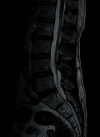
Preoperative magnetic resonance imaging (MRI) scan. This sagittal T2-weighted MRI image shows a hyperintense cystic lesion on the left side at the L4–5 level.
Figure 2
Figure 2
Preoperative magnetic resonance imaging (MRI) scan. This coronal T2-weighted MRI image shows cystic lesions on both sides, with each lesion arising from the right L4–5 facet joint.
Figure 3
Figure 3
Preoperative magnetic resonance imaging (MRI) scan. This axial T2-weighted MRI image shows a cystic lesion (red arrow) arising from the right L4–5 facet joint, causing the narrowing of the right neural foramina. Bilateral facet synovial cysts are also evident.
Figure 4
Figure 4
Preoperative magnetic resonance imaging (MRI) scan. This sagittal T2-weighted MRI image shows a hyperintense cystic lesion on the right side at the L4–5 level.
Figure 5
Figure 5
Preoperative X-ray film. These anteroposterior and lateral views reveal grade 1 spondylolisthesis of L4 over L5.
Figure 6
Figure 6
Preoperative X-ray films. No further instability of L4 over L5 is evident in upright lateral flexion and extension views.
Figure 7
Figure 7
(a) Intraoperative image. It shows the removal of the facet cyst (black arrow) alongside the neural structure (▲). (b) Intraoperative image. No neural structure compression remains after cyst removal (∎).
Figure 8
Figure 8
Postoperative X-ray films at the 1-year follow-up. Increased instability is not apparent in either the anteroposterior or the lateral view.
Figure 9
Figure 9
Postoperative magnetic resonance imaging (MRI) scan at the 1-year follow-up. This axial T2-weighted MRI image confirms the absence of bilateral cysts.
Figure 10
Figure 10
Postoperative magnetic resonance imaging (MRI) scan at the 1-year follow-up. This sagittal T2-weighted MRI image confirms the absence of bilateral cysts.

References

    1. Kouyialis A. T., Boviatsis E. J., Korfias S., Sakas D. E. Lumbar synovial cyst as a cause of low back pain and acute radiculopathy: a case report. Southern Medical Journal . 2005;98(2):223–225. doi: 10.1097/01.SMJ.0000129792.92433.B6. - DOI - PubMed
    1. Trummer M., Flaschka G., Tillich M., Homann C. N., Unger F., Eustacchio S. Diagnosis and surgical management of intraspinal synovial cysts: report of 19 cases. Journal of Neurology, Neurosurgery & Psychiatry . 2001;70(1):74–77. doi: 10.1136/jnnp.70.1.74. - DOI - PMC - PubMed
    1. Ahn Y., Oh H. K., Kim H., Lee S. H., Lee H. N. Percutaneous endoscopic lumbar foraminotomy: an advanced surgical technique and clinical outcomes. Neurosurgery . 2014;75(2):124–133. doi: 10.1227/NEU.0000000000000361. - DOI - PMC - PubMed
    1. Ruetten S., Komp M., Merk H., Godolias G. Full-endoscopic interlaminar and transforaminal lumbar discectomy versus conventional microsurgical technique: a prospective, randomized, controlled study. LWW . 2008;33(9):931–939. doi: 10.1097/BRS.0b013e31816c8af7. - DOI - PubMed
    1. Gadgil A. A., Eisenstein S. M., Darby A., Cassar Pullicino V. Bilateral symptomatic synovial cysts of the lumbar spine caused by calcium pyrophosphate deposition disease: a case report. Spine . 2002;27(19):E428–E431. doi: 10.1097/00007632-200210010-00024. - DOI - PubMed

Publication types

LinkOut - more resources