. 2022 Oct 7;25(11):105308.
doi: 10.1016/j.isci.2022.105308. eCollection 2022 Nov 18.

Transplanted human induced pluripotent stem cells- derived retinal ganglion cells embed within mouse retinas and are electrophysiologically functional

Affiliations

Transplanted human induced pluripotent stem cells- derived retinal ganglion cells embed within mouse retinas and are electrophysiologically functional

Vrathasha Vrathasha et al. iScience. .

Abstract

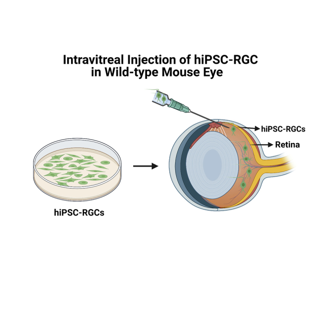
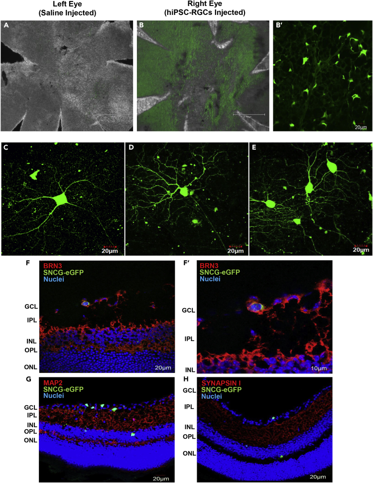
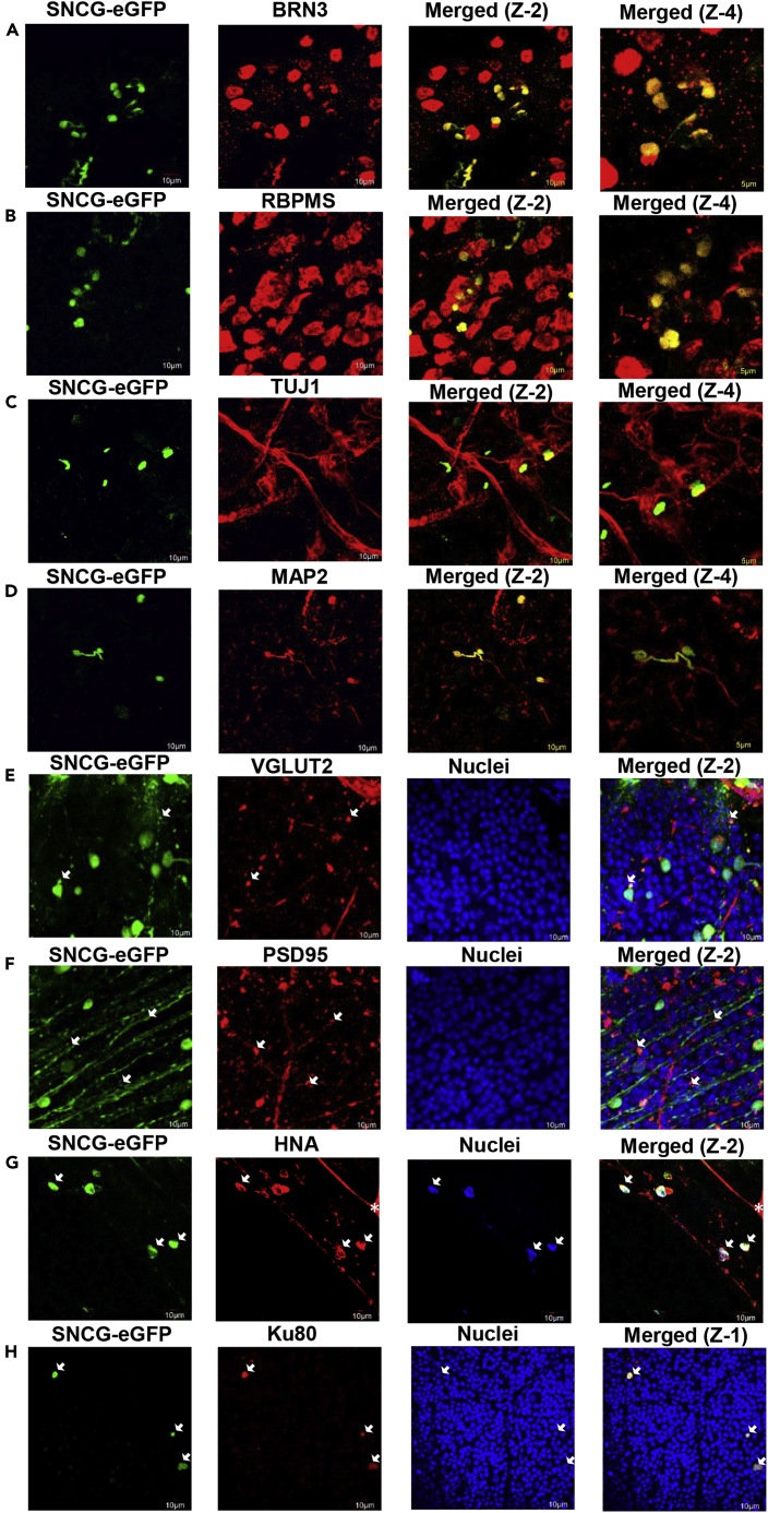
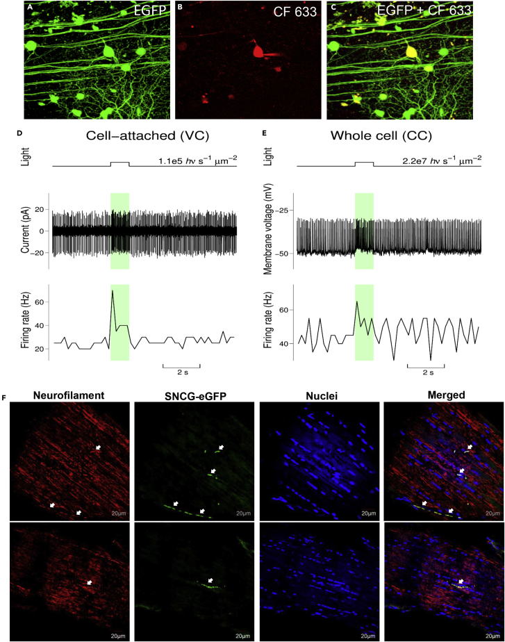
Glaucoma is an optic neuropathy characterized by permanent visual field loss caused by the death of retinal ganglion cells (RGCs) and it is the leading cause of irreversible blindness worldwide. Consequently, there is an unmet need for the development of new strategies for its treatment. We investigated RGC replacement therapy as a treatment for ganglion cell loss. Human-induced pluripotent stem cells (hiPSCs) were differentiated into mature, functional RGCs in vitro, labeled with AAV2.7m8-SNCG-eGFP, and transplanted intravitreally in wild-type 4-month-old C57BL/6J mice. Survival of the transplanted hiPSC-RGCs was assessed by color fundus photography and histological studies confirmed the localization of the transplanted hiPSC-RGCs within the retina. Two-photon live imaging of retinal explants and electrophysiological studies confirmed that the morphology and function of the transplanted hiPSC-RGCs were similar to native RGCs. These experiments will provide key strategies to enhance the efficiency of stem cell replacement therapy for neurodegenerative diseases, including glaucoma.

Keywords: Bioengineering; Biological sciences; Biotechnology; Cell biology; Stem cells research; Tissue engineering.


Conflict of interest statement

All affiliations are listed on the title page of the article. All funding sources for this study are listed in the “acknowledgments” section of the article. We, the authors and our immediate family members have no financial interests to declare. We, the authors and our immediate family members, have no positions to declare and are not members of the journal’s advisory board. We, the authors, have a patent related to this work, which is noted in the “declaration of interests” section of the article and on this form below, and we have noted the patents of immediate family members. “There are restrictions to the availability of the hiPSC-RGCs owing to patent pending on the differentiation protocol.”

Figures

None
Graphical abstract
Figure 1
Figure 1
Images of hiPSC-RGCs expressing SNCG-eGFP after intravitreal injection (A) Fluorescent images of the hiPSC-RGCs transduced with AAV2.7m8 SNCG-eGFP (4X (scale bar is 750 μm) and 10X (scale bar is 300 μm) magnification). (B and C) hiPSC-RGCs transduced with SNCG-eGFP vector were injected into the vitreous space of wild-type C57BL/6J mice. Fundus photography of the transplanted hiPSC-RGCs was taken using BAF-cSLO (scale bar is 200 μm) and SD-OCT at 2-, 4-, and 6-weeks post-transplantation. See Table S2.
Figure 2
Figure 2
Images of transplanted hiPSC-RGCs in vivo (A and B) Fluorescent images of the flattened whole-mount retina of saline-injected left eye and hiPSC-RGCs injected right eye (B = 4X (scale bar is 750 μm), B’ = 10X (scale bar is 20 μm)). (C–E) Neurite outgrowth of transplanted hiPSC-RGCs taken as two-photon images acquired from live retinal samples. Cryosection images of transplanted hiPSC-RGCs (n = 3) were detected in the GCL and INL layer of the murine retina and co-stained for (F) Brn3, (G) MAP2, and (H) Synapsin I. The scale bar of images is 20 μm.
Figure 3
Figure 3
Immuno-staining of the transplanted hiPSC-RGCs in mouse retina with RGC-specific markers (A–H) Flattened whole-mount images of transplanted hiPSC-RGCs (n = 8) were co-stained with RGC-specific markers (A) BRN3, (B) RBPMS, (C) TUJ1, (D) MAP2, (E) VGLUT2, and (F) PSD95. Donor hiPSC-RGCs were also co-stained with (G) human nuclear antigen (HNA) and (H) Ku80 to confirm the donor origin of SNCG-eGFP + cells. ∗ Since the human nuclear antigen antibody was raised in mice, some cross-reactivity with the murine retina was observed. Z-1: Zoom-1 (scale bar is 10 μm), Z-2: Zoom-2 (scale bar is 10 μm), and Z-4: Zoom-4 (scale bar is 5 μm).
Figure 4
Figure 4
Injected hiPSCs integrate into the host retina and generate light responses Two-photon images at the top row show targeted hiPSC-RGCs (centered) before, during, and after recording its light responses. (A) Image shows eGFP fluorescence observed from hiPSC-RGCs in the retinal sample before any attempt to approach it with the recording pipettes. The image is a projection of the Z-stack consisting of 234 optical slices acquired with a 0.5 μm step. (B) The red fluorescence from the CF 633 filled pipette and targeted cell is shown. This image comes from the single optical slice acquired at the end of the patch-clamp recording session. The patch pipette can be seen on the right side of the cell. A slight repositioning of the targeted cell was owing to its re-centering for the IR video system used to control pipette manipulations. (C) The Z-stack projection (151 optical slices with 0.5 μm step) shows combined eGFP and CF 633 fluorescence after the completion of the pipette recording and withdrawal of the patch-pipette. The targeted cell has a bright yellow color. Note that eGFP + hiPSC-RGC processes observed before the patch-clamp recording (panel A) can still be readily identified after its completion (panel C). The scale bar is 50 μm. (D and E) Light responses of the targeted cell recorded in the cell-attached configuration (voltage-clamp mode) and whole-cell configuration (current-clamp mode) are illustrated (n = 4). Here upper graphs give the time course of light stimuli and indicate flash intensities, the middle graphs plot current vs. time and membrane voltage vs. time traces (panels D and E respectively), and the bottom graphs plot firing rate vs time traces. The current vs. time trace from panel D has been high pass filtered, and the voltage vs. time trace from panel E has not been corrected for the LJP, the correction can be done by subtracting 15 mV from the reported values. The firing rate traces were calculated using a 200 ms time bin. The colored light bars indicate light stimulation events. See also Figures S1–S4. (F) Detection of transplanted hiPSC-RGCs in the optic nerve of the mouse and its expression co-localized with the neurofilament marker (n = 2). The scale bar is 20 μm.

References

    1. Aoki H., Hara A., Niwa M., Motohashi T., Suzuki T., Kunisada T. An in vitro mouse model for retinal ganglion cell replacement therapy using eye-like structures differentiated from ES cells. Exp. Eye Res. 2007;84:868–875. - PubMed
    1. Banin E., Obolensky A., Idelson M., Hemo I., Reinhardtz E., Pikarsky E., Ben-Hur T., Reubinoff B. Retinal incorporation and differentiation of neural precursors derived from human embryonic stem cells. Stem Cells (Dayton, Ohio) 2006;24:246–257. - PubMed
    1. Bellocchio E.E., Reimer R.J., Fremeau R.T., Jr., Edwards R.H. Uptake of glutamate into synaptic vesicles by an inorganic phosphate transporter. Science (New York, N.Y.) 2000;289:957–960. - PubMed
    1. Berg S., Kutra D., Kroeger T., Straehle C.N., Kausler B.X., Haubold C., Schiegg M., Ales J., Beier T., Rudy M., Eren K., Cervantes J.I., Xu B., Beuttenmueller F., Wolny A., Zhang C., Koethe U., Hamprecht F.A., Kreshuk A. Ilastik: interactive machine learning for (Bio)image analysis. Nat. Methods. 2019;16:1226–1232. - PubMed
    1. Chaffiol A., Caplette R., Jaillard C., Brazhnikova E., Desrosiers M., Dubus E., Duhamel L., Macé E., Marre O., Benoit P., Hantraye P., Bemelmans A.P., Bamberg E., Duebel J., Sahel J.A., Picaud S., Dalkara D. A new promoter allows optogenetic vision restoration with enhanced sensitivity in macaque retina. Mol. Ther. 2017;25:2546–2560. - PMC - PubMed