

The value of dynamic MRI in cervical spondylotic myelopathy: About 24 cases
- PMID: 36389194
- PMCID: PMC9661660
- DOI: 10.1016/j.amsu.2022.104717
The value of dynamic MRI in cervical spondylotic myelopathy: About 24 cases
Abstract
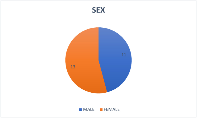
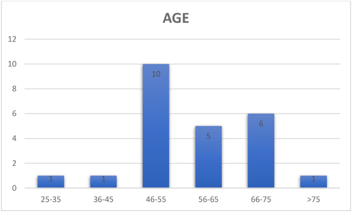
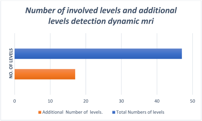
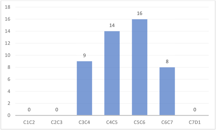
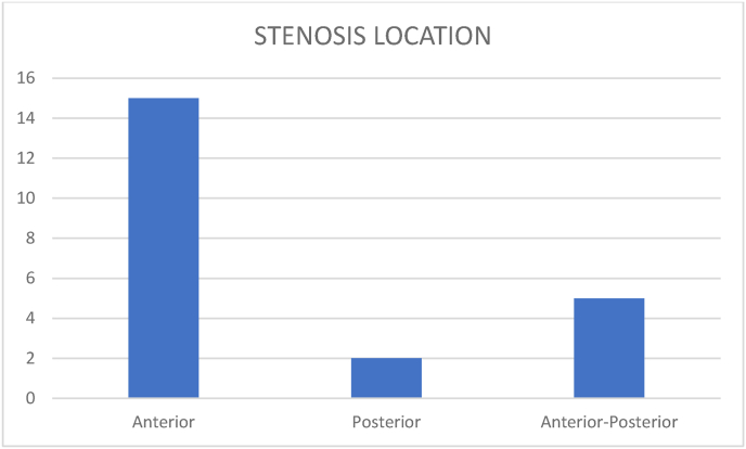
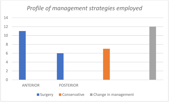
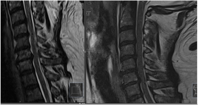
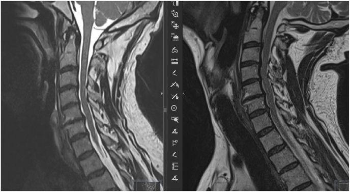
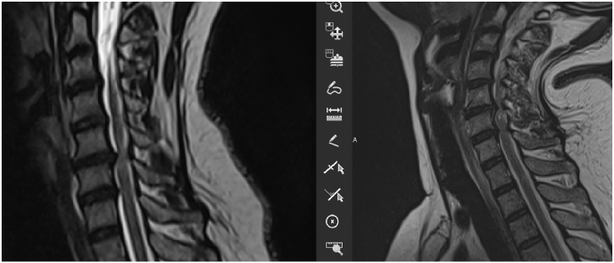
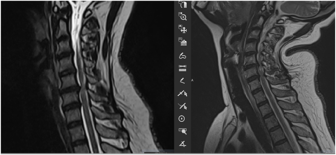
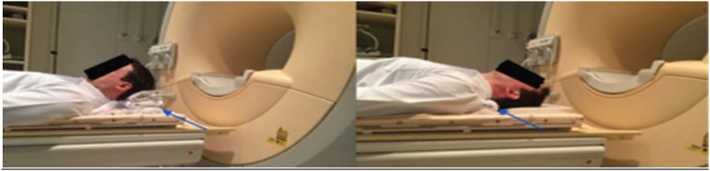
Dynamic magnetic resonance imaging (MRI) of the cervical spine is extremely useful in assessing pathological changes at the spinal cord, vertebrae, discs, ligaments and facet joints. We attempted to document the radiological changes that the cervical spine undergoes during dynamic maneuvers and the effects of Dynamic MRI in management of cervical myelopathy, emphasizing on the changes in treatment protocol effected by the new findings discovered. Our work is based on 24 consecutive patients with cervical spondylotic myelopathy had cervical MR imaging in neutral position, in flexion and extension of the cervical spine between January 2021 and December 2021. The result found the mean age was 57.9 years (range 26-85 years). Among these 24 patients, there were 11 males and 13 females. Total number of levels of compression were 47 and the additional levels of involvement were 17. Additional levels of compression were noted in 12 patients, among these 17 new levels, 7 were in the posterior and 10 in the anterior. The most affected level was C5C6 with 16 cases. All additional levels of compression were noted in extension; Reduction of the cervical canal was observed in 20 patients only in extension. In the bending sequences we have noticed an increase of the canal diameter in 3 patients. The location of the compression is in 15 cases anterior, 2 cases posterior and 5 cases are mixed anterior and posterior Surgery was considered in 17 patients. Anterior procedures were 11 (ACDF/corpectomy and fusion) and Posterior surgeries were 6 (laminoplasty/laminectomy), and. The rest of the patients did not require surgery and was conservatively treated. A change of the signal was found in 3 patients during the acquisition in extension position a. Most studies have shown a reduction of the root canal with an increase of the compression level, which was the case in our study. MRI is a useful tool for diagnosis of CM, it does not give an exact idea as to which is the offending level in a multilevel compression that requires surgery. Even the approach and procedure cannot be decided on a static examination and hence are subject to significant interpractitioner the role of extension MRI in determining cervical compression levels. Thus, dynamic cervical spine MRI should be an important investigation before we decide to write off surgical treatment in patients with cervical myelopathy and cord signal changes without definitive compression on static MRI. Flexion and extension MRI is an important tool for decision making and planning appropriate management in cervical compressive myelopathy.
Keywords: Case series; Cervical myelopathy; Cervical spondylosis; Dynamic; Magnetic resonance imaging; Spinal canal.
© 2022 Published by Elsevier Ltd on behalf of IJS Publishing Group Ltd.
Conflict of interest statement
The authors declare having no conflicts of interest for this article.
Figures
References
-
- Sibhi Ganapathy*, Venkataramakrishna Tukapuram, Nikunj Godhani, Swaroop Gopal. Dynamic mri of the cervical spine – an important tool in planning surgical treatment of cervical compressive Myelopathy International Journal of Neurosurgery2018; 2(1): 17-22.
-
- Lei Zhang, Delphine Zeitoun, Alfonso Rangel, Jean Yves Lazennec, Yves Catonné, and Hugues Pascal-Moussellard. Preoperative Evaluation of the Cervical Spondylotic Myelopathy with Flexion-Extension Magnetic Resonance Imaging SPINE Volume vol. 36, Number 17, pp E1134–E1139. - PubMed
-
- Dalbayrak Sedat, Yaman Onur, Fırıdı Mustafa Nevzat, Yılmaz Tevfik, Yılmaz Mesut. The contribution of cervical dynamic magnetic resonance imaging to the surgical treatment of cervical spondylotic myelopathy. Turk Neurosurg. 2015;25(1):36–42. - PubMed
Further reading
-
- Raphael R. Pratali, Justin S. Smith,y Bruno C. Ancheschi, Daniel A. Maranho, ,z Aniello Savarese, z Marcello H. Nogueira-Barbosa, and Carlos Fernando P.S. Herrero. A technique for dynamic cervical magnetic resonance imaging applied to cervical spondylotic myelopathy. Spine Vol 44, Number 1, pp E26–E32. - PubMed
-
- Delphine Zeitoun , Firass El Hajj , Elhadi Sariali , Yves Catonné , Hugues Pascal- Moussellard. Evaluation of spinal cord compression and hyperintense intramedullary lesions on T2- weighted sequences in patients with cervical spondylotic myelopathy using flexion-extension MRI protocol. The Spine Journal S1529 9430(14)01771-01779 DOI: 10.1016/j.spinee.2014.12.001. - PubMed
LinkOut - more resources
Full Text Sources
