. 2022 Dec;74(6):623-634.
doi: 10.1007/s10616-022-00549-9. Epub 2022 Sep 30.

Reduced immunogenicity of β-lactoglobulin by single amino acid substitution

Affiliations

Reduced immunogenicity of β-lactoglobulin by single amino acid substitution

Tadashi Yoshida et al. Cytotechnology. 2022 Dec.

Abstract

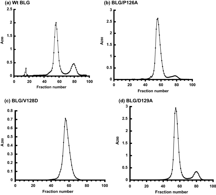
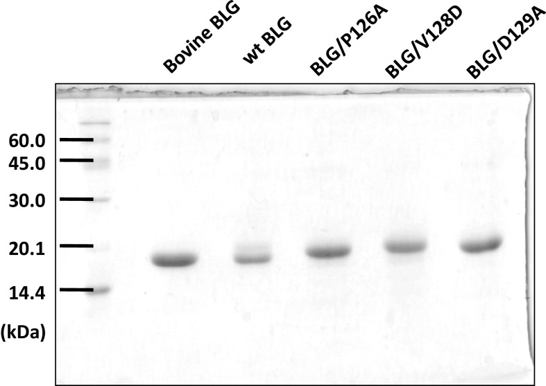
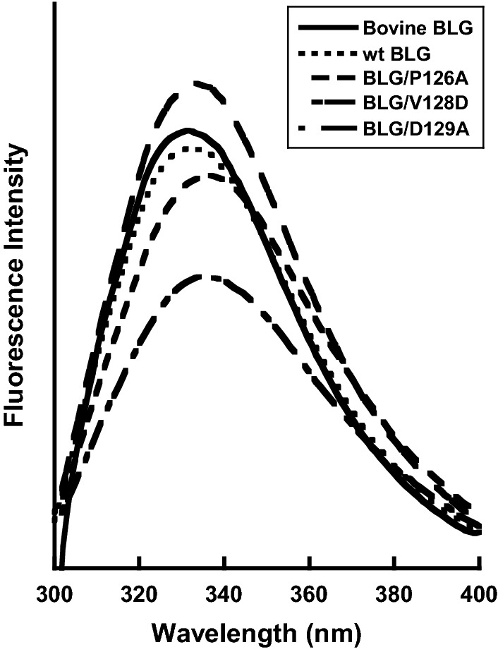
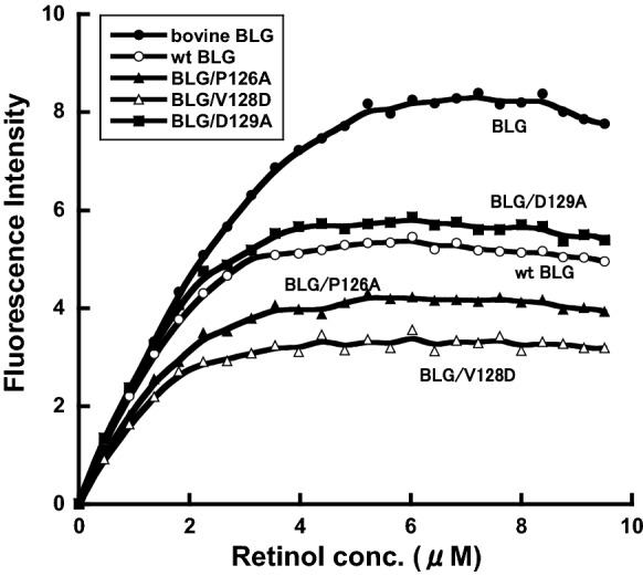
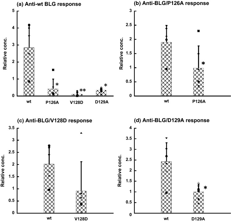
To reduce the immunogenicity of β-lactoglobulin (BLG), we prepared single amino acid substituted recombinant BLG mutants (BLG/P126A, BLG/V128D and BLG/D129A) in the methylotrophic yeast Pichia Pastris by fusion of the cDNA to the sequence coding for the α-factor signal peptide from Saccharomyces cerevisiae. Isoelectric points of single amino acid substituted BLGs were lower than that of native BLG. CD spectra indicated that the secondary structure of BLG had maintained native structure in single amino acid substituted BLGs. Fluorescence studies indicated that the conformation around Trp had not changed in single amino acid substituted BLGs. Anti-BLG antibody response was evaluated after immunization to C57BL/6 mice. Antibody response was reduced after immunization with BLG/P126A, BLG/V128D and BLG/D129A. And novel immunogenicity was not observed in the experiments. T cell proliferative response was evaluated in C57BL/6 mice, and it was clarified that BLG mutants also showed low response. Methods employed in this study was considered to be very effective to reduce immunogenicity of BLG.

Keywords: Protein engineering; Reduced immunogenicity; Single amino acid substitution; β-Lactoglobulin.


Conflict of interest statement

Conflict of interestThe authors have no conflicts of interest to declare.

Figures

Fig. 1
Fig. 1
Anion-exchange chromatogram of the mutant BLGs. a wt BLG, b BLG/P126A, c BLG/V128D, d BLG/D129A. Conditions: column, DEAE Sepharose Fast Flow (GE Healthcare, Buckinghamshire, UK); column size, 2.5 ID × 50 cm; flow rate, 5 mL/min; elution, 0–1.0 M NaCl. To detect the recombinant protein, the absorbance was monitored at 280 nm
Fig. 2
Fig. 2
SDS–PAGE analysis of the purified mutant BLGs. The mutant BLGs (wt, BLG/P126A, BLG/V128D, BLG/D129A) were stained by Coomassie blue. (Color figure online)
Fig. 3
Fig. 3
IEF pattern of mutant BLGs. IEF was carried out with Phast Gel System (GE Healthcare, Buckinghamshire, UK) and Phast Gel IEF 4–6.5 (GE Healthcare, Buckinghamshire, UK). Lane 1 and 7, Isoelectric Focusing Calibration Broad pI (pI 3–10); lane 2, BLG/D129A; lane 3, BLG/V128D; lane 4, BLG/P126A; lane 5, wt BLG; lane 6, bovine BLG
Fig. 4
Fig. 4
CD spectra of the mutant BLGs. CD spectra of BLG, wt, BLG/P126A, BLG/V128D, BLG/D129A were measured with a spectropolarimeter, using a cell with a 1.0 mm path length. Protein concentration was 0.01% in PBS
Fig. 5
Fig. 5
Intrinsic fluorescence spectra of the mutant BLGs. The intrinsic fluorescence spectra of BLG, wt, BLG/P126A, BLG/V128D, BLG/D129A were measured under an excitation wavelength of 283 nm. Protein concentration was 0.01% in PBS
Fig. 6
Fig. 6
Retinol-binding activity of the mutant BLGs. The retinol-binding activities of BLG, wt, Fig. 3 CD spectra of the mutant BLGs
Fig. 7
Fig. 7
Immunogenicity of the mutant BLGs in C57BL/6 mice. The anti-wtBLG (a), anti-BLG/P126A (b), anti-BLG/V128D (c), anti-BLG/D129A (d) response after the secondary immunization were evaluated by noncompetitive ELISA. Markers indicate individual data and bars indicate average values. A significant difference compared with anti wt serum as determined by Student’s t-test is indicated by single asterisk (p < 0.05) and two asterisk (p < 0.01)
Fig. 8
Fig. 8
Proliferative response to BLG of lymph node cells from C57BL/6 mice immunized with BLG (●), BLG/P126A (▲), BLG/V128D (△) or BLG/D129A (■). A significant difference compared with anti wt response as determined by Student’s t-test is indicated by single asterisk (p < 0.05) and two asterisk (p < 0.01)

References

    1. Armstrong JM, McKenzie HA, Sawyer WH. On the fractionation of beta-lactoglobulin and alpha-lactalbumin. Biochim Biophys Acta. 1967;147:60–72. doi: 10.1016/0005-2795(67)90090-6. - DOI - PubMed
    1. Brownlow S, Cabral JH, Cooper R, Flower DR, Yewdall SJ, Polikarpov I, North AC, Sawyer L. Bovine β-lactoglobulin at 1.8 Å resolution–still an enigmatic lipocalin. Structure. 1997;5:481–495. doi: 10.1016/s0969-2126(97)00205-0. - DOI - PubMed
    1. Chen FM, Lee JH, Yang YH, Lin YT, Wang LC, Yu HH, Chiang BL. Analysis of α-lactalbumin-, β-lactoglobulin-, and casein-specific IgE among children with atopic diseases in a tertiary medical center in northern Taiwan. J Microbiol Immunol Infect. 2014;47:130–136. doi: 10.1016/j.jmii.2012.08.009. - DOI - PubMed
    1. Cogan U, Kopelman M, Mokady S, Shintzky M. Binding affinities of retinol and related compounds to retinol binding proteins. Eur J Biochem. 1976;65:71–78. doi: 10.1111/j.1432-1033.1976.tb10390.x. - DOI - PubMed
    1. Flower DR. The lipocalin protein family: structure and function. Biochem J. 1996;318:1–14. doi: 10.1042/bj3180001. - DOI - PMC - PubMed

LinkOut - more resources