Review
. 2022 Nov;12(11):e949.
doi: 10.1002/ctm2.949.

When the clock ticks wrong with COVID-19

Affiliations
Review

When the clock ticks wrong with COVID-19

Silvana Papagerakis et al. Clin Transl Med. 2022 Nov.

Abstract

Severe acute respiratory syndrome coronavirus 2 (SARS-CoV-2) is a member of the coronavirus family that causes the novel coronavirus disease first diagnosed in 2019 (COVID-19). Although many studies have been carried out in recent months to determine why the disease clinical presentations and outcomes can vary significantly from asymptomatic to severe or lethal, the underlying mechanisms are not fully understood. It is likely that unique individual characteristics can strongly influence the broad disease variability; thus, tailored diagnostic and therapeutic approaches are needed to improve clinical outcomes. The circadian clock is a critical regulatory mechanism orchestrating major physiological and pathological processes. It is generally accepted that more than half of the cell-specific genes in any given organ are under circadian control. Although it is known that a specific role of the circadian clock is to coordinate the immune system's steady-state function and response to infectious threats, the links between the circadian clock and SARS-CoV-2 infection are only now emerging. How inter-individual variability of the circadian profile and its dysregulation may play a role in the differences noted in the COVID-19-related disease presentations, and outcome remains largely underinvestigated. This review summarizes the current evidence on the potential links between circadian clock dysregulation and SARS-CoV-2 infection susceptibility, disease presentation and progression, and clinical outcomes. Further research in this area may contribute towards novel circadian-centred prognostic, diagnostic and therapeutic approaches for COVID-19 in the era of precision health.

Keywords: COVID-19; SARS-CoV-2 infection; circadian clock; clinical outcomes; epigenetics; microRNAs; oral and systemic precision health; personalized medicine.


Conflict of interest statement

The authors declare no conflict of interest.

Figures

FIGURE 1
FIGURE 1
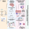
Circadian clock – a determinant of immune defence and its links to coronavirus disease 2019 (COVID‐19). Both innate and adaptive immune systems are modulated by the circadian clock at almost every level. This is a schematic representation of the known aspects of monocyte and macrophage (A), lymphocyte (B) and neutrophil (C) that are regulated by the circadian clock. (A) Macrophages and monocytes have been the most studied cell types in the context of circadian rhythms. Macrophages exhibit a high amplitude of clock gene expression that appear to modulate several macrophage functions, including phagocytosis, the expression of pattern‐recognition receptors (PRRs), recruitment to tissues, mitochondrial dynamics and cytokines in response to a challenge48, , , , , suggesting that circadian misalignment is involved in the acquiring of the hyperactivated pulmonary macrophage phenotype observed in COVID‐19 patients resulting in a damaging loop of pro‐inflammatory cytokine release and recruitment of other cytotoxic effector cells thereby exacerbating tissue damage. (B) Both B and T cells in mouse lymph nodes express several clock in a rhythmic manner. In human blood, T‐ and B‐cell numbers varied throughout the day and were found to be higher at night and then declined in the morning. A circadian rhythm of cytokine production (IL‐2, IFN‐γ, IL‐10 and TNF‐α) was also observed after the TCR stimulation of T helper cells in vitro. Both CD4+ and CD8+ T‐cell proliferation after antigenic stimulation showed circadian rhythms with stronger proliferation during the late subjective day and during the subjective night. The circadian control of lymphocyte proliferation and cytokine production may be involved in the lymphocytopenia observed in patients with COVID‐19 where there is a marked reduction in the CD4+ T lymphocyte number and elevated levels of TNF‐α and IL‐6 that correlate with the severity of COVID‐19 disease. (C) Peripheral neutrophils also express components of the molecular clock, and endotoxin administration was found to downregulate clock genes (Clock, Per3, Cry1‐2, Rev‐erb and Rora) expression in human neutrophils. Daily variations in neutrophil count and functions, such as superoxide production (i.e. rhythmic Gp91phox expression), phagocytosis and expression of cell adhesion molecules (e.g. l‐selectin, ICAM1 and LFA‐1), have been described in several studies, suggesting a significant impact of the circadian clock on neutrophil activity. The previous description suggests that the circadian disruption may exacerbate the altered neutrophil abundance, phenotype and functionality observed in COVID‐19 patients that include elevated neutrophil levels, increased degranulation reduced reactive oxygen species release and heightened capacity for neutrophil associated extracellular trap formation 249 ). Some disagreement regarding the role of circadian clock in immunity are raised and more studies are needed. Source: BioRender.com
FIGURE 2
FIGURE 2
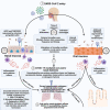
Reciprocal links between coronavirus disease 2019 (COVID‐19) and different melatonergic pathways suggestive of melatonin as a viable therapeutic target and chronomodulator of COVID‐19 infection. (1) Severe acute respiratory syndrome coronavirus 2 (SARS‐CoV‐2) entry into host cells results in a pro‐inflammatory cytokine storm that is further exacerbated by the constitutive activation of the renin–angiotensin activating system along with the hypothalamic pituitary adrenal axis due to angiotensin II (Ang II) accumulation and the loss of surface angiotensin converting enzyme. (2) The pro‐inflammatory cytokines may suppress melatonin production in the pineal gland via the immune pineal axis and increased cytokine levels lead to the activation of the indoleamine 2,3‐dioxygenase (IDO) and tryptophan 2,3‐dioxygenase (TDO) enzymes. IDO/TDO activation promotes the degradation of tryptophan into kynurenine and reduces the rate of tryptophan conversion into melatonin. Melatonin metabolism will be also affected by cytokine‐mediated gut dysbiosis and impaired gut permeability, which may lead to reduced uptake of tryptophan and the microbiome‐derived short chain fatty acid butyrate accompanied by increased permeability to lipopolysaccharide (LPS) which can induce further cytokine production. (3) This results in a marked decrease in melatonin levels and the corresponding loss of its anti‐inflammatory, anti‐oxidative, immune enhancing and cytoprotective effects, which are largely mediated via the induction of Bmal1 disinhibition of the mitochondrial pyruvate decarboxylase–acetyl CoA and inhibition of the pro‐inflammatory NF‐κB pathway. Moreover, the altered melatonin levels will disrupt the sleep/wake cycles of COVID‐19 patients which will further increase their susceptibility. All the previously mentioned developments are expected to increase the severity of inflammation and worsen disease progression. ACE2, angiotensin converting enzyme 2; BMAL1, brain and muscle ARNT‐like 1; HPA, hypothalamic pituitary adrenal; MTN, melatonin; MTN‐R, melatonin receptors; NF‐κB, nuclear factor kappa‐light‐chain‐enhancer of activated B cells; PDC, pyruvate decarboxylase; RAAS, renin–angiotensin aldosterone system; Sirt, Sirtuin. Source: BioRender.com
FIGURE 3
FIGURE 3
Potential links between angiotensin‐converting enzyme (ACE)/angiotensin‐converting enzyme 2 (ACE2) expression in epithelial cells and circadian clock gene expression in the context of coronavirus disease 2019 (COVID‐19). Targeting the circadian regulator Bmal1 by genetic silencing or by treating lung epithelial cells with the REV‐ERB agonist SR9009 or Cry stabilizer KL001 reduces ACE2 expression and inhibits severe acute respiratory syndrome coronavirus 2 (SARS‐CoV‐2) entry and replication. Binding of the surface S glycoprotein of SARS‐CoV‐2 to ACE2 may directly compete with the processing of the pro‐inflammatory angiotensin II (Ang II) to the anti‐inflammatory Ang (1–7). Ang II is known to affect the central clock system in the suprachiasmatic nuclei (SCN) through its effect on Per2 levels which may in turn affects the expression of BMAL1 in peripheral tissues, including the pulmonary epithelium. Source: BioRender.com
FIGURE 4
FIGURE 4
Circadian clock, coronavirus disease 2019 (COVID‐19) and the microbiome. (1) Severe acute respiratory syndrome coronavirus 2 (SARS‐CoV‐2) entry may happen through the oral and nasal cavities. The oral microbiota has been shown to exhibit a strong circadian behaviour influenced by multiple internal and external circadian factors, whereas the circadian behaviour of the nasal microbiome is still understudied. It has been reported that the metabolic activity of the majority of oral microbes exhibits a clear diurnal rhythm that corresponds to the aerobicity of the microbes. Thus, different exposure times to the virus may result in different susceptibilities to viral entry as the microbiotal profile changes during the day. (2) The viral entry may cause oral and/or microbiotal dysbiosis. Several other risk factors are thought to facilitate oral pathogens’ entrance into the lower respiratory tree through the anatomical oral/nasal/lung axis such as poor oral hygiene, periodontal disease, respiratory health status, environmental and/or iatrogenic factors. Although little is known about the link between oral microbiota and the severity of COVID‐19 infection, microbial sequencing studies of the bronchoalveolar fluid (BALF) of COVID‐19 patients revealed that several oral opportunistic pathogens such as Capnocytophaga and Veillonella were found in the lung of COVID‐19 patients, suggesting that the oral cavity could be the source of the lung co‐infections observed in COVID‐19 patients. It has also been suggested that the bidirectional interactions between gut microbiota and the respiratory mucosa through the physiological gut–lung axis may be involved in regulating the immune responses to SARS‐CoV‐2, which may involve the circadian clock. Indeed, changes in the host microbiome can be integrated into the host circadian via several pathways, including changes in microbial pattern‐recognition receptors (PRR) signalling. Gut dysbiosis due to viral infections can affect the diurnal release and absorption of microbiome‐derived metabolites, including free fatty acids that will consequently affect the circadian metabolic activity of peripheral organs, including the immune system. Furthermore, it has been shown that the microbiome may influence the circadian clock via epigenetic modification through the action of microbial histone deacetylase. Finally, the microbiome greatly affects the diurnal release of several hormones, including growth hormones and sex hormones. (3) Oral and/or nasal dysbiosis may affect COVID‐19 susceptibility, severity and outcomes. ACE‐2, angiotensin converting enzyme 2; FFA, free fatty acids; HDAC, histone deacetylase; TLR, toll‐like receptor; TMPRSS2, transmembrane protease serine 2. Source: BioRender.com
FIGURE 5
FIGURE 5
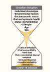
Determinants of the individual circadian clock profile and coronavirus disease 2019 (COVID‐19) disease heterogeneity. Misalignment of the behavioural (fasting/feeding and sleep/wake pattern) and light/dark cycles can lead to circadian disruption; the circadian profile may display a high individual heterogeneity reflective of a combination of various determinants (including age, sex, socioeconomic status), genetic heterogeneity, individual chronotype, oral and systemic health status (e.g. comorbidities, microbiome composition), lifestyle, geographical location and environmental factors and so on that can impact the susceptibility, severity and prognosis of COVID‐19‐related disease. Source: BioRender.com

References

    1. Sanders JM, Monogue ML, Jodlowski TZ, Cutrell JB. Pharmacologic treatments for coronavirus disease 2019 (COVID‐19): a review. JAMA – J Am Med Assoc. 2020;323:1824‐1836. 10.1001/jama.2020.6019 - DOI - PubMed
    1. Dong E, Du H, Gardner L. An interactive web‐based dashboard to track COVID‐19 in real time. Lancet Infect Dis. 2020;20:533‐534. 10.1016/S1473-3099(20)30120-1/ATTACHMENT/9DE52FFD-61D4-4C3F-826F-841D978D469D/MMC1.PDF - DOI - PMC - PubMed
    1. Dong Y, Mo X, Hu Y, et al. Epidemiology of COVID‐19 among children in China. Pediatrics. 2020;145:e20200702. - PubMed
    1. Hou Y, Zhao J, Martin W, et al. New insights into genetic susceptibility of COVID‐19: an ACE2 and TMPRSS2 polymorphism analysis. BMC Med. 2020;18:1‐8. 10.1186/s12916-020-01673-z - DOI - PMC - PubMed
    1. Ray S, Reddy AB. COVID‐19 management in light of the circadian clock. Nat Rev Mol Cell Biol. 2020;21:494‐495. 10.1038/s41580-020-0275-3 - DOI - PMC - PubMed

Publication types