. 2022 Dec 20;204(12):e0029022.
doi: 10.1128/jb.00290-22. Epub 2022 Nov 17.

Roles and Organization of BxpB (ExsFA) and ExsFB in the Exosporium Outer Basal Layer of Bacillus anthracis

Affiliations

Roles and Organization of BxpB (ExsFA) and ExsFB in the Exosporium Outer Basal Layer of Bacillus anthracis

Jorge Durand-Heredia et al. J Bacteriol. .

Abstract

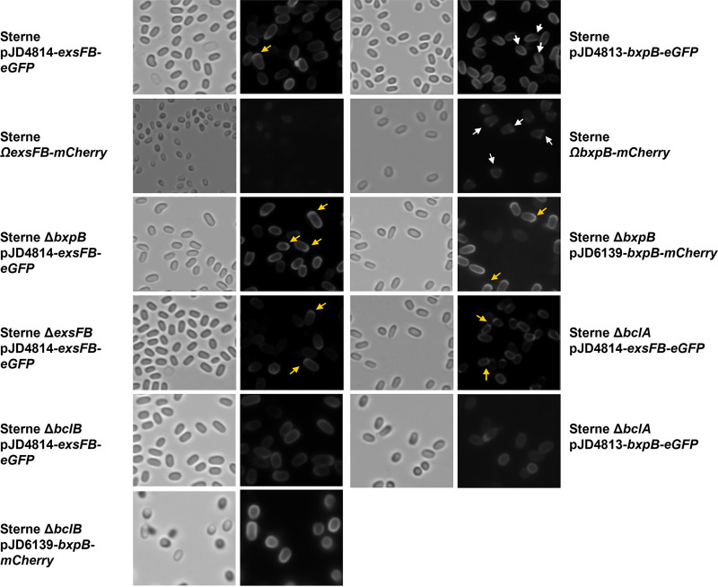
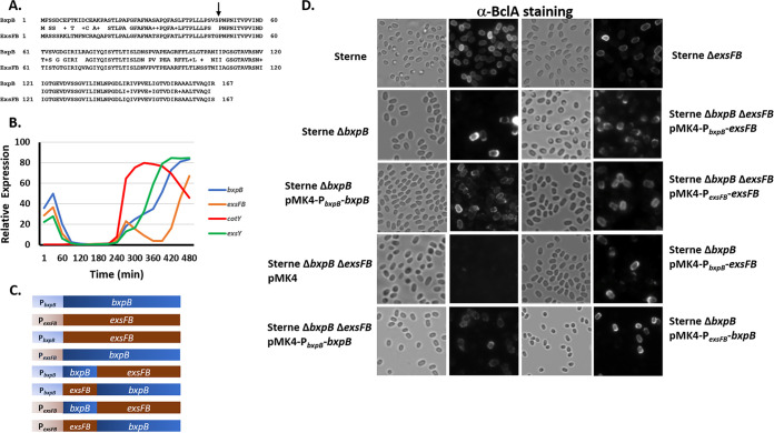
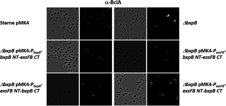
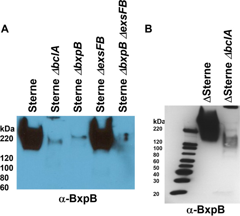
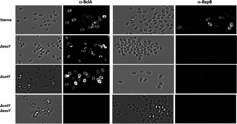
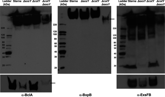
BxpB (also known as ExsFA) and ExsFB are an exosporium basal layer structural protein and a putative interspace protein of Bacillus anthracis that are known to be required for proper incorporation of the BclA collagen-like glycoprotein on the spore surface. Despite extensive similarity of the two proteins, their distribution in the spore is markedly different. We utilized a fluorescent fusion approach to examine features of the two genes that affect spore localization. The timing of expression of the bxpB and exsFB genes and their distinct N-terminal sequences were both found to be important for proper assembly into the exosporium basal layer. Results of this study provided evidence that the BclA nap glycoprotein is not covalently attached to BxpB protein despite the key role that the latter plays in BclA incorporation. Assembly of the BxpB- and ExsFB-containing outer basal layer appears not to be completely abolished in mutants lacking the ExsY and CotY basal layer structural proteins despite these spores lacking a visible exosporium. The BxpB and, to a lesser extent, the ExsFB proteins, were found to be capable of self-assembly in vitro into higher-molecular-weight forms that are stable to boiling in SDS under reducing conditions. IMPORTANCE The genus Bacillus consists of spore-forming bacteria. Some species of this genus, especially those that are pathogens of animals or insects, contain an outermost spore layer called the exosporium. The zoonotic pathogen B. anthracis is an example of this group. The exosporium likely contributes to virulence and environmental persistence of these pathogens. This work provides important new insights into the exosporium assembly process and the interplay between BclA and BxpB in this process.

Keywords: Bacillus anthracis; BclA; exosporium; immunofluorescence; promoters; protein assembly; spore.


Conflict of interest statement

The authors declare no conflict of interest.

Figures

FIG 1
FIG 1
Bright-field and epifluorescence micrographs of spores from Sterne, ΔbxpB, ΔexsFB, and ΔbclA cells producing the BxpB-mCherry, BxpB-eGFP, or the ExsFB-eGFP fusion proteins expressed from plasmids or in a single copy at the amyS locus (denoted by Ω). Yellow arrows indicate examples of enhanced fluorescence at a spore pole, and white arrows indicate examples of reduced fluorescence at a spore pole.
FIG 2
FIG 2
Effect of expression of BxpB and ExsFB on BclA spore surface distribution. (A) Amino acid sequence alignment of BxpB and ExsFB with sequence identities and similarities (denoted by plus sign) indicated. The arrow denotes the position where the chimeric proteins were fused. (B) Microarray-based RNA expression profiles of the exosporium determinants bxpB, cotY, exsY, and exsFB during the in vitro growth and sporulation cycle. Microarray data were from Bergman et al. (43). (C) Representations of the full-length and chimeric bxpB and exsFB determinants expressed from either the bxpB or exsFB promoters. (D) Bright-field and anti-BclA fluorescence of spores of the Sterne parent strain and the ΔbxpB-, ΔexsFB-, and ΔbxpB ΔexsFB-null mutants bearing the plasmid-borne bxpB or exsFB determinants expressed from their native or swapped promoter sequences where indicated. Spores were reacted with anti-BclA polyclonal serum and goat anti-rabbit-Alexa Fluor 568 secondary antibodies.
FIG 3
FIG 3
Bright-field and anti-BclA fluorescence of spores of the Sterne parent strain and the ΔbxpB mutant bearing the plasmid-borne bxpB or exsFB chimeric determinants expressed from the bxpB or exsFB promoter elements. Spores were reacted with anti-BclA polyclonal serum and goat anti-rabbit-Alexa Fluor 568 secondary antibodies.
FIG 4
FIG 4
(A) Western blotting of spore extracts probed with antiserum against mCherry (left) or BxpB (right). The blot was probed with anti-mCherry and then stripped and reprobed with anti-BxpB. The arrow indicates the position of the larger anti-mCherry-reactive species in the Sterne ΔbclA pMK4 bclA-NTD-mcherry lane. (B) Strategy for bclA-NTD-his12 labeling of the BclA partner protein. The BclA NTD sequence (red bar represents residues 1 to 19, and black bar represents residues 20 to 35) with His12 residues at the C terminus was expressed in B. anthracis. During assembly around the spore (20), a proteolytic cleavage event removes the N-terminal 19 residues, with the remaining NTD sequences being stably attached to a partner protein in the exosporium (represented by the blue circle). (C) Western blotting of spore extracts probed with antiserum against the His tag. The pGS4900 clones 1 and 2 refer to two independent isolates bearing the plasmid. The size of the BclA-NTD-His12 protein is 5,287 daltons (Da) (3,290 Da when cleaved). A 35-kDa species was detected with the anti-His tag antiserum, and higher-MW SDS-resistant complexes are evident as well.
FIG 5
FIG 5
Impact of BclA on BxpB incorporation into the exosporium basal layer. (A) Western blot of Sterne spore extracts probed with antiserum against BxpB. (B) Western blot of Delta Sterne spore extracts probed with antiserum against BxpB. The faint band in the Delta bxpB lanes is due to cross reactivity with ExsFB.
FIG 6
FIG 6
Immunofluorescence of spores from Sterne, Sterne ΔexsY, Sterne ΔcotY, and Sterne ΔcotY ΔexsY treated with antiserum against BclA or BxpB. Left-hand images are bright-field images, and the right-hand images of each pair are fluorescent images of the same field.
FIG 7
FIG 7
Western blotting of spore extracts probed with rabbit polyclonal antiserum against rBclA (left), rBxpB (middle), and rExsFB (right). Strain designations of the spore extracts are indicated above the image of the blot. The bottom panels are from the high-molecular-weight region of the blots from a shorter exposure time of a comparable gel. The arrows in the bottom anti-BclA and the anti-BxpB panels indicate the position of the reactive species in the ΔcotY ΔexsY spore extract lane, which migrate differently from the high-MW complexes seen in the extracts from the single-mutant spores.
FIG 8
FIG 8
Assembly of the outer basal layer in the absence of the ExsY-CotY layer. (A) Spores from a triple null mutant of B. anthracis that lack CotE, CotY, and ExsY were reacted with anti-BclA, anti-BxpB, and anti-ExsFB antiserum. Fluorescent, bright-field, and merged images are shown. Red arrows in the bright-field image indicate positions of the putative basal layer casings that reacted with the antiserum. Blue arrows denote spores that reacted with the antiserum. (B) Transmission electron micrographs of the spore preparations from the cotE cotY exsY triple deletion mutant illustrating the presence of the empty vesicles (examples indicated with purple arrows). (C) Western blotting of spore extracts probed sequentially with antiserum directed against rBclA, rBxpB, and rExsFB.
FIG 9
FIG 9
Bacterial two-hybrid analysis of BxpB and ExsFB interactions. β-Galactosidase assays were conducted in E. coli bearing compatible plasmids harboring the T18 and T25 adenylate cyclase domains (44). The fusions were to BxpB (designated B) and ExsFB (designated EF). B-T25 signifies a BxpB fusion with the T25 domain fused at its C terminus, whereas T18-EF indicates ExsFB with the T18 domain fused at its N terminus. The positive control (+) was a strain containing plasmids with the zip sequence fused to each of the adenylate cyclase domains (44), and the negative control (−) was a strain bearing two fusions with B. anthracis proteins that we have shown not to be interactive in this assay (the exosporium protein ExsY and a putative nucleotide sugar dehydrogenase subfamily protein encoded on the pXO1 plasmid; locus tag AW20_5669). β-Galactosidase activity is expressed in Miller units. Controls are shown as gray bars, BxpB-BxpB interactions as blue bars, ExsFB-ExsFB as red bars, and BxpB-ExsFB as blue-and-red checkerboard bars. The error bars signify the standard deviation of ≥3 independent assays. An asterisk denotes the statistical significance (P < 0.05) for the sample relative to the negative control.
FIG 10
FIG 10
Western blotting of recombinant BxpB and ExsFB isolated from E. coli and stored in PBS. (A) rBxpB incubated at 4°C (lane 1) and −80°C (lane 2) overnight was subjected to SDS-PAGE, transferred to immobilon filters, and probed sequentially with anti-6×His tag and anti-rBxpB antisera. (B) His-tagged BxpB and ExsFB were purified from E. coli and incubated in PBS at 4°C for 2 weeks. The samples were subjected to SDS-PAGE and blotted with anti-His tag antiserum. Positions of faint bands are denoted by an asterisk. (C) BxpB sample prepared by a different author in a different year, indicative of the reproducibility of this process.

Similar articles

Cited by

References

    1. Tan IS, Ramamurthi KS. 2014. Spore formation in Bacillus subtilis. Environ Microbiol Rep 6:212–225. 10.1111/1758-2229.12130. - DOI - PMC - PubMed
    1. Ehling-Schulz M, Lereclus D, Koehler TM. 2019. The Bacillus cereus group: Bacillus species with pathogenic potential. Microbiol Spectr 7. 10.1128/microbiolspec.GPP3-0032-2018. - DOI - PMC - PubMed
    1. Stewart GC. 2015. The exosporium layer of bacterial spores: a connection to the environment and the infected host. Microbiol Mol Biol Rev 79:437–457. 10.1128/MMBR.00050-15. - DOI - PMC - PubMed
    1. Gerhardt P. 1967. Cytology of Bacillus anthracis. Fed Proc 26:1504–1517. - PubMed
    1. Gerhardt P, Black SH. 1961. Permeability of bacterial spores. II. Molecular variables affecting solute permeation. J Bacteriol 82:750–760. 10.1128/jb.82.5.750-760.1961. - DOI - PMC - PubMed

Publication types

MeSH terms