. 2022 Dec 14;96(23):e0149822.
doi: 10.1128/jvi.01498-22. Epub 2022 Nov 17.

Transcription Properties of Beta-HPV8 and HPV38 Genomes in Human Keratinocytes

Affiliations

Transcription Properties of Beta-HPV8 and HPV38 Genomes in Human Keratinocytes

Tina M Rehm et al. J Virol. .

Abstract

Persistent infections with high-risk human papillomaviruses (HR-HPV) from the genus alpha are established risk factors for the development of anogenital and oropharyngeal cancers. In contrast, HPV from the genus beta have been implicated in the development of cutaneous squamous cell cancer (cSCC) in epidermodysplasia verruciformis (EV) patients and organ transplant recipients. Keratinocytes are the in vivo target cells for HPV, but keratinocyte models to investigate the replication and oncogenic activities of beta-HPV genomes have not been established. A recent study revealed, that beta-HPV49 immortalizes normal human keratinocytes (NHK) only, when the viral E8^E2 repressor (E8-) is inactivated (T. M. Rehm, E. Straub, T. Iftner, and F. Stubenrauch, Proc Natl Acad Sci U S A 119:e2118930119, 2022, https://doi.org/10.1073/pnas.2118930119). We now demonstrate that beta-HPV8 and HPV38 wild-type or E8- genomes are unable to immortalize NHK. Nevertheless, HPV8 and HPV38 express E6 and E7 oncogenes and other transcripts in transfected NHK. Inactivation of the conserved E1 and E2 replication genes reduces viral transcription, whereas E8- genomes display enhanced viral transcription, suggesting that beta-HPV genomes replicate in NHK. Furthermore, growth of HPV8- or HPV38-transfected NHK in organotypic cultures, which are routinely used to analyze the productive replication cycle of HR-HPV, induces transcripts encoding the L1 capsid gene, suggesting that the productive cycle is initiated. In addition, transcription patterns in HPV8 organotypic cultures and in an HPV8-positive lesion from an EV patient show similarities. Taken together, these data indicate that NHK are a suitable system to analyze beta-HPV8 and HPV38 replication. IMPORTANCE High-risk HPV, from the genus alpha, can cause anogenital or oropharyngeal malignancies. The oncogenic properties of high-risk HPV are important for their differentiation-dependent replication in human keratinocytes, the natural target cell for HPV. HPV from the genus beta have been implicated in the development of cutaneous squamous cell cancer in epidermodysplasia verruciformis (EV) patients and organ transplant recipients. Currently, the replication cycle of beta-HPV has not been studied in human keratinocytes. We now provide evidence that beta-HPV8 and 38 are transcriptionally active in human keratinocytes. Inactivation of the viral E8^E2 repressor protein greatly increases genome replication and transcription of the E6 and E7 oncogenes, but surprisingly, this does not result in immortalization of keratinocytes. Differentiation of HPV8- or HPV38-transfected keratinocytes in organotypic cultures induces transcripts encoding the L1 capsid gene, suggesting that productive replication is initiated. This indicates that human keratinocytes are suited as a model to investigate beta-HPV replication.

Keywords: HPV38; HPV8; beta-human papillomavirus; epidermodysplasia verruciformis; gene expression.


Conflict of interest statement

The authors declare no conflict of interest.

Figures
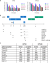
FIG 1
FIG 1
(A) qPCR analysis of HPV8 or HPV38 wt genome transfected into NHK after 3, 6, and 9 days p.t. of NHK. PGK1 was used as a reference transcript. Data are derived from four independent experiments using cells from different donors. (B) Transcript maps of HPV8 and HPV38. The genome is represented in a linear fashion. Shown are the upstream regulatory region (URR), the early genes (E1 to E8) and the late genes L1 and L2. Nucleotide positions in the table refer to the last exon nucleotide for splice donors (D) and the first exon nucleotide for splice acceptors (A). Transcripts identified by qPCR and sequencing are indicated as gray bars. The putative extension of these transcripts to the early polyadenylation site is indicated by dashed lines. (C) Nucleotide positions of splice donors and acceptors of HPV8 and 38. The positions of splice donors refer to the last nucleotide in the exon and of splice acceptors to the first nucleotide of the exon.
FIG 2
FIG 2
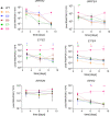
qPCR analyses of spliced HPV8 transcripts expressed 3, 6, and 9 days p.t. of NHK with HPV8 wt E1, E2, E6, E7, or E8 genomes. PGK1 was used as a reference transcript. Experiment was repeated four times independently with cells from different donors. Error bars indicate the standard error of the mean (SEM). Statistical significance was determined using a ratio-paired t test using the wt as a reference (*, P < 0.05; **, P < 0.01).
FIG 3
FIG 3
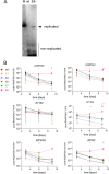
(A) Southern blot analysis of low-molecular weight DNA, harvested 6 days p.t. of NHK transiently transfected with HPV38 wt and E8 genomes. The DNA was digested with DpnI to remove nonreplicated input DNA and the single-cutter Eco105I to linearize the viral genome. As a marker (M), 100 pg linearized HPV38 genome was used. The arrow indicates the position of replicated genomes. (B) qPCR analyses of spliced HPV38 transcripts expressed 3, 6, and 9 days p.t. of NHK with HPV38 wt E1, E2, E6, E7, or E8 genomes. PGK1 was used as a reference transcript. The experiment was repeated four times independently using cells from different donors. Errors bars indicate the SEM. Statistical significance was determined using a ratio-paired t test using the wt as a reference (*, P < 0.05; **, P < 0.01; ***, P < 0.001).
FIG 4
FIG 4
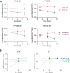
(A) qPCR analysis of HPV8 and HPV38 E6 and E7 transcripts in polyA-enriched RNA in NHK from four different donors transfected independently (3, 6, and 9 days) with wt or E8- genomes. Error bars indicate the SEM. PGK1 was used as a reference transcript. Statistical significance was determined using a ratio-paired t test using the wt as a reference (*, P < 0.05; **, P < 0.01; ***, P < 0.001). (B) Comparison of qPCR analyses of HPV8, 38, and 49 E6 and E7 transcripts in polyA-enriched RNA from NHK transfected with E8 genomes. PGK1 was used as a reference transcript. Error bars indicate the SEM.
FIG 5
FIG 5
qPCR analysis of organotypic raft cultures (3D) consisting of NHK transiently transfected with HPV16, HPV8, or HPV38 wt genomes and corresponding monolayer cells (2D). Data are derived from three (HPV16), five (HPV38), or six (HPV8) independent transfections using cells from different donors. PGK1 was used as a reference transcript. In addition to the viral transcripts, the keratinocyte differentiation marker KRT10 (keratin 10) was determined and is expressed relative to cells maintained in monolayer (2D) culture. Statistical significance was determined using a ratio-paired t test using monolayer cultures as a reference (*, P < 0.05; **, P < 0.01).
FIG 6
FIG 6
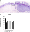
(A) H&E staining of the paraffin-embedded lesion (right) is a zoom-in of the image on the left. The arrow indicates a cell in the apical stratum spinosum with EV-typical cytopathic effect. Scale bars are as follows: (left) 500 μm, (right) 100 μm. (B) qPCR analysis of RNA from a HPV8-positive lesion of an EV patient. PGK1 was used as a reference transcript.

References

    1. de Martel C, Plummer M, Vignat J, Franceschi S. 2017. Worldwide burden of cancer attributable to HPV by site, country and HPV type. Int J Cancer 141:664–670. 10.1002/ijc.30716. - DOI - PMC - PubMed
    1. Howley PM, Pfister HJ. 2015. Beta genus papillomaviruses and skin cancer. Virology 479–480:290–296. 10.1016/j.virol.2015.02.004. - DOI - PMC - PubMed
    1. Quint KD, Genders RE, de Koning MN, Borgogna C, Gariglio M, Bouwes Bavinck JN, Doorbar J, Feltkamp MC. 2015. Human beta-papillomavirus infection and keratinocyte carcinomas. J Pathol 235:342–354. 10.1002/path.4425. - DOI - PubMed
    1. Hartevelt MM, Bavinck JN, Kootte AM, Vermeer BJ, Vandenbroucke JP. 1990. Incidence of skin cancer after renal transplantation in The Netherlands. Transplantation 49:506–509. 10.1097/00007890-199003000-00006. - DOI - PubMed
    1. Orth G. 2006. Genetics of epidermodysplasia verruciformis: insights into host defense against papillomaviruses. Semin Immunol 18:362–374. 10.1016/j.smim.2006.07.008. - DOI - PubMed

Publication types

MeSH terms

Substances

LinkOut - more resources