Review
. 2023:1410:35-95.
doi: 10.1007/5584_2022_742.

HSF1 and Its Role in Huntington's Disease Pathology

Affiliations
Review

HSF1 and Its Role in Huntington's Disease Pathology

Hyuck Kim et al. Adv Exp Med Biol. 2023.

Abstract

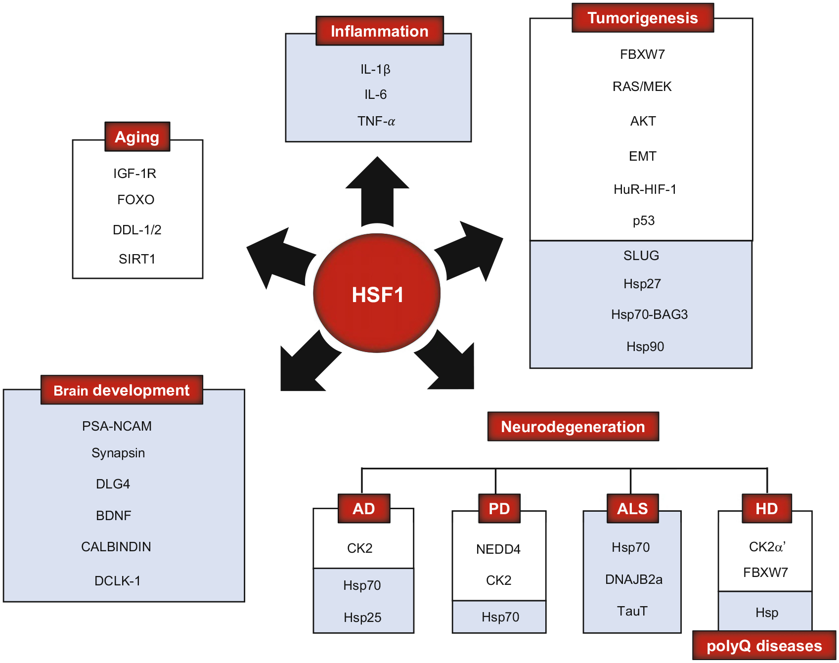
Purpose of review: Heat shock factor 1 (HSF1) is the master transcriptional regulator of the heat shock response (HSR) in mammalian cells and is a critical element in maintaining protein homeostasis. HSF1 functions at the center of many physiological processes like embryogenesis, metabolism, immune response, aging, cancer, and neurodegeneration. However, the mechanisms that allow HSF1 to control these different biological and pathophysiological processes are not fully understood. This review focuses on Huntington's disease (HD), a neurodegenerative disease characterized by severe protein aggregation of the huntingtin (HTT) protein. The aggregation of HTT, in turn, leads to a halt in the function of HSF1. Understanding the pathways that regulate HSF1 in different contexts like HD may hold the key to understanding the pathomechanisms underlying other proteinopathies. We provide the most current information on HSF1 structure, function, and regulation, emphasizing HD, and discussing its potential as a biological target for therapy.

Data sources: We performed PubMed search to find established and recent reports in HSF1, heat shock proteins (Hsp), HD, Hsp inhibitors, HSF1 activators, and HSF1 in aging, inflammation, cancer, brain development, mitochondria, synaptic plasticity, polyglutamine (polyQ) diseases, and HD.

Study selections: Research and review articles that described the mechanisms of action of HSF1 were selected based on terms used in PubMed search.

Results: HSF1 plays a crucial role in the progression of HD and other protein-misfolding related neurodegenerative diseases. Different animal models of HD, as well as postmortem brains of patients with HD, reveal a connection between the levels of HSF1 and HSF1 dysfunction to mutant HTT (mHTT)-induced toxicity and protein aggregation, dysregulation of the ubiquitin-proteasome system (UPS), oxidative stress, mitochondrial dysfunction, and disruption of the structural and functional integrity of synaptic connections, which eventually leads to neuronal loss. These features are shared with other neurodegenerative diseases (NDs). Currently, several inhibitors against negative regulators of HSF1, as well as HSF1 activators, are developed and hold promise to prevent neurodegeneration in HD and other NDs.

Conclusion: Understanding the role of HSF1 during protein aggregation and neurodegeneration in HD may help to develop therapeutic strategies that could be effective across different NDs.

Keywords: Aggregation; Heat shock factor (HSF1); Heat shock proteins (Hsp); Huntington’s diseases (HD); Mitochondria.


Conflict of interest statement

Disclosure of Interests All authors declare they have no conflict of interest.

Figures

Fig. 1
Fig. 1
Diagram of structural domains, regulatory enzymes, and PTMs of human HSF1. HSF1 can be divided into different structural domains: DBD (DNA binding domain), HR-A/B (heptad repeat-A/B), RD (regulatory domain), and TAD (transactivation domain). PTMs located at the top part of HSF1 are modifications with positive regulatory properties (red arrow), whereas PTMs located at the (bottom) represent modifications with repressive properties (black arrow). The different enzymes responsible for positive PTMs are SIRT1 (sirtuin 1), PIM2 (proviral integrations of Moloney virus 2, Pim-2 proto-oncogene, Ser/Thr kinase), CK2 (casein kinase holoenzyme), p300 (histone acetyltransferase p300), MEK (mitogen-activated protein kinase kinase), AKT (protein kinase B), and PLK1 (polo-like kinase 1). The enzymes responsive for the repressive PTMs are p300, GCN5 (general control non-repressed protein 5 histone acetyltransferase), MK2 (MAPK activated protein kinase 2), AMPK (5′-AMP-activated protein kinase), PLK1, SAE1/2 (SUMO-activating enzyme), UBC9 (RING-type E3 SUMO transferase), GSK3β (glycogen synthase kinase 3β), CK2, CK2α’ (catalytic subunit CK2 holoenzyme), and ERK (extracellular signal-regulated kinase). Ac acetylation, P phosphorylation, and S sumoylation
Fig. 2
Fig. 2
Structural insights into HSF-DNA interaction topology. (a, b) Crystal structure of the DNA binding domain (DBD) of HSF1 and HSF2 bound to a two-site HSE as a dimer. These independently solved structures revealed that a previously unknown carboxy-terminal helix (red) that is conserved in both HSF1 and HSF2 directs these HSFs to wrap around the HSE DNA, resulting in a topology where the DBD and the remainder of the HSF1 protein (not present in the crystal structure) occupy opposite sides of the DNA. (c) A new model of the HSF-DNA interaction. Structural studies support a model in contrast with the previous model for the topology of DNA-bound HSF oligomers. In the old model (left), the oligomerization domains (light blue) were positioned on top of the DBD, such that the rest of the protein buried the free surface of the DBD (shown in red, in contrast to the DNA-bound portion of the DBD shown in blue). In the new model (right), this free surface of the DBD is solvent-exposed, making it available for interactions with regulatory proteins and accepting PTMs. (Figure adapted from (Gomez-Pastor et al. 2018) with authors’ permission)
Fig. 3
Fig. 3
HSF1 activation/attenuation cycle. In response to proteotoxic stress conditions, HSF1 is subject to a multi-step activation and attenuation cycle. Inactive HSF1 monomer is kept in the cytoplasm in a complex with regulatory proteins such as Hsp 40, 70, and 90, as well as the cytosolic chaperonin TCP1 ring complex (TRiC). Upon stress sensing, HSF1 is modified by several activating PTMs that promote DNA binding and transcriptional activation of target genes in concert with cofactor recruitment. HSF1 is then modified by different inhibitory PTMs and by p23 causing DNA dissociation, HSF1 inactivation, and HSF1 degradation (see Fig. 1) for PTMs details). It is currently unknown where HSF1 degradation occurs and the extent to which HSF1 is newly synthesized or recycled into the cytoplasm. Ultimately, after attenuation, HSF1 is maintained in the cytoplasm by an inhibitory protein complex in a negative feedback mechanism. Color code: DNA-binding domain (dark blue), leucine zipper oligomerization domain LZ1–3 (light blue), regulatory domain (gray-white), LZ4 (yellow), and activation domain (orange). (Figure adapted from (Gomez-Pastor et al. 2018) with authors’ permission)
Fig. 4
Fig. 4
HSF1-regulatory enzymes and their contribution in HSF1 activation cycle. Upon proteotoxic stress, misfolded proteins titrate away the Hsp repressive complex, allowing HSF1 trimerization and nuclear accumulation. The HSF1 trimer binds to HSE in the promoter region of HSF1 target genes (old model of HSF-DNA binding: see Fig. 2). HSF1 activation is modified by several PTMs (see Fig. 1). Enzymes responsible for controlling nuclear translocation and activation include MEK and AMPK (with opposite functions). PGC-1α and different co-activators modulate HSF1 transcriptional capacity. Enzymes like HDAC7, HDAC9, and SIRT1 prolong HSF1 binding to the DNA, while acetylation by p300/CBP has an opposite effect and mediate DNA dissociation. Ubiquitin proteasome-dependent HSF1 degradation occurs by E3 ligases FBXW7 and NEDD4. FBXW7 is recruited by phosphorylation of HSF1 mediated by GSK3β, ERK, and CK2α’, whereas NEDD4 is accessed by p300/CBP-mediated acetylation (Figure reprinted from “Rethinking HSF1 in Stress, Development, and Organismal Health” by Li et al. 2017, with copyright permission from Elsevier. Figure legend has been modified accordingly for this publication)
Fig. 5
Fig. 5
Relationship between HSF1 and different physiological and pathological processes. Changes in activity and stability of HSF1 are responsible for regulating different cellular processes that are essential for life, including brain development, immune response, metabolism, and cell growth. Dysregulation of HSF1 contributes to different diseases like cancer and neurodegeneration. For each process, we included different proteins related to HSF1 and that contribute to the regulation of such a process. Proteins shown in a white box correspond to proteins that directly or indirectly regulate HSF1 activity or stability. In contrast, proteins shown in a blue box are proteins whose expression is influenced by HSF1
Fig. 6
Fig. 6
Diagram of human wild-type HTT relative to its known domains and post-translational modifications. HTT is composed of different domains; N17 (containing the first 17 amino acids of the protein), polyQ tract (whose expansion is responsible for HD), polyP repeats, and five helical clusters of HEAT repeats. Several PTMs (e.g., acetylation [Ac, pink], phosphorylation [P, orange], sumoylation [S, green], and ubiquitination [Ub, blue]) alter HTT’s cell biology and toxic properties. Within the N17 region, we find (P1) P-Ser13 and P-Ser16, (Ac1) Ac-Lys9, S-Lys6, 9 and 13, and (S1) S-Lys9. Other PTMs can also be detected along with the protein (P2) P-Ser116, (P3) Ser-419, 421, 434, 533, 535, and 536, (P4) P-Ser1181 and 1,201, (P5) P-Ser2076, P(6) P-Ser2653 and 2,657, (Ac2) Ac-Lys178 and 236, (Ac3) Ac-Lys 345, (Ac4) Ac-Lys444, and (S2) S-Lys444. For a detailed list of all the PTMs identified in vivo and in vitro, see (Arbez et al. 2017). The corresponding amino acid sequence from exon 1 is shown in blue for the N17, in purple for the polyQ repeat, and in green for the polyP-rich region. Sequences were obtained from the NCBI protein database (Human: NP_002102.4)
Fig. 7
Fig. 7
Model for p53-HSF1-PGC-1α integrated responses in HD. Crosstalk between the transcription factors p53, HSF1, and PGC-1α in regulating transcription, protein homeostasis, mitochondrial function, and apoptosis. There are alterations in different pathways (CREB/TAF4, CK2α’/FBXW7, and MDM2) in the presence of mHTT, which independently leads to the deregulation of the levels and functions of all three transcription factors. However, HSF1 becomes a key player in the subsequent regulation of the levels of p53 and PGC-1α by directly regulating the transcription of PGC-1α and controlling p53 protein stability in HD. The potential role of p53 in regulating the HSF1 degradation pathway in HD would add a positive feedback loop into the p53-HSF1-PGC-1α axis, which triggers mitochondrial dysfunction and neuronal death. Reprinted and modified from “Mitochondrial Dysfunction in Huntington’s Disease; Interplay Between HSF1, p53 and PGC-1α Transcription Factors” by Intihar et al. 2019, with permission from the original authors
Fig. 8
Fig. 8
Working model for the connection between HSF1, mitochondrial impairment, and synapse dysfunction in HD. mHTT alters excitatory synaptic transmission and mitochondrial function by altering Ca2+ buffering capacity and PGC-1α and p53 levels. mHTT is also responsible for promoting HSF1 degradation, which results in downregulation of Hsp and synaptic proteins. The effects caused by HSF1 depletion then further disrupt glutamate release and uptake in neurons, resulting in excitotoxicity. (Reprinted from “Excitatory synapse impairment and mitochondrial dysfunction in Huntington’s disease: heat shock factor 1 (HSF1) converging mechanisms” by (Zarate and Gomez-Pastor 2020), with permission from Neural Regeneration Research)

Similar articles

Cited by

References

    1. Acebron SP, Martin I, del Castillo U, Moro F, Muga A (2009) DnaK-mediated association of ClpB to protein aggregates. A bichaperone network at the aggregate surface. FEBS Lett 583:2991–2996 - PubMed
    1. Acevedo-Torres K, Berrios L, Rosario N et al. (2009) Mitochondrial DNA damage is a hallmark of chemically induced and the R6/2 transgenic model of Huntington’s disease. DNA Repair (Amst) 8:126–136 - PMC - PubMed
    1. Adachi H, Katsuno M, Minamiyama M et al. (2003) Heat shock protein 70 chaperone overexpression ameliorates phenotypes of the spinal and bulbar muscular atrophy transgenic mouse model by reducing nuclear-localized mutant androgen receptor protein. J Neurosci 23:2203–2211 - PMC - PubMed
    1. Adachi H, Katsuno M, Minamiyama M et al. (2005) Wide-spread nuclear and cytoplasmic accumulation of mutant androgen receptor in SBMA patients. Brain 128:659–670 - PubMed
    1. Ahn SG, Thiele DJ (2003) Redox regulation of mammalian heat shock factor 1 is essential for Hsp gene activation and protection from stress. Genes Dev 17: 516–528 - PMC - PubMed

MeSH terms