. 2022 Nov 17;13(1):7057.
doi: 10.1038/s41467-022-34776-7.

Transplantable human thyroid organoids generated from embryonic stem cells to rescue hypothyroidism

Affiliations

Transplantable human thyroid organoids generated from embryonic stem cells to rescue hypothyroidism

Mírian Romitti et al. Nat Commun. .

Abstract

The thyroid gland captures iodide in order to synthesize hormones that act on almost all tissues and are essential for normal growth and metabolism. Low plasma levels of thyroid hormones lead to hypothyroidism, which is one of the most common disorder in humans and is not always satisfactorily treated by lifelong hormone replacement. Therefore, in addition to the lack of in vitro tractable models to study human thyroid development, differentiation and maturation, functional human thyroid organoids could pave the way to explore new therapeutic approaches. Here we report the generation of transplantable thyroid organoids derived from human embryonic stem cells capable of restoring plasma thyroid hormone in athyreotic mice as a proof of concept for future therapeutic development.


Conflict of interest statement

S.C. and M.R. have filed EU patent regarding the hESC-thyroid derived organoids. All other authors declare no competing interests.

Figures
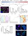
Fig. 1
Fig. 1. Transient overexpression of NKX2-1 and PAX8 promotes differentiation of human ESCs into thyroid follicular cells.
a Schematic representation of the protocol leading to thyroid follicle differentiation from human ESCs. b NKX2-1 and PAX8 co-staining at day 9. c Quantification by flow cytometry of NKX2-1GFP and PAX8 expressing cells after Dox stimulation, at days 9 and 16 (n = 4). d qRT-PCR analysis of exogenous NKX2-1 and PAX8 and endogenous PAX8, FOXE1, TG, and TSHR genes after Dox stimulation (day 9) (n = 5 for NKX2-1 exog, PAX8 exog and PAX8 endo; n = 3 for FOXE1, TG and TSHR). e Proportion of NKX2-1GFP cells expressing BrdU during the differentiation protocol (n = 3 per time point). f Quantification by flow cytometry of NKX2-1GFP cells during the differentiation protocol (n = 3 for −Dox, Day 23, 45 and 58; n = 4 for Day 9, 11, 14, 16, 30, and 37). g Heatmap of normalized bulk RNA-Seq of thyroid genes expression among NKX2-1GFP cells at different stages of the thyroid differentiation protocol. Rows represent markers and columns represent specific time points. Color values in the heatmap represent mean expression levels. h qRT-PCR analysis of PAX8, NIS, TSHR, TG, and TPO at thyroid organoids from day 45 of differentiation protocol compared to un-induced control (−Dox) (n = 3). i Immunostaining for NKX2-1 and TG at D30 and TG at day 37, 45, and D58 showing thyroid differentiation and cell organization overtime. Data from at least three independent experiments are shown. Statistical analyses were performed using two-sided unpaired Mann–Whitney (p values are presented in the graphs; data presented as median (IQR)). The experiment was performed at least three times with similar results (b, i). Scale bars 20 μm. Source data are provided as a Source Data file.
Fig. 2
Fig. 2. Immunostaining and Single-cell RNA-seq characterization of human ESC-derived thyroid cells at day 45.
ad Confocal immunofluorescence images of the three-dimensional follicular structures co-expressing NKX2-1 and (a) E-CADHERIN, (b) TG, (c) TPO (d) TG-I storage in the lumenal compartment. e Single-cell RNA-Seq unsupervised clustering of in vitro hESC-derived human thyroid organoid model cells. Each cluster is represented by a specific color. f Heatmap showing normalized expression of selected marker genes with rows representing cell clusters, while columns represent genes. The intensity of the color in each square indicates the mean expression within the cluster. g UMAP overlaid with gene expression plots for thyrocyte markers. Color indicates normalized expression. h Diffusion analysis of thyrocyte lineage with thyroid progenitor cells as root cells. UMAP overlaid with pseudotime. Color in pseudotime plot indicates order of cell progression. i Expression trends of thyroid genes along the pseudotime trajectory. The experiment was performed at least three times with similar results (ad). ad Scale bars, 20 μm.
Fig. 3
Fig. 3. Single-cell RNA-seq and immunostaining characterization of human ESC-derived thyroid cells at day 58.
a Single-cell RNA-Seq unsupervised clustering of in vitro hESC-derived human thyroid organoid model cells. Each cluster is represented by a specific color. b Heatmap showing normalized expression of selected marker genes with rows representing cell clusters, while columns represent genes. The intensity of the color in each square indicates the mean expression within the cluster. c UMAP overlaid with gene expression plots for thyrocyte markers. Color indicates normalized expression. d UMAP showing integration analysis of single-cell RNA-seq data from thyroid organoids at day 45 and 58. e Single-cell RNA-Seq unsupervised clustering of publicly available human adult thyroid tissue dataset. Each cluster is represented by a specific color. f UMAP showing integration analysis of single-cell RNA-seq data from human adult thyroid tissue and hESC-derived thyroid organoids at days 45 and 58. g qRT-PCR analysis of PAX8, NIS, TSHR, TG, and TPO at thyroid organoids from day 58 of differentiation protocol compared to un-induced control (−Dox) (n = 4). hk Confocal immunofluorescence images at day 58 of the differentiation protocol. h Three-dimensional follicular structures co-expressing NKX2-1 and E-CADHERIN. i Intra-lumenal F-actin (Phalloidin) and TG; (j) NKX2-1 cells with TPO cytoplasmic and apical membrane accumulation. k NKX2-1 cells showing TG-I and T4 stored in the lumenal compartment. Data from at least three independent experiments are shown. Statistical analyses were performed using two-sided unpaired Mann–Whitney (p values are presented in the graphs; data presented as median (IQR)). The experiment was performed at least three times with similar results (hk). Scale bars, 20 μm and 10 μm for high magnification follicles. Source data are provided as a Source Data file.
Fig. 4
Fig. 4. In vivo functionality of transplanted human ESC-derived thyroid follicles.
a Schematic representation of the hESC-derived thyroid organoids transplantation protocol under the kidney capsule of NOD-SCID thyroid RAI-ablated mice. b, c Histological analysis of the grafted tissue 5 weeks after transplantation. b Hematoxylin and eosin staining shows human thyroid follicles located in the cortical region of the host kidney. Red arrows show the monolayer epithelium of the transplanted tissue surrounded by stromal cells (green arrows). c Confocal images show co-expression of NKX2-1 and E-CADHERIN in the monolayered follicles. The grafted tissue shows TG mainly accumulated in the luminal compartment, whereas TPO is strongly expressed in the apical membrane. d NKX2-1, E-CADHERIN and T4 immunostaining demonstrate hESC-derived follicles with specific accumulation of T4 within the lumen of several structures as observed in human adult thyroid tissue. e qRT-PCR analysis of FOXE1, NIS, TSHR, TG and TPO expression from human adult thyroid tissue sample compared to graft tissue and/or hESC-derived thyroid cells from day 58 (normalized by PAX8 levels; n = 3, 4 and 4, respectively). f Maximum intensity projections generated from SPEC/CT images of controls (left), RAI-ablated and non-transplanted (middle), and RAI-ablated and transplanted (right) mice. Images were obtained four weeks after organoids transplantation. The 123I uptake in the mouse thyroid tissue is shown by the orange arrow, while the signal from the human thyroid tissue (graft) is highlighted by the green arrow. g, h Plasma levels of (g) T4 and (h) T3 in controls (n = 6), RAI-ablated (RAI; n = 6) and RAI-ablated/grafted mice (graft; n = 10). i Correlation of plasma TSH and T4 levels among grafted animals (n = 10). j Comparison of hepatic Dio1 mRNA levels in controls (n = 6), RAI-ablated (n = 6) and RAI-ablated/transplanted mice (n = 10). All measurements were performed five weeks after transplantation. Two-sided unpaired Mann–Whitney (g, h, j) and Spearman Correlation (i) tests were used for statistical analysis (r and p values are presented in the graphs; ns not significant; data are presented as median (IQR)). Similar results were obtained from all grafted samples (c, d). Scale bars, 50 μm and 20 μm for zoomed area. Source data are provided as a Source Data file.

Similar articles

Cited by

References

    1. Vanderpump MPJ. The epidemiology of thyroid disease. Br. Med. Bull. 2011;99:39–51. doi: 10.1093/bmb/ldr030. - DOI - PubMed
    1. Garmendia Madariaga A, Santos Palacios S, Guillén-Grima F, Galofré JC. The incidence and prevalence of thyroid dysfunction in Europe: a meta-analysis. J. Clin. Endocrinol. Metab. 2014;99:923–931. doi: 10.1210/jc.2013-2409. - DOI - PubMed
    1. Taylor PN, et al. Global epidemiology of hyperthyroidism and hypothyroidism. Nat. Rev. Endocrinol. 2018;14:301–316. doi: 10.1038/nrendo.2018.18. - DOI - PubMed
    1. Dew R, et al. Clinical, behavioural and pharmacogenomic factors influencing the response to levothyroxine therapy in patients with primary hypothyroidism-protocol for a systematic review. Syst. Rev. 2017;6:60. doi: 10.1186/s13643-017-0457-z. - DOI - PMC - PubMed
    1. Kraut, E. & Farahani, P. A systematic review of clinical practice guidelines’ recommendations on levothyroxine therapy alone versus combination therapy (LT4 plus LT3) for hypothyroidism. Clin. Investigative Med.38, E305-E313 (2015). - PubMed

Publication types

Substances