

Healthy adults with Streptococcus pneumoniae meningitis and Streptococcus pneumoniae subdural abscess: two case reports and a literature review
- PMID: 36396983
- PMCID: PMC9679341
- DOI: 10.1177/03000605221137470
Healthy adults with Streptococcus pneumoniae meningitis and Streptococcus pneumoniae subdural abscess: two case reports and a literature review
Abstract
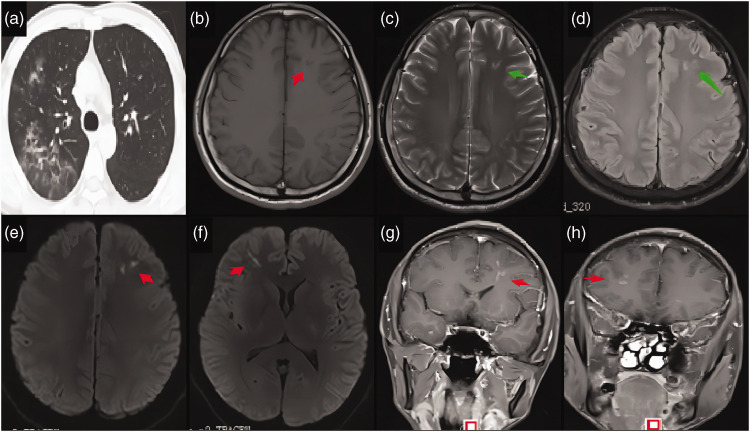
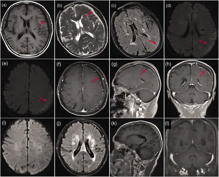
We present the cases of two otherwise healthy adults, one with meningitis and another with a subdural abscess, with both conditions attributable to Streptococcus pneumoniae. A 31-year-old man was admitted with a 3-day history of fever, headache, and vomiting. Physical examination revealed intermittent confusion, irritability, and neck stiffness. Cerebrospinal fluid (CSF) culture was positive for S. pneumoniae. Contrast-enhanced magnetic resonance imaging (C-MRI) revealed multiple small lesions on the bilateral frontal lobes. Intravenous ceftriaxone and vancomycin were administered, followed by intravenous moxifloxacin. His symptoms resolved within 3 months. Additionally, a 66-year-old man was admitted for acute fever with confusion, abnormal behavior, and a recent history of acute respiratory infection. Physical examination revealed confusion, neck stiffness, and a positive right Babinski sign. CSF metagenomic analysis detected S. pneumoniae. C-MRI disclosed left occipitotemporal meningoencephalitis with subdural abscesses. Intravenous ceftriaxone was administered for 3 weeks. His condition gradually improved, with resorbed lesions detected on repeat MRI. This study expanded the clinical and imaging spectra of S. pneumoniae meningitis. In healthy adults, S. pneumoniae can invade the brain, but subdural abscess is a rare neuroimaging manifestation. Early diagnosis of S. pneumoniae meningitis by high-throughput sequencing and flexible treatment strategies are necessary for satisfactory outcomes.
Keywords: Streptococcus pneumoniae meningitis; case report; healthy adult; magnetic resonance imaging; metagenomic analysis; subdural abscess.
Figures
References
-
- Thigpen MC, Whitney CG, Messonnier NE, et al. Bacterial meningitis in the United States, 1998–2007. N Engl J Med 2011; 21364: 2016–2025. doi: 10.1056/NEJMoa1005384. - PubMed
-
- Samakoses R, Suwanpakdee D, Watanaveeradej V, et al. Cerebrospinal fluid lymphocytosis in an infant with acute Streptococcus pnuemoniae meningitis: a case report. J Med Assoc Thai 2010; 93: S49–S52. - PubMed
Publication types
MeSH terms
Substances
LinkOut - more resources
Full Text Sources
