. 2022 Nov 18;17(1):74.
doi: 10.1186/s13024-022-00576-2.

Finding memo: versatile interactions of the VPS10p-Domain receptors in Alzheimer's disease

Affiliations

Finding memo: versatile interactions of the VPS10p-Domain receptors in Alzheimer's disease

Alena Salasova et al. Mol Neurodegener. .

Abstract

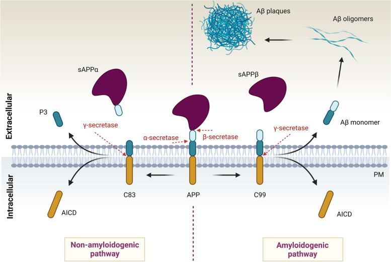
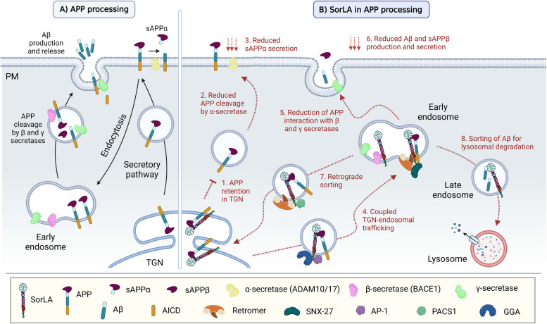
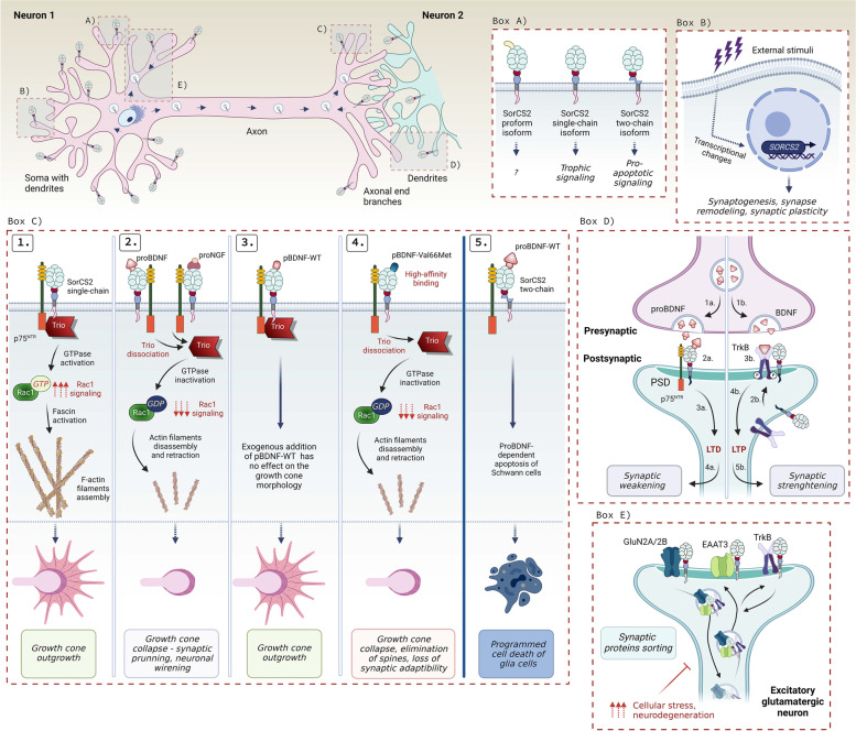
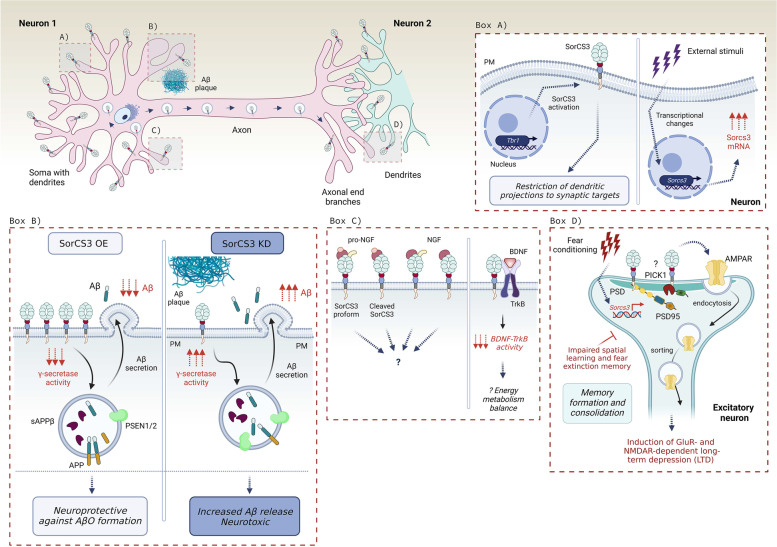
The family of VPS10p-Domain (D) receptors comprises five members named SorLA, Sortilin, SorCS1, SorCS2 and SorCS3. While their physiological roles remain incompletely resolved, they have been recognized for their signaling engagements and trafficking abilities, navigating a number of molecules between endosome, Golgi compartments, and the cell surface. Strikingly, recent studies connected all the VPS10p-D receptors to Alzheimer's disease (AD) development. In addition, they have been also associated with diseases comorbid with AD such as diabetes mellitus and major depressive disorder. This systematic review elaborates on genetic, functional, and mechanistic insights into how dysfunction in VPS10p-D receptors may contribute to AD etiology, AD onset diversity, and AD comorbidities. Starting with their functions in controlling cellular trafficking of amyloid precursor protein and the metabolism of the amyloid beta peptide, we present and exemplify how these receptors, despite being structurally similar, regulate various and distinct cellular events involved in AD. This includes a plethora of signaling crosstalks that impact on neuronal survival, neuronal wiring, neuronal polarity, and synaptic plasticity. Signaling activities of the VPS10p-D receptors are especially linked, but not limited to, the regulation of neuronal fitness and apoptosis via their physical interaction with pro- and mature neurotrophins and their receptors. By compiling the functional versatility of VPS10p-D receptors and their interactions with AD-related pathways, we aim to further propel the AD research towards VPS10p-D receptor family, knowledge that may lead to new diagnostic markers and therapeutic strategies for AD patients.

Keywords: Alzheimer’s disease; Comorbidity; Neurotrophins; SorCS1; SorCS2; SorCS3; SorLA; Sortilin.


Conflict of interest statement

The authors declare that they have no competing interests.

Figures

Fig. 1
Fig. 1
A simplified scheme of APP proteolytic processing and the origin of Aβ plaques. APP is a type I transmembrane receptor that contains Aβ peptide within its sequence. α-secretases such as ADAM10/17 cleave APP inside the Aβ peptide, which is disrupting, and produces soluble, secreted sAPPα fragment. sAPPα is neuroprotective, and thus this cleavage is called non-amyloidogenic pathway. The C83 peptide can be further cleaved by γ-secretase producing soluble P3 peptide. In contrast, APP can be cleaved by β-secretase, for example BACE1, creating a cytotoxic, soluble sAPPβ. The proteolysis by β-secretase exposes Aβ peptide, which is further cleaved by γ-secretase. This cleavage results in the release of Aβ monomers into the extracellular space, where they can further polymerize forming Aβ oligomers, and later Aβ plaques. This pathway is neurotoxic and is called amyloidogenic pathway
Fig. 2
Fig. 2
VPS10p-D receptors – their structure, and genetic and transcriptional relations to AD. The VPS10-D receptors are produced as proforms containing a propeptide which is cleaved by Furin. Except for SorLA, the receptors exhibit similar structure, mostly differing in the sequence of their intracellular cytoplasmic tails. SorLA can dimerize at neutral pH while Sortilin forms dimers only at acidic pH, for example in lysosomes. SorCS1-3 are paralogs that tend to form stable homodimers. All the receptors have been genetically linked to AD. Independently from the genetic background, AD patients display changes in the receptors’ expression levels within the brain parenchyma. These are predominantly diminished, and likely contribute to the disease progression such as decreased neuroprotection
Fig. 3
Fig. 3
The role of SorLA in APP processing. A APP is directed from the trans-Golgi network (TGN) to the plasma membrane via the secretory pathway. APP molecules are either cleaved by α-secretase at the plasma membrane or recycled through endocytosis, and trafficked by early endosomes. There, APP is sequentially cleaved by β- and γ-secretases, thus generating Aβ monomers that are secreted to the extracellular space. B A model of SorLA involvement in the amyloid cascade. 1.-3. SorLA interacts with APP in TGN acting as a retention factor, which reduces α-secretase cleavage and secretion of sAPPα from the cell surface. 4.-7. In addition, SorLA forms a complex with APP that shuttles between the TGN and endosomes. The anterograde transport is dependent on SorLA’s interaction with AP-1 and GGA (4.), while the retrograde transport is determined by its binding to retromer or PACS1 (7.). SorLA’s interaction with retromer and SNX-27 in early endosomes additionally enables the sorting of APP along the recycling pathway to the plasma membrane (5.). This way, the sorting receptor is responsible for reducing the interaction between APP and β- and γ- secretases (5.). Importantly, binding of SorLA to BACE1 blocks the APP-BACE1 interaction, which reduces the production of secreted Aβ peptides (6.). Last but not least, SorLA also engages with Aβ peptides in endosomes and navigates them for the lysosomal degradation (8.)
Fig. 4
Fig. 4
SorLA localization within a neuron and its signaling in AD. SorLA predominantly localizes in neural soma and dendrites, either in sorting vesicles or at the plasma membrane. Box A) The presence of extracellular BDNF in human brain mediates expression of SORL1, which increases SORLA protein levels attenuating the production and secretion of Aβ. Box B) A scheme of how SorLA regulates EphA4 signaling. Under physiological conditions (left panel), EphA4 binds its juxtapositioned ligand EphA1 which triggers clustering of EphA4 receptors, and their subsequent phosphorylation. EphA4 activation triggers disassembly and retraction of F-actin filaments causing growth cone collapse crucial e.g. for dendritic spine pruning. AD patients (right panel) show increased levels of EphA4 in close proximity to Aβ plaques. Moreover, EphA4 binds Aβ oligomers which results in increased AphA4 activation and abnormal actin filaments retraction causing dendritic spine retraction and synaptic loss. SorLA (middle panel) binds EphA4, which prevents EphA4 clustering. Increased SorLA levels thus diminish the EphA4 activation, which lowers the responsiveness of the neurons to growth cone retraction even in presence of AβO, thus protecting the neurons against synaptotoxicity. Box C) SorLA binds and traffics TrkB receptor towards the synapse where they remain as a receptor complex. Upon BDNF release and subsequent activation of TrkB, SorLA further drives TrkB internalization, which is a critical step for the subsequent BDNF-dependent neurotrophic response and synaptic plasticity
Fig. 5
Fig. 5
Functional involvement of Sortilin in AD-related signaling. Sortilin localizes in sorting vesicles and on the plasma membrane (PM) in neuronal somas, dendrites and axons. Box A) Sortilin undergoes ectodomain shedding by ADAM10/17, which produces soluble Sortilin fragments. In humans, C-terminal fragments are found within the Aβ plaques, however, their precise origin and trafficking route is unknown (marked as “?”). Box B) Sortilin binds BACE1 in TGN and facilitates its intracellular trafficking via anterograde and retrograde pathways, the later directed either towards the recycling pathway or for the lysosomal degradation. Box C) Sortilin binds APP at PM; however, its involvement in APP processing is controversial. Left panel—Sortilin binds APP at axonal PM where they undergo internalization. Sortilin either traffics APP for its lysosomal degradation (a.) or engages in amyloidogenic pathway by enhancing APP cleavage by BACE1 (b.), subsequently causing an increased formation and secretion of sAPPβ and Aβ. Sortilin is also a PSEN1/2 substrate. Right panel – Sortilin has a neuroprotective role as it mediates the uptake of soluble APP from the extracellular space (1.) for lysosomal degradation thus decreasing their extracellular concentration. Moreover, Sortilin binds APP in neurites where it drives its preferential cleavage by ADAM10/17 (2.), thus elevating sAPPα levels. Consequently, there is less APP internalized (3.) prior the sequential cleavage by β- and γ-secretases (4.), resulting in decreased production of sAPPβ and Aβ (4.-5.). However, the molecular mechanisms are rather unknown (marked with “?”). Box D) Upon proNGF binding, Sortilin forms a complex with p75NTR receptor, which mediates pro-apoptotic cell responses (left). The presence of AβO increases Sortilin expression, which likely enhances the formation and activity of Sortilin-p75NTR complexes. Sortilin-p75.NTR complex binds and internalizes AβO leading to increased intracellular neurotoxicity, and later cell death (middle). Sortilin can also bind and sequester extracellular ApoE, subsequently facilitating its lysosomal degradation, which has a neuroprotective effect (right). It is not clear if Sortilin sequesters ApoE-Aβ complexes (marked as “?”)
Fig. 6
Fig. 6
SorCS1 localization and signaling relevant to AD. SorCS1 localization is restricted to cell soma and dendrites. Box A) SorCS1 forms homodimers, but also heterodimers with Sortilin via SorCS1 prodomain, and with SorCS2/3. Box B) SorCS1 binds APP in vesicular compartments; however, SorCS1 variants control APP sorting in different manner. While SorCS1b mediates APP trafficking towards PM, SorCS1c blocks it. Box C) SorCS1 binds TrkB, which inhibits TrkB activation by BDNF stimulation. SorCS1 might be responsible (marked “?”) for TrkB sorting between TGN, PM and recycling pathway. Box D) This figure schematizes the possible consequences of SorCS1 loss and gain of function. In homeostatic state (middle panel), SorCS1 binds APP and the retromer complex via its VPS35 subunit. This protein complex is internalized and later recycled into TGN. SorCS1c remains in complex with APP and retromer, which retains APP in TGN, and subsequently regulates its cleavage by BACE1 and γ-secretase. This way SorCS1 could control the physiological levels of secreted sAPPβ and Aβ. The abortion of SorCS1c-VPS35 interaction (left panel) enhances the APP anterograde trafficking causing an increased production and release of neurotoxic Aβ and sAPPβ. SorCS1 overexpression (OE; right panel) seems to strengthen the APP retention in TGN, thus significantly reducing the production and secretion of Aβ and sAPPβ, which has a neuroprotective effect against the formation of Aβ oligomers. Box E) SorCS1 is a substrate for PSEN1/2 and ADAM17, which attenuates its protein levels. However, molecular mechanisms involved in this regulation are unknown. Box F) SorCS1 sorts and recycles a number of synaptic receptors including Neurexin, AMPAR or Neuroligin at the postsynaptic side, by which it establishes the correct axon-to-dendrite polarization of synaptic proteins, processes critical for correct neurotransmission, connectivity, and synaptic plasticity
Fig. 7
Fig. 7
SorCS2 signaling in neuronal networks relevant for AD. SorCS2 is found in neural soma, dendrites and axons. Box A) SorCS2 exists in three isoforms that have different signaling profiles. SorCS2 is initially produced as a proform, which can be cleaved by Furin from its propeptide, giving rise to a single-chain receptor. The single-chain can be further cleaved within the leucine-rich domain, producing a two-chain isoform. Box B) SorCS2 expression changes upon external stimuli, which affects synaptic plasticity. Box C) SorCS2 interactions with neurotrophins. 1. SorCS2 single-chain binds p75NTR and Trio, which mediates Rac1 and Fascin signaling. Fascin activation leads to F-actin filaments assembly and growth cone outgrowth. 2. ProBDNF or proNGF binding to SorCS2 leads to dissociation of Trio causing Rac1 signaling inactivation, actin filaments disassembly and retraction, and grow cone collapse, which is important for synaptic pruning and neuronal wirening. 3. Propeptide of BDNF-WT binding to SorCS2 does not affect the growth cone outgrowth. 4. Propeptide of BDNF with Val66Met mutation exhibits high binding affinity to SorCS2, subsequently dissociating Trio, and inhibiting Rac1 signaling. This pathway promotes elimination of spines and loss of synaptic adaptability. 5. SorCS2 two-chain binds p75NTR, which mediates proBDNF-dependent apoptosis. Box D) SorCS2 controls synaptic plasticity. Upon proBDNF release (1a.), SorCS2 mediates synaptic weakening (4a.) via its interaction with proBDNF and p75NTR (2a.), which induces long-term potentiation (LTD; 3a.). SorCS2 and the BDNF receptor TrkB are located outside of the postsynaptic density (PSD). Upon BDNF release (1b.), SorCS2 and TrkB relocate to the synapse (2b.) where they interact. TrkB binds BDNF, and undergoes phosphorylation and activation (3b.), subsequently inducing LTP (4b.) and synaptic strengthening (5b.). Box E) SorCS2 interacts with synaptic receptors GluN2A/2B, EAAT3, and TrkB at PSD of glutamergic neurons, and regulates their anterograde and retrograde trafficking. Impairments in these processes lead to increased cellular stress and neurodegeneration
Fig. 8
Fig. 8
SorCS3 localization and AD-related signaling. SorCS3 predominantly displays somatodendritic localization, especially at the plasma membrane where it binds its ligands. It is also found in axons but at minor extent. Box A) SorCS3 is a downstream effector of transcription factor Tbr1, which restricts dendritic projections to their synaptic targets during development (left panel). Moreover, SorCS3 transcription is inducible upon various external stimuli such as LTP, seizures or fear conditioning (right panel). Box B) SorCS3 signaling is involved in amoyloidogenic pathway. SorCS3 overexpression (OE) reduces γ-secretase activity and APP processing resulting in lower production and secretion of Aβ. SorCS3 gain of function is thus neuroprotective against accumulation of Aβ oligomers. In contrast, SorCS3 downregulation (KD) results in an enhanced γ-secretase activity and thus larger formation and secretion of Aβ into the extracellular space. Thus, SorCS3 loss of function is neurotoxic as it promotes the amyloidogenic pathway. However, molecular mechanisms behind these observations are unknown. Box C) SorCS3 can signal upon its ligands’ binding already as a proform. Both SorCS3 isoforms bind proNGF and NGF, however the functional consequences remain unknown. SorCS3 can bind TrkB, by which it blunts BDNF-mediated TrkB activation. It is believed that this signaling axis is important for energy metabolism balance. Box D) SorCS3 signaling at the synapse is critical for memory formation and consolidation of excitatory neurons. SorCS3 resides at the postsynaptic side of excitatory neurons where it interacts with PSD95 and probably with PICK1 (the interaction was shown only in the HEK293 cells), which was suggested to mediate the endocytosis and sorting of AMPA receptors, a critical step for GluR- and NMDAR-dependent long-term depression (LTD), spatial learning and fear extinction memory. Notably, Sorcs3 expression is upregulated in engram neurons during memory formation

References

    1. 2020 Alzheimer's disease facts and figures. Alzheimer's & Dementia 2020;16(3):391-460. 10.1002/alz.12068. - PubMed
    1. Scheltens P, De Strooper B, Kivipelto M, Holstege H, Chételat G, Teunissen CE, Cummings J, van der Flier WM. Alzheimer’s disease. Lancet. 2021;397:1577–1590. - PMC - PubMed
    1. Wang J, Gu BJ, Masters CL, Wang Y-J. A systemic view of Alzheimer disease — insights from amyloid-β metabolism beyond the brain. Nat Rev Neurol. 2017;13:612. - PubMed
    1. Busche MA, Hyman BT. Synergy between amyloid-β and tau in Alzheimer’s disease. Nat Neurosci. 2020;23:1183–1193. - PMC - PubMed
    1. Panza F, Lozupone M, Logroscino G, Imbimbo BP. A critical appraisal of amyloid-β-targeting therapies for Alzheimer disease. Nat Rev Neurol. 2019;15:73–88. - PubMed

Publication types