. 2022 Nov 17;53(1):95.
doi: 10.1186/s13567-022-01109-x.

Genotyping and biofilm formation of Mycoplasma hyopneumoniae and their association with virulence

Affiliations

Genotyping and biofilm formation of Mycoplasma hyopneumoniae and their association with virulence

Yuzi Wu et al. Vet Res. .

Abstract

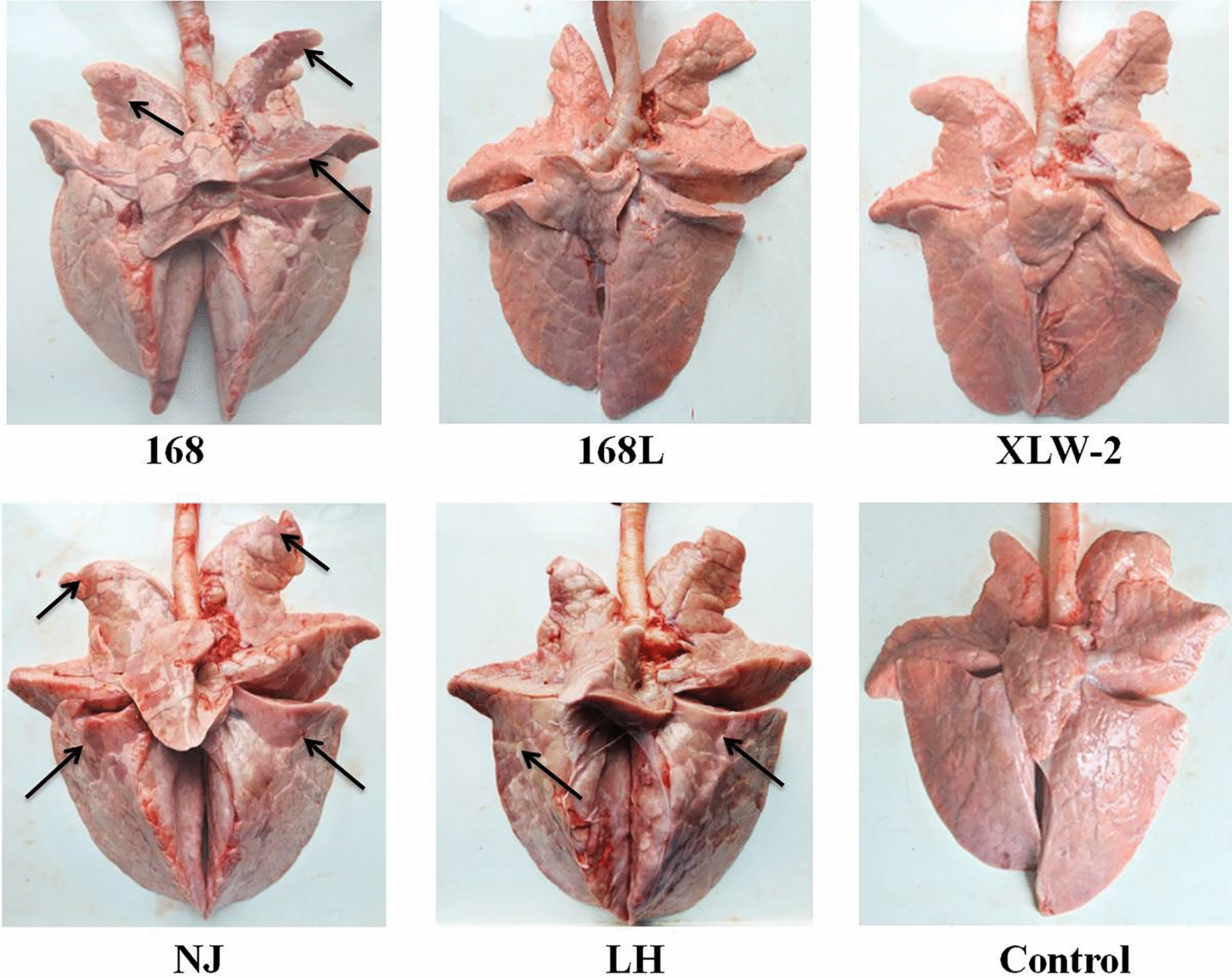
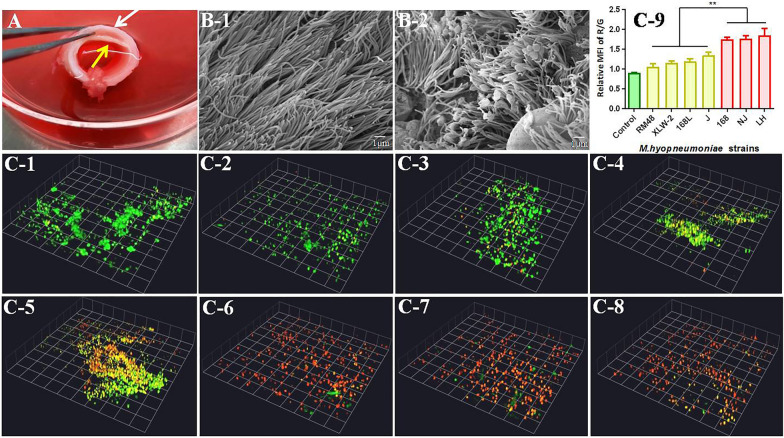
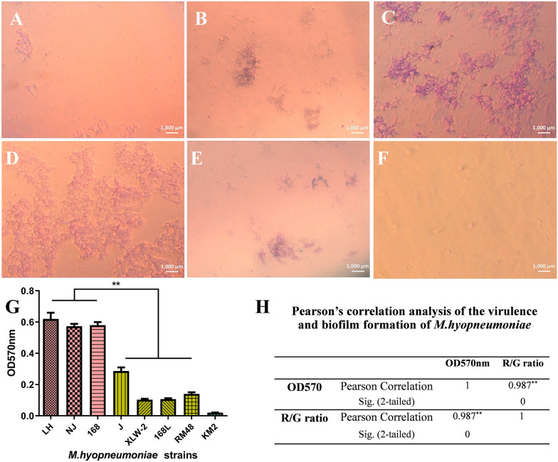
Mycoplasma hyopneumoniae, the causative agent of swine respiratory disease, demonstrates differences in virulence. However, factors associated with this variation remain unknown. We herein evaluated the association between differences in virulence and genotypes as well as phenotype (i.e., biofilm formation ability). Strains 168 L, RM48, XLW-2, and J show low virulence and strains 232, 7448, 7422, 168, NJ, and LH show high virulence, as determined through animal challenge experiments, complemented with in vitro tracheal mucosa infection tests. These 10 strains with known virulence were then subjected to classification via multilocus sequence typing (MLST) with three housekeeping genes, P146-based genotyping, and multilocus variable-number tandem-repeat analysis (MLVA) of 13 loci. MLST and P146-based genotyping identified 168, 168 L, NJ, and RM48 as the same type and clustered them in a single branch. MLVA assigned a different sequence type to each strain. Simpson's index of diversity indicates a higher discriminatory ability for MLVA. However, no statistically significant correlation was found between genotypes and virulence. Furthermore, we investigated the correlation between virulence and biofilm formation ability. The strains showing high virulence demonstrate strong biofilm formation ability, while attenuated strains show low biofilm formation ability. Pearson correlation analysis revealed a significant positive correlation between biofilm formation ability and virulence. To conclude, there was no association between virulence and our genotyping data, but virulence was found to be significantly associated with the biofilm formation ability of M. hyopneumoniae.

Keywords: Mycoplasma hyopneumoniae; biofilm; genotyping; virulence.


Conflict of interest statement

The authors declare that they have no competing interests.

Figures

Figure 1
Figure 1
Anatomical observation of pneumonic lung lesions in M. hyopneumoniae-infected pigs. Lungs lesions on infection by strains 168, 168 L, XLW-2, NJ, and LH are shown. Lung lesions with clear boundaries are indicated by black arrows. Control: lungs without any infection.
Figure 2
Figure 2
HE staining showing pathological changes in M. hyopneumoniae-infected pig lung tissues. 168, 168 L, XLW2, NJ, and LH: pathological changes on infection with the corresponding strain. Control: negative control. The swelling and hyperplasia of alveolar epithelial cells are indicated by black arrows. Smaller or occluded alveolar lumen and narrowed bronchial lumen infiltrated with inflammatory and deciduous epithelial cells are indicated by yellow arrows. Congestion or hemorrhage around alveoli and bronchi is indicated by white arrows.
Figure 3
Figure 3
Damage caused by M. hyopneumoniae infection to porcine tracheal epithelial cells. A Tracheal mucosa preparation. The cartilage (white arrow) was excised to reserve the inner tracheal mucosa (yellow arrow) for in vitro tracheal mucosa infection tests. B Scanning electron microscopy showing damage to porcine tracheal epithelial cilia. B Panel 1: negative control, with no damage. B Panel 2: Porcine tracheal epithelial cells after infection with strain 168 for 24 h. C Laser scanning confocal microscopy showing viability of tracheal epithelial cells on infection with different M. hyopneumoniae strains for 24 h. Dead cells emitted red fluorescence, while live cells emitted green fluorescence. The thickness of the vertical structure of cells was analyzed by the x-y-z axis scanning with confocal microscopy. Three-dimensional diagrams of M. hyopneumoniae-infected and control cells are shown. C panel 1: negative control, C panel 2: RM48, C panel 3: XLW-2, C, panel 4: 168 L, C, panel 5: J, C, panel 6: 168, C panel 7: NJ, and C panel 8: LH. C panel 9: comparison of cell viability. Values represent the ratio of mean fluorescence intensity of red fluorescence relative to green fluorescence.
Figure 4
Figure 4
Agarose gel electrophoresis of amplicons. The agarose gel electrophoresis of amplicons was obtained on using primers targeting three MLST housekeeping genes (adk, rpoB, and tpiA), P146 gene, and 13 MLVA loci (P97 R1; P97 R2; H2 R2; H3; H5R1; H5R2; H6R3; CH; P95; P146 R1; P146 R2; P146 R3; and P216R1). Lane M: DL 2000 DNA marker; lanes 1–7: strains J, 168, 168 L, NJ, XLW-2, LH, and RM48, respectively; lane 8: negative control.
Figure 5
Figure 5
MLST-, P146-, and MLVA-based dendrograms. A MLST- and P146-based dendrograms. The dendrogram was derived from combining individual distance matrices of adk, rpoB, and tpiA sequences. Strains 168, 168 L, NJ, and RM48 show identical ST (green node in B and D), and the remaining six strains were clustered into a different ST. P146 ST are appended to the right and demonstrate consistency in allele differentiation. B Minimal spanning tree calculated with P146 allelic profiles. Samples in the same color belong to the same P146-based genotype. The numbers around the nodes indicate ST derived by MLST. The allelic profiles and P146-based genotyping classified the 10 strains into seven ST. C Dendrogram derived from UPGMA cluster analysis of MLVA profiles obtained using 13 loci. Strains could be delineated into two major clusters, with < 30% similarity. ST and P146-based genotypes are appended to the right and demonstrate consistency in allele differentiation. D Minimal spanning tree of all samples with a complete MLVA profile. Genetic distances among strains NJ, RM48, 168 L, and 168 based on MLVA data were closer than among 7422, XLW-2, and J. Strains LH, 7448, and 232 show relatively large genetic distances with other strains. Samples in one color belong to the same P146-based genotype. MLST data were consistent with P146-based genotyping data.
Figure 6
Figure 6
Biofilm formation ability of M. hyopneumoniae strains. AF Biofilms formed by strain NJ, which were stained with crystal violet and observed under a microscope after A 1 day, B 2 days, C 3 days, D 4 days, and E 5 days of incubation. F negative control. GH Comparison of biofilms formed by seven M. hyopneumoniae strains. Biofilm formation ability of strains was determined and identified by crystal violet staining after 4 days of incubation. Bars represent mean ± SD for three independent replicate experiments. KM2 represents control (uninoculated broth).

References

    1. Leal Zimmer FMA, Paes JA, Zaha A, Ferreira HB. Pathogenicity & virulence of Mycoplasma hyopneumoniae. Virulence. 2020;11:1600–1622. - PMC - PubMed
    1. Xiong Q, Wei Y, Feng Z, Gan Y, Liu Z, Liu M, Bai F, Shao G. Protective efficacy of a live attenuated Mycoplasma hyopneumoniae vaccine with an ISCOM-matrix adjuvant in pigs. Vet J. 2014;199:268–274. - PubMed
    1. Yu Y, Liu M, Hua L, Qiu M, Zhang W, Wei Y, Gan Y, Feng Z, Shao G, Xiong Q. Fructose-1,6-bisphosphate aldolase encoded by a core gene of Mycoplasma hyopneumoniae contributes to host cell adhesion. Vet Res. 2018;49:114. - PMC - PubMed
    1. Dos Santos LF, Sreevatsan S, Torremorell M, Moreira MA, Sibila M, Pieters M. Genotype distribution of Mycoplasma hyopneumoniae in swine herds from different geographical regions. Vet Microbiol. 2015;175:374–381. - PubMed
    1. Raymond BBA, Jenkins C, Turnbull L, Whitchurch CB, Djordjevic SP. Extracellular DNA release from the genome-reduced pathogen Mycoplasma hyopneumoniae is essential for biofilm formation on abiotic surfaces. Sci Rep. 2018;8:10373. - PMC - PubMed

LinkOut - more resources