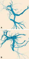
Anomalous duplication of the portal vein with prepancreatic postduodenal portal vein
- PMID: 36397802
- PMCID: PMC9613369
- DOI: 10.2185/jrm.2022-009
Anomalous duplication of the portal vein with prepancreatic postduodenal portal vein
Abstract
Objective: We report a case of unusual anomalous duplication of the portal vein. Patient: A 40-year-old man with portal vein duplication. One portal vein is derived from the superior mesenteric vein and splenic vein and enters the caudate lobe of the liver. Another portal vein, known as the prepancreatic postduodenal portal vein, is derived from the superior mesenteric vein and courses anterior to the pancreas and posterior to the duodenum. Conclusion: Duplication of the portal vein is an extremely rare developmental anomaly, and in previous reports, the superior mesenteric and splenic veins entered the liver separately. We present a previously unreported case of anomalous duplication of the portal vein, one of which was the prepancreatic postduodenal portal vein.
Keywords: anatomic variant; duplication of the portal vein; portal vein; prepancreatic postduodenal portal vein.
©2022 The Japanese Association of Rural Medicine.
Figures


References
-
- Ozbülbül NI. Congenital and acquired abnormalities of the portal venous system: multidetector CT appearances. Diagn Interv Radiol 2011; 17: 135–142. - PubMed

