

Rathke's Cleft Cyst Abscess with a Very Unusual Course
- PMID: 36398168
- PMCID: PMC9665969
- DOI: 10.1055/s-0042-1750798
Rathke's Cleft Cyst Abscess with a Very Unusual Course
Abstract
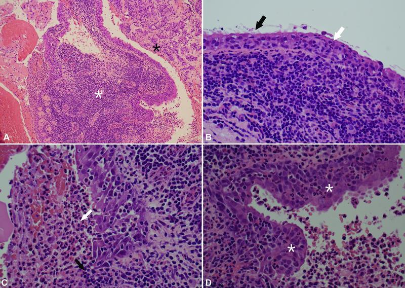
Infected Rathke's cleft cysts (RCC) are extremely rare with only a few published cases. We report the case of a 31-year-old man who presented with headaches, visual disturbance, and hypopituitarism secondary to an infected RCC with extension of abscesses along the optic tract. Magnetic resonance imaging showed ring enhancing cystic lesions within an expanded sella with suprasellar and intraparenchymal extension. The radiological appearance suggested a high-grade optic glioma, but an endoscopic transsphenoidal biopsy revealed frank pus in the pituitary fossa, which subsequently grew Staphylococcus aureus . Pathological examination of the cyst wall showed an inflamed RCC. Following a prolonged course of intravenous antibiotics, the infection resolved and vision improved. RCC abscesses are rare and the intracranial extension of the infection in our case makes it unique.
Keywords: Rathke's cleft cyst; pituitary abscess; transphenoidal surgery.
Asian Congress of Neurological Surgeons. This is an open access article published by Thieme under the terms of the Creative Commons Attribution-NonDerivative-NonCommercial License, permitting copying and reproduction so long as the original work is given appropriate credit. Contents may not be used for commercial purposes, or adapted, remixed, transformed or built upon. ( https://creativecommons.org/licenses/by-nc-nd/4.0/ ).
Conflict of interest statement
Conflict of Interest None declared.
Figures


References
-
- Jain K C, Varma A, Mahapatra A K. Pituitary abscess: a series of six cases. Br J Neurosurg. 1997;11(02):139–143. - PubMed
-
- Li Z, Yang C, Bao X. Clinical features and treatment of secondary pituitary abscess after transsphenoidal surgery: a retrospective study of 23 cases. World Neurosurg. 2018;113:e138–e145. - PubMed
-
- Aranda F, García R, Guarda F J. Rathke's cleft cyst infections and pituitary abscesses: case series and review of the literature. Pituitary. 2021;24(03):374–383. - PubMed
-
- Gomez Perun J, Eiras J, Carcavilla L I. [Intrasellar abscess into a cyst of the Rathke's pouch (authors' translation)] Neurochirurgie. 1981;27(03):201–205. - PubMed
-
- Kimura H, Fukushima T, Matsuda T, Tomonaga M. Abscess formation in a Rathke's cleft cyst [in Japanese] No To Shinkei. 1994;46(04):392–395. - PubMed
Publication types
LinkOut - more resources
Full Text Sources