

Exosomes loaded with miR-665 inhibit the progression of osteosarcoma in vivo and in vitro
- PMID: 36398229
- PMCID: PMC9641455
Exosomes loaded with miR-665 inhibit the progression of osteosarcoma in vivo and in vitro
Abstract
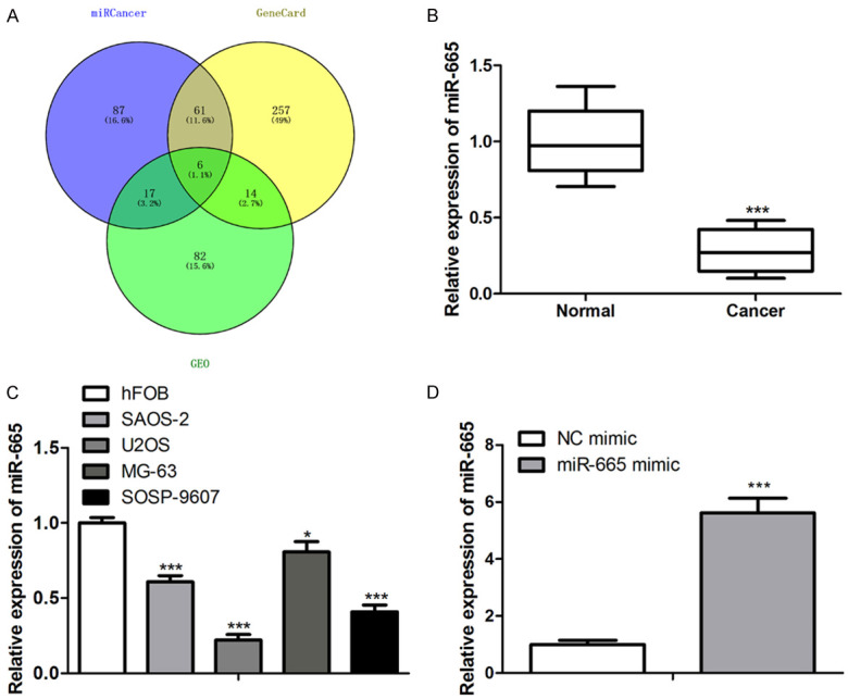
Objective: Osteosarcoma (OS) is the most common primary malignant bone tumor and has a poor prognosis. Recent research has suggested that miR-665 affects the progression of OS. Moreover, an exosome delivery system presents better targeting effects, higher permeability, and lower immunogenicity than other nano-delivery systems do. The purpose of this study is to explore whether an exosome loaded with the miR-665 delivery system can inhibit OS development.
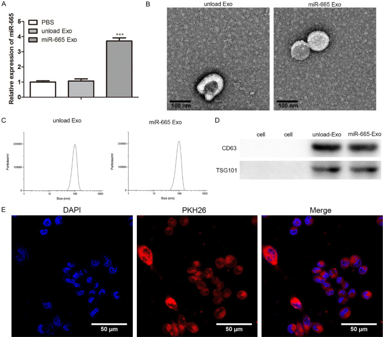
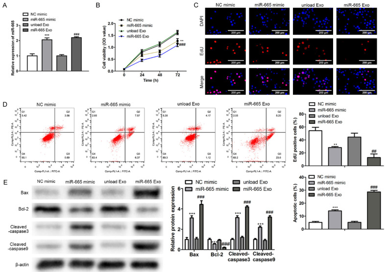
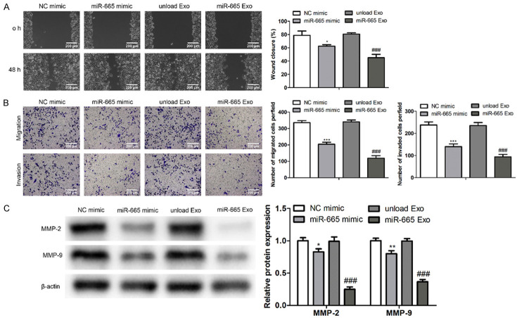
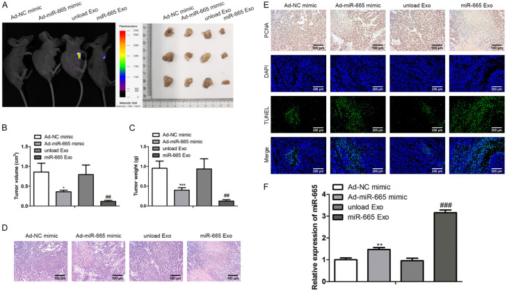
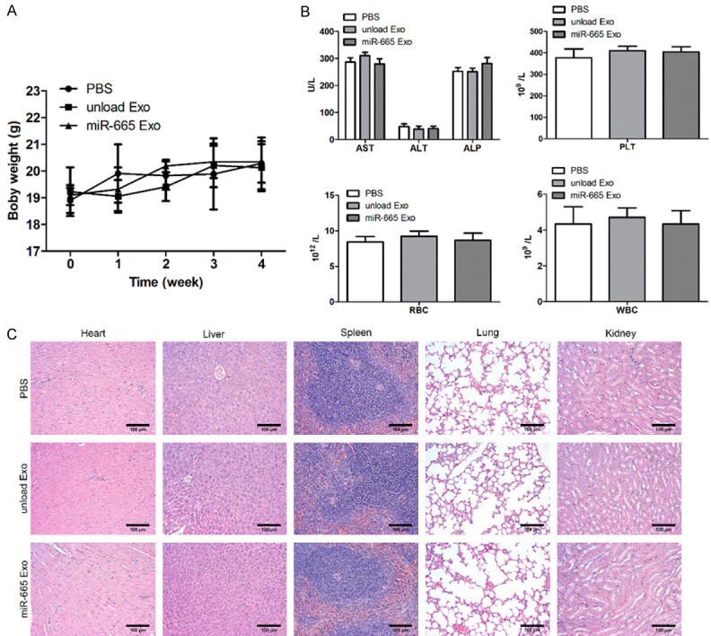
Methods: The miR-665 expression was detected through a quantitative real-time polymerase chain reaction assay. Transmission electron microscopy, nano-particle size analysis, and fluorescence microscope were utilized to observe exosomes. Cell growth was estimated by cell counting kit 8 and ethynyl deoxyuridine analyses. Assays of flow cytometry and Terminal-deoxynucleotidyl Transferase Mediated Nick End Labeling were introduced to test apoptosis in vitro or in vivo, respectively. Cell migration and invasion were measured using scratch and transwell assays. Engineered exosomes were prepared using electroporation. H&E staining was employed to observe necrotic cells and the function of heart, liver, spleen, lung and kidney. The expression of proteins was estimated by immunoblot analysis.
Results: This work documented that the expression of miR-665 was down-regulated in OS tissues. Additionally, we proved that the over-expression of miR-665 inhibited OS proliferation. Besides, we found that exosomes loaded with miR-665 had similar tumor-inhibiting effects in vivo and in vitro. Furthermore, we verified that the exosome delivery system exhibited good safety and target efficiency.
Conclusion: This work proved that exosomes loaded with miR-665 inhibited the progression of OS in vivo and in vitro in a safe manner.
Keywords: Osteosarcoma; exosomes; miR-665.
AJTR Copyright © 2022.
Conflict of interest statement
None.
Figures
Similar articles
-
Exosomes and osteosarcoma drug resistance.Front Oncol. 2023 Mar 17;13:1133726. doi: 10.3389/fonc.2023.1133726. eCollection 2023. Front Oncol. 2023. PMID: 37007086 Free PMC article. Review.
-
miRNA-221-3p derived from M2-polarized tumor-associated macrophage exosomes aggravates the growth and metastasis of osteosarcoma through SOCS3/JAK2/STAT3 axis.Aging (Albany NY). 2021 Aug 13;13(15):19760-19775. doi: 10.18632/aging.203388. Epub 2021 Aug 13. Aging (Albany NY). 2021. PMID: 34388111 Free PMC article.
-
Circ-FOXM1 promotes the proliferation, migration and EMT process of osteosarcoma cells through FOXM1-mediated Wnt pathway activation.J Orthop Surg Res. 2022 Jul 7;17(1):344. doi: 10.1186/s13018-022-03207-0. J Orthop Surg Res. 2022. PMID: 35799265 Free PMC article.
-
Ginsenoside Rg3 inhibits osteosarcoma progression by reducing circ_0003074 expression in a miR-516b-5p/KPNA4-dependent manner.J Orthop Surg Res. 2021 Dec 20;16(1):724. doi: 10.1186/s13018-021-02868-7. J Orthop Surg Res. 2021. PMID: 34930332 Free PMC article.
-
Engineered Exosomes Loaded with miR-563 Inhibit Lung Cancer Growth.J Oncol. 2022 Jun 6;2022:6141857. doi: 10.1155/2022/6141857. eCollection 2022. J Oncol. 2022. PMID: 36090893 Free PMC article.
Cited by
-
Research Advances of Engineered Exosomes as Drug Delivery Carrier.ACS Omega. 2023 Nov 9;8(46):43374-43387. doi: 10.1021/acsomega.3c04479. eCollection 2023 Nov 21. ACS Omega. 2023. PMID: 38027310 Free PMC article. Review.
-
Proof-of-Concept Study on the Use of Tangerine-Derived Nanovesicles as siRNA Delivery Vehicles toward Colorectal Cancer Cell Line SW480.Int J Mol Sci. 2023 Dec 30;25(1):546. doi: 10.3390/ijms25010546. Int J Mol Sci. 2023. PMID: 38203716 Free PMC article.
-
Bioinspired nanomedicines for the management of osteosarcoma: Recent progress and perspectives.Mater Today Bio. 2025 Mar 5;32:101607. doi: 10.1016/j.mtbio.2025.101607. eCollection 2025 Jun. Mater Today Bio. 2025. PMID: 40151805 Free PMC article. Review.
-
Visualization of microRNA therapy in cancers delivered by small extracellular vesicles.J Nanobiotechnology. 2023 Nov 29;21(1):457. doi: 10.1186/s12951-023-02187-5. J Nanobiotechnology. 2023. PMID: 38031152 Free PMC article.
-
Exosomes and osteosarcoma drug resistance.Front Oncol. 2023 Mar 17;13:1133726. doi: 10.3389/fonc.2023.1133726. eCollection 2023. Front Oncol. 2023. PMID: 37007086 Free PMC article. Review.
References
-
- Briccoli A, Rocca M, Salone M, Guzzardella GA, Balladelli A, Bacci G. High grade osteosarcoma of the extremities metastatic to the lung: long-term results in 323 patients treated combining surgery and chemotherapy, 1985-2005. Surg Oncol. 2010;19:193–199. - PubMed
-
- Ottaviani G, Jaffe N. The epidemiology of osteosarcoma. Cancer Treat Res. 2009;152:3–13. - PubMed
-
- Mortus JR, Zhang Y, Hughes DP. Developmental pathways hijacked by osteosarcoma. Adv Exp Med Biol. 2014;804:93–118. - PubMed
-
- Powers M, Zhang W, Lopez-Terrada D, Czerniak BA, Lazar AJ. The molecular pathology of sarcomas. Cancer Biomark. 2010;9:475–91. - PubMed
-
- Gangi A, Chung A, Mirocha J, Liou DZ, Leong T, Giuliano AE. Breast-conserving therapy for triple-negative breast cancer. JAMA Surg. 2014;149:252–258. - PubMed
LinkOut - more resources
Full Text Sources