. 2022 Dec 7;33(12):2263-2272.
doi: 10.1021/jasms.2c00226. Epub 2022 Nov 18.

Exploring New Methods to Study and Moderate Proton Beam Damage for Multimodal Imaging on a Single Tissue Section

Affiliations

Exploring New Methods to Study and Moderate Proton Beam Damage for Multimodal Imaging on a Single Tissue Section

Catia Costa et al. J Am Soc Mass Spectrom. .

Abstract

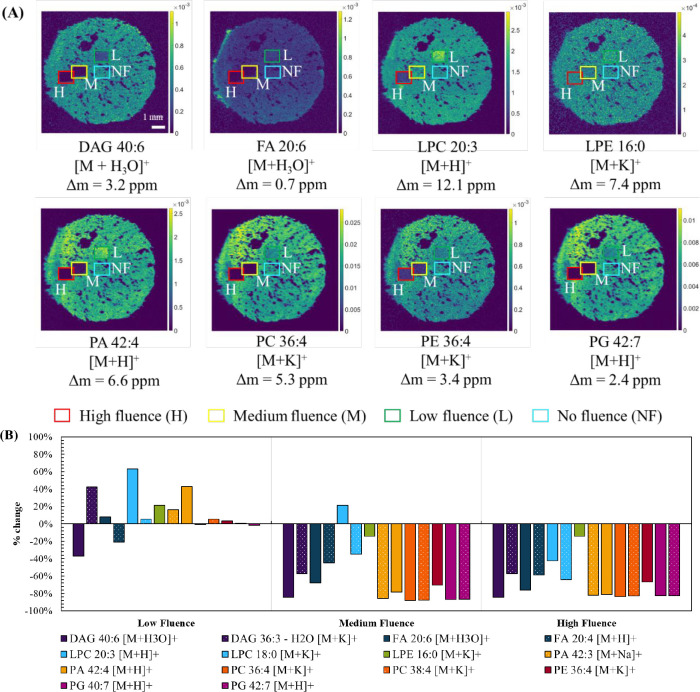
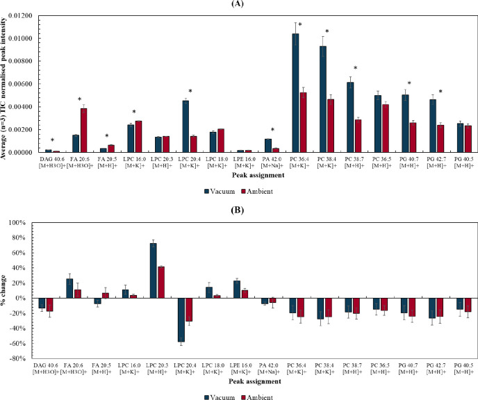
Characterizing proton beam damage in biological materials is of interest to enable the integration of proton microprobe elemental mapping techniques with other imaging modalities. It is also of relevance to obtain a deeper understanding of mechanical damage to lipids in tissues during proton beam cancer therapy. We have developed a novel strategy to characterize proton beam damage to lipids in biological tissues based on mass spectrometry imaging. This methodology is applied to characterize changes to lipids in tissues ex vivo, irradiated under different conditions designed to mitigate beam damage. This work shows that performing proton beam irradiation at ambient pressure, as well as including the application of an organic matrix prior to irradiation, can reduce damage to lipids in tissues. We also discovered that, irrespective of proton beam irradiation, placing a sample in a vacuum prior to desorption electrospray ionization imaging can enhance lipid signals, a conclusion that may be of future benefit to the mass spectrometry imaging community.


Conflict of interest statement

The authors declare no competing financial interest.

Figures

Figure 1
Figure 1
(A) Total ion count (TIC) image for the three sequential tissue sections and respective regions of interest (ROI) selected. (B) Relative standard deviations (RSD %) of normalized (to TIC) data of three ROIs taken on three tissue homogenates and across all three sections (n = 9). *Indicates compounds with m/z < 700.
Figure 2
Figure 2
(A) Extracted ion maps obtained using DESI following irradiation with a 2.5 MeV proton beam under vacuum, irradiated at % low, medium, and high fluences as described in Table 1. (B) Percentage change (% change) in TIC-normalized peak intensity between a low, medium, or high fluence area and a nonirradiated area (no fluence).
Figure 3
Figure 3
(A) Average (n = 3) normalized (to TIC) peak intensity for a range of lipid species taken from unirradiated regions of interest (approximately 1 × 1 mm areas) of tissue sections irradiated under ambient or vacuum conditions and sequentially imaged using DESI. * is used to show differences with p < 0.05 (Table S3, Supporting Information). (B) % change in normalized (to TIC) peak intensity between “low fluence” and “no irradiation” ROIs on tissue sections irradiated under ambient or vacuum conditions. Error bars represent the RSD% of the normalized peak intensities taken across the 3 regions of interest.
Figure 4
Figure 4
(A) ROIs for irradiated and nonirradiated areas. (B) DESI ion maps of selected lipid peaks showing the irradiated areas. (C) % change between the “low fluence” and no irradiation areas at different scan speeds and pattern.
Figure 5
Figure 5
(A) Extracted ion maps of lipid signals obtained using DESI of tissue homogenates coated with matrices (DHB, CHCA, 9-AA) and a control (no matrix) after proton beam irradiation. (B) Normalized (to TIC) peak intensity taken from nonirradiated areas (n = 3) in each of the tissue sections. Lipids marked with an γ, δ, and θ present statistically different levels (p < 0.05) between the control and DHB, control, and CHCA and control and 9-AA, respectively (see Table 4, Supporting Information). (C) Percentage change between low fluence and no fluence ROIs in the same sample.
Figure 6
Figure 6
Percentage change between medium fluence and no fluence in the “ambient irradiation” and “irradiation in the presence of DHB matrix” experiments.

References

    1. Neumann E. K.; Djambazova K. V.; Caprioli R. M.; Spraggins J. M. Multimodal Imaging Mass Spectrometry: Next Generation Molecular Mapping in Biology and Medicine. J. Am. Soc. Mass Spectrom. 2020, 31 (12), 2401–2415. 10.1021/jasms.0c00232. - DOI - PMC - PubMed
    1. Iakab S. A.; Ràfols P.; Correig-Blanchar X.; García-Altares M. Perspective on Multimodal Imaging Techniques Coupling Mass Spectrometry and Vibrational Spectroscopy: Picturing the Best of Both Worlds. Anal. Chem. 2021, 93 (16), 6301–6310. 10.1021/acs.analchem.0c04986. - DOI - PMC - PubMed
    1. de Jesus J. M.; Costa C.; Burton A.; Palitsin V.; Webb R.; Taylor A.; Nikula C.; Dexter A.; Kaya F.; Chambers M.; Dartois V.; Goodwin R. J. A.; Bunch J.; Bailey M. J. Correlative Imaging of Trace Elements and Intact Molecular Species in a Single-Tissue Sample at the 50 μm Scale. Anal. Chem. 2021, 93 (40), 13450–13458. 10.1021/acs.analchem.1c01927. - DOI - PubMed
    1. Perry W. J.; Weiss A.; Van de Plas R.; Spraggins J. M.; Caprioli R. M.; Skaar E. P. Integrated molecular imaging technologies for investigation of metals in biological systems: A brief review. Curr. Opin. Chem. Biol. 2020, 55, 127–135. 10.1016/j.cbpa.2020.01.008. - DOI - PMC - PubMed
    1. Sturm G.; List M.; Zhang J. D., Tissue heterogeneity is prevalent in gene expression studies. NAR: Genomics Bioinf. 2021, 3 ( (3), ).10.1093/nargab/lqab077 - DOI - PMC - PubMed