Randomized Controlled Trial
. 2022 Nov;4(6):e220073.
doi: 10.1148/rycan.220073.

Prospective Multireader Evaluation of Photon-counting CT for Multiple Myeloma Screening

Affiliations
Randomized Controlled Trial

Prospective Multireader Evaluation of Photon-counting CT for Multiple Myeloma Screening

Fides R Schwartz et al. Radiol Imaging Cancer. 2022 Nov.

Abstract

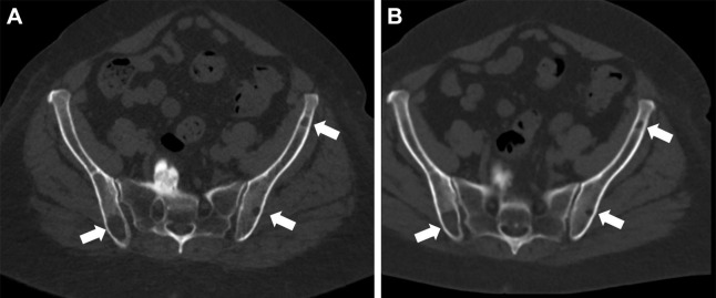
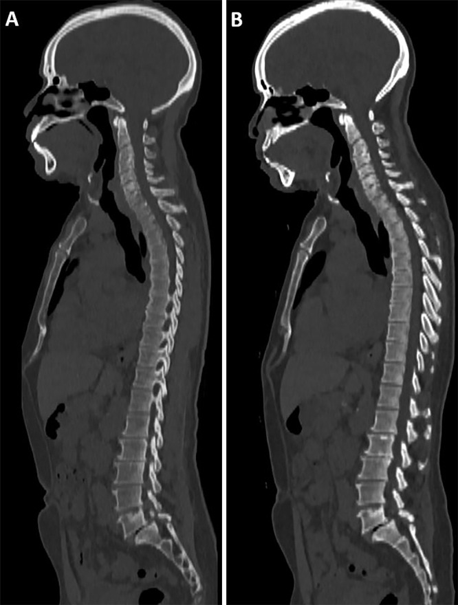
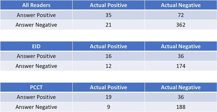
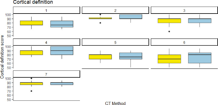
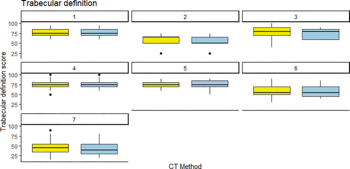
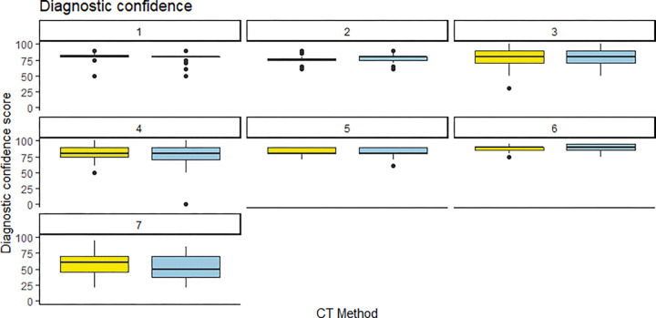
Purpose To determine whether photon-counting CT (PCCT) acquisition of whole-body CT images provides similar quantitative image quality and reader satisfaction for multiple myeloma screening at lower radiation doses than does standard energy-integrating detector (EID) CT. Materials and Methods Patients with monoclonal gammopathy of undetermined significance prospectively underwent clinical noncontrast whole-body CT with EID and same-day PCCT (August-December 2021). Five axial scan locations were evaluated by seven radiologists, with 11% (eight of 70) of images including osteolytic lesions. Images were shown in randomized order, and each reader rated the following: discernibility of the osseous cortex and osseous trabeculae, perceived image noise level, and diagnostic confidence. Presence of lytic osseous lesions was indicated. Contrast-to-noise ratio (CNR) and signal-to-noise ratio (SNR) were calculated. Comparisons were made using paired t tests and mixed linear effects models. Results Seven participants (four women) were included (mean age, 66 years ± 9 [SD]; body mass index, 30.1 kg/m2 ± 5.2). Mean cortical definition, trabecular definition, image noise, and image quality scores were 83, 67, 75, and 78 versus 84, 66, 74, and 76 for EID and PCCT, respectively (P = .65, .11, .26, and .11, respectively). PCCT helped identify more lesions (79% [22 of 28]) than did EID (64% [18 of 28]). CNRs and SNRs were similar between modalities. PCCT had lower radiation doses than EID (volume CT dose index: EID, 11.37 ± 2.8 vs PCCT, 1.8 ± 0.6 [P = .06]; dose-length product: EID, 1654.1 ± 409.6 vs PCCT, 253.4 ± 89.6 [P = .05]). Conclusion This pilot investigation suggests that PCCT affords similar quantitative and qualitative scores as EID at significantly lower radiation doses. Keywords: CT, CT-Spectral, Skeletal-Axial, Spine, Hematologic Diseases, Whole-Body Imaging, Comparative Studies Supplemental material is available for this article. © RSNA, 2022.

Keywords: CT; CT-Spectral; Comparative Studies; Hematologic Diseases; Skeletal-Axial; Spine; Whole-Body Imaging.


Conflict of interest statement

Disclosures of conflicts of interest: F.R.S. Payment or honoraria from Siemens Healthineers for educational luncheon presentation at Society of Thoracic Radiology (STR) March 2022, Las Vegas; support from Siemens Healthineers for travel to STR 2022; participation on a photon-counting CT advisory board for Siemens Healthineers (no financial compensation). E.N.V. No relevant relationships. C.E.S. Royalties or licenses from Now UptoDate for the "Imaging of osteomyelitis" section. R.C. No relevant relationships. E.S. Research funding for this work from Siemens Healthineers; grants or contracts from GE and Siemens; royalties or licenses from GE, SunNuclear, XCAT, and Metis Health Analytics; consulting fees from Nanox; payment or honoraria for lectures, presentations, speakers bureaus, manuscript writing, or education events from Cambridge University Press and Wiley and Sons; payment from Rubin Anders for expert testimony; participation on a DataSafety Monitoring Board or Advisory Board for Imalogix, Metis Health Analytics, GE, and Siemens; leadership or fiduciary role in the American Association of Physicists in Medicine (AAPM). R.J.F. No relevant relationships. N.S. No relevant relationships. L.W. No relevant relationships. E.M. No relevant relationships.

Figures

(A) Energy-integrating detector (EID) CT and (B) photon-counting CT
(PCCT) images in a 71-year-old woman with a body mass index of 33 kg/m2 and
multiple lytic multiple myeloma lesions (white arrows). Dose-length product
for this study was 1608 mGy·cm for EID and 257 mGy·cm for
PCCT. Volume CT dose index was 11.3 mGy for EID and 1.8 mGy for
PCCT.
Figure 1:
(A) Energy-integrating detector (EID) CT and (B) photon-counting CT (PCCT) images in a 71-year-old woman with a body mass index of 33 kg/m2 and multiple lytic multiple myeloma lesions (white arrows). Dose-length product for this study was 1608 mGy·cm for EID and 257 mGy·cm for PCCT. Volume CT dose index was 11.3 mGy for EID and 1.8 mGy for PCCT.
(A) Energy-integrating detector (EID) CT and (B) photon-counting CT
(PCCT) images in a 61-year-old man with a body mass index of 28 kg/m2 and no
multiple myeloma lesions. Dose-length product for this study was 1421
mGy·cm for EID and 213 mGy·cm for PCCT. Volume CT dose index
was 9.04 mGy for EID and 1.47 mGy for PCCT.
Figure 2:
(A) Energy-integrating detector (EID) CT and (B) photon-counting CT (PCCT) images in a 61-year-old man with a body mass index of 28 kg/m2 and no multiple myeloma lesions. Dose-length product for this study was 1421 mGy·cm for EID and 213 mGy·cm for PCCT. Volume CT dose index was 9.04 mGy for EID and 1.47 mGy for PCCT.
Confusion matrices for all images and readers combined and separated
out according to imaging modality. There were more lesions detected and
fewer missed lesions with PCCT than with EID CT. EID = energy-integrating
detector, PCCT = photon-counting CT.
Figure 3:
Confusion matrices for all images and readers combined and separated out according to imaging modality. There were more lesions detected and fewer missed lesions with PCCT than with EID CT. EID = energy-integrating detector, PCCT = photon-counting CT.
Individual reader scoring differences between energy-integrating
detector CT (EID, yellow) and photon-counting CT (PCCT, blue) for cortical
definition. Note that the median score was almost identical for both CT
methods, save for one reader (reader 6) who rated the PCCT images slightly
higher than the EID images.
Figure 4:
Individual reader scoring differences between energy-integrating detector CT (EID, yellow) and photon-counting CT (PCCT, blue) for cortical definition. Note that the median score was almost identical for both CT methods, save for one reader (reader 6) who rated the PCCT images slightly higher than the EID images.
Individual reader scoring differences between energy-integrating
detector CT (EID, yellow) and photon-counting CT (PCCT, blue) for trabecular
definition. Note that the median score was almost identical for both CT
methods, save for one reader (reader 2) who rated the PCCT images slightly
lower than the EID images.
Figure 5:
Individual reader scoring differences between energy-integrating detector CT (EID, yellow) and photon-counting CT (PCCT, blue) for trabecular definition. Note that the median score was almost identical for both CT methods, save for one reader (reader 2) who rated the PCCT images slightly lower than the EID images.
Individual reader scoring differences between energy-integrating
detector CT (EID, yellow) and photon-counting CT (PCCT, blue) for diagnostic
confidence. Note that the median score was almost identical for both CT
methods, save for one reader (reader 6) who rated the PCCT images slightly
higher than the EID images.
Figure 6:
Individual reader scoring differences between energy-integrating detector CT (EID, yellow) and photon-counting CT (PCCT, blue) for diagnostic confidence. Note that the median score was almost identical for both CT methods, save for one reader (reader 6) who rated the PCCT images slightly higher than the EID images.

References

    1. Terpos E , Kleber M , Engelhardt M , et al. ; European Myeloma Network . European Myeloma Network guidelines for the management of multiple myeloma-related complications . Haematologica 2015. ; 100 ( 10 ): 1254 – 1266 . - PMC - PubMed
    1. Moreau P , San Miguel J , Sonneveld P , et al. ; ESMO Guidelines Committee . Multiple myeloma: ESMO Clinical Practice Guidelines for diagnosis, treatment and follow-up . Ann Oncol 2017. ; 28 ( suppl_4 ): iv52 – iv61 . - PubMed
    1. Ormond Filho AG , Carneiro BC , Pastore D , et al. . Whole-body imaging of multiple myeloma: diagnostic criteria . RadioGraphics 2019. ; 39 ( 4 ): 1077 – 1097 . - PubMed
    1. Cowan AJ , Green DJ , Kwok M , et al. . Diagnosis and management of multiple myeloma: a review . JAMA 2022. ; 327 ( 5 ): 464 – 477 . - PubMed
    1. van de Donk NWCJ , Pawlyn C , Yong KL . Multiple myeloma . Lancet 2021. ; 397 ( 10272 ): 410 – 427 . - PubMed

Publication types