. 2023 Mar 1;13(3):616-631.
doi: 10.1158/2159-8290.CD-22-0659.

Detecting Liver Cancer Using Cell-Free DNA Fragmentomes

Affiliations

Detecting Liver Cancer Using Cell-Free DNA Fragmentomes

Zachariah H Foda et al. Cancer Discov. .

Abstract

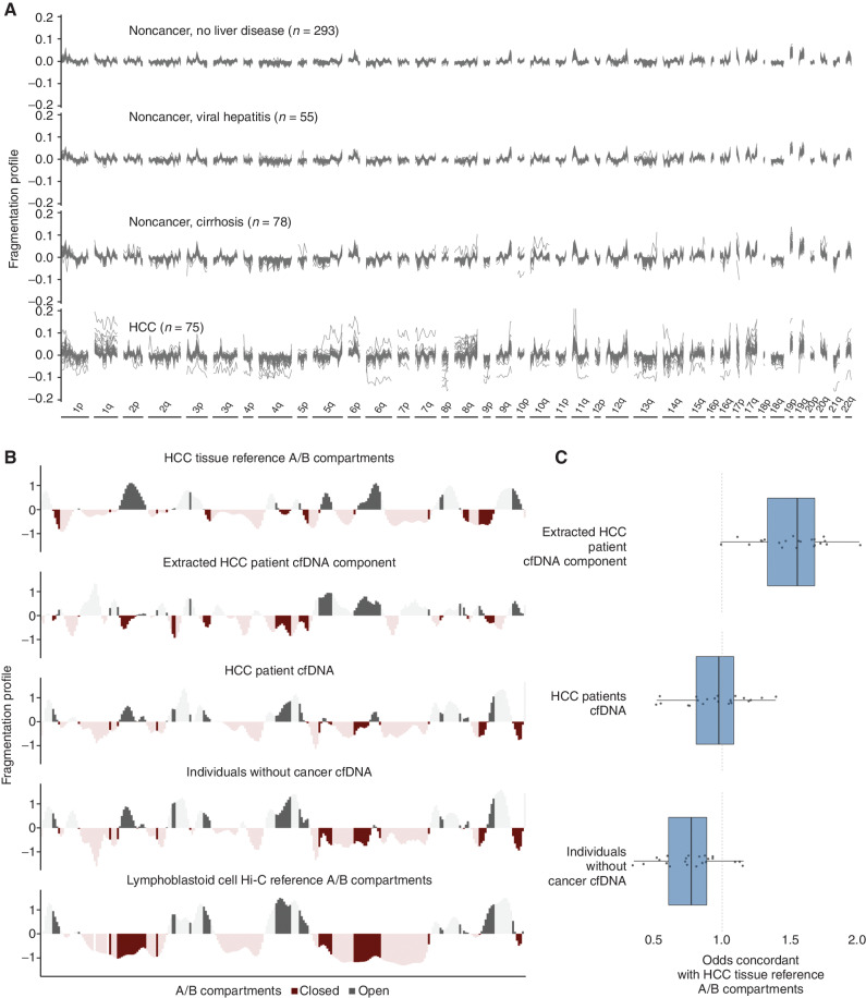
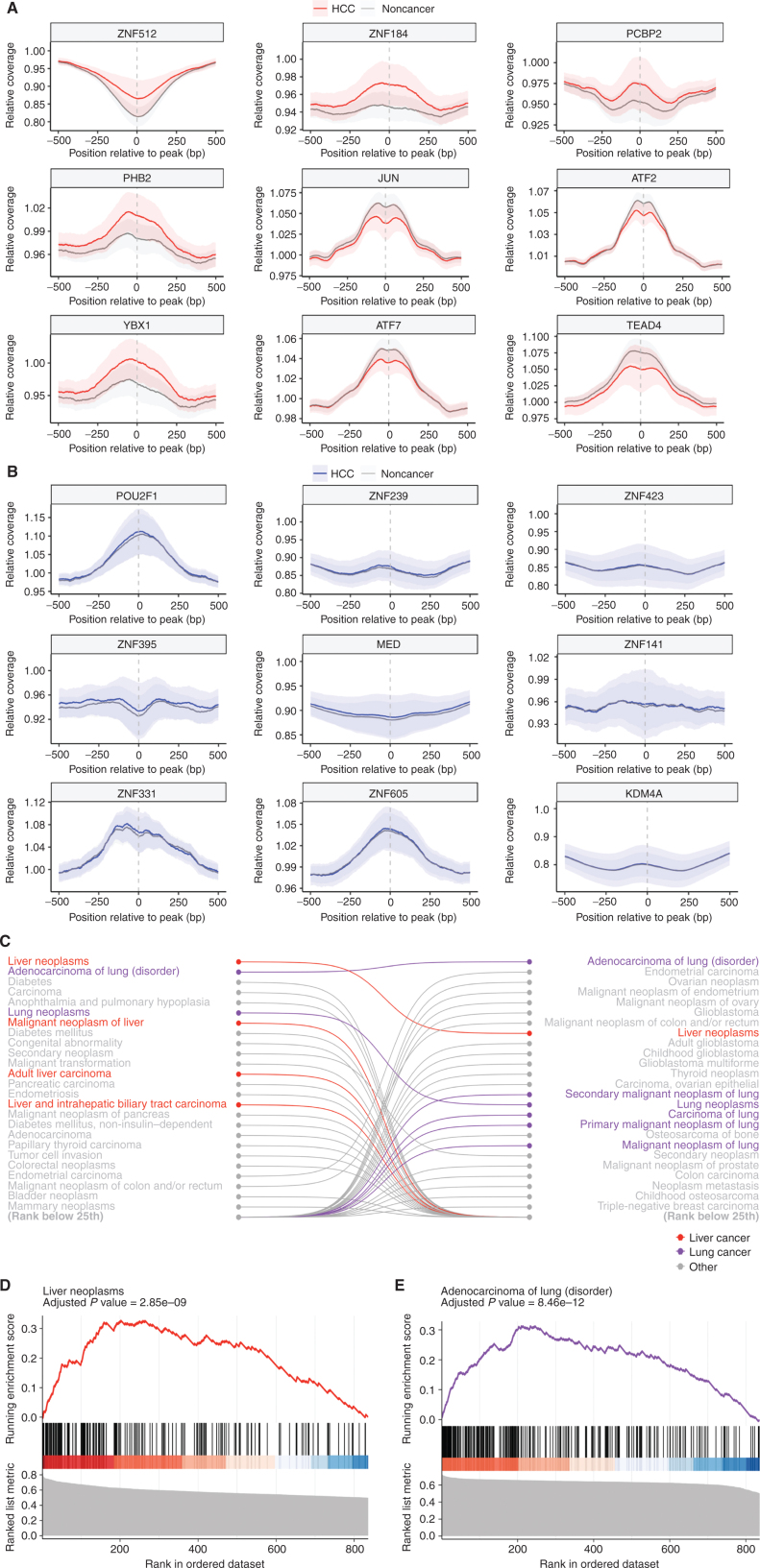
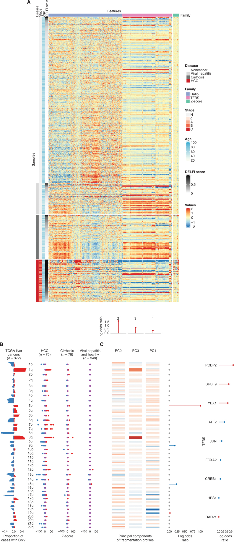
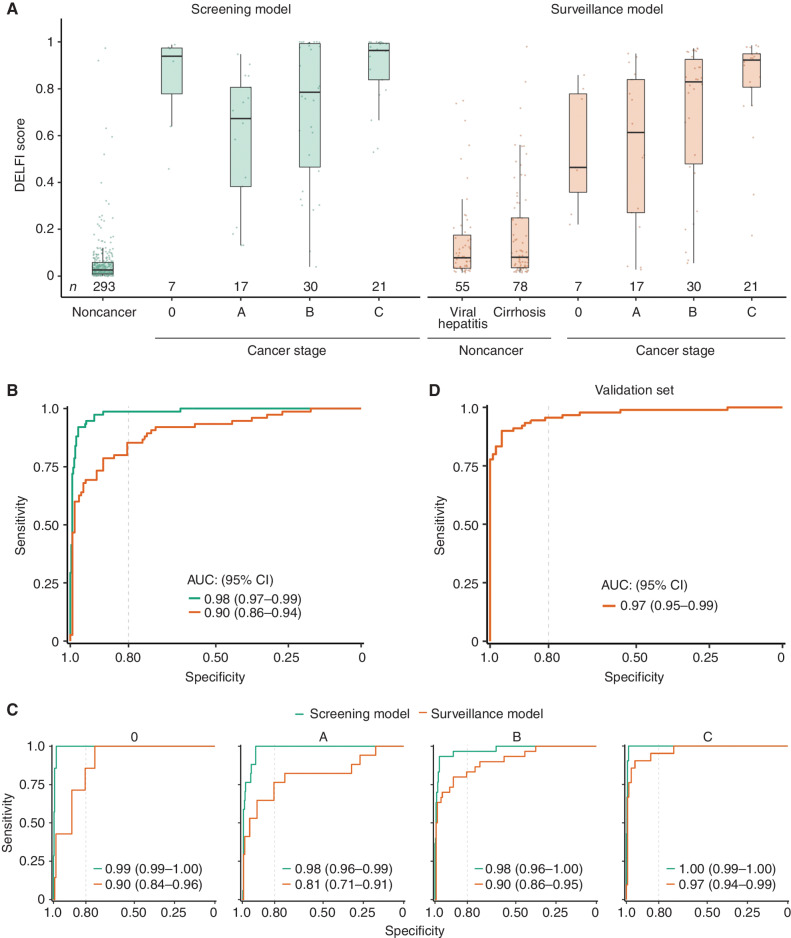
Liver cancer is a major cause of cancer mortality worldwide. Screening individuals at high risk, including those with cirrhosis and viral hepatitis, provides an avenue for improved survival, but current screening methods are inadequate. In this study, we used whole-genome cell-free DNA (cfDNA) fragmentome analyses to evaluate 724 individuals from the United States, the European Union, or Hong Kong with hepatocellular carcinoma (HCC) or who were at average or high-risk for HCC. Using a machine learning model that incorporated multifeature fragmentome data, the sensitivity for detecting cancer was 88% in an average-risk population at 98% specificity and 85% among high-risk individuals at 80% specificity. We validated these results in an independent population. cfDNA fragmentation changes reflected genomic and chromatin changes in liver cancer, including from transcription factor binding sites. These findings provide a biological basis for changes in cfDNA fragmentation in patients with liver cancer and provide an accessible approach for noninvasive cancer detection.

Significance: There is a great need for accessible and sensitive screening approaches for HCC worldwide. We have developed an approach for examining genome-wide cfDNA fragmentation features to provide a high-performing and cost-effective approach for liver cancer detection. See related commentary Rolfo and Russo, p. 532. This article is highlighted in the In This Issue feature, p. 517.


Figures

Figure 1. Genome-wide fragmentation profiles reflect underlying chromatin structure. A, Fragmentation profiles of 501 individuals in 473 nonoverlapping 5 mb genomic regions. Fragmentation profiles for cancer individuals show marked heterogeneity as compared with noncancer individuals with and without liver disease. B, Comparison of plasma fragmentation features to reference A/B compartments. Track 1 shows A/B compartments extracted from liver cancer tissue (28). Track 2 shows a median liver cancer component extracted from the HCC plasma samples of 10 liver patients with high tumor fraction by ichor CNA (56). Track 3 shows the median fragmentation profile in the plasma for these 10 HCC samples and track 4 shows the median profile for 10 healthy plasma samples. Track 5 shows A/B compartments for lymphoblast cells (28). These five tracks show chromosome 22 as an example, with darker shading indicating informative regions of the genome where the two reference tracks differ in domain (open/closed) or magnitude. C, among these informative bins, for each chromosome, the log odds of the plasma component matching the HCC reference track in domain. Log odds greater than 1 indicate more similarity to the HCC reference track, whereas log odds less than 1 indicate more similarity to the lymphoblast reference track. The extracted HCC component has the greatest similarity to the HCC reference track and the noncancer plasma has the greatest similarity to the lymphoblast reference track; the HCC plasma track is intermediate to the two.
Figure 1.
Genome-wide fragmentation profiles reflect underlying chromatin structure. A, Fragmentation profiles of 501 individuals in 473 nonoverlapping 5-Mb genomic regions. Fragmentation profiles for individuals with cancer show marked heterogeneity as compared with noncancer individuals with and without liver disease. B, Comparison of plasma fragmentation features to reference A/B compartments. Track 1 shows A/B compartments extracted from liver cancer tissue (28). Track 2 shows a median liver cancer component extracted from the HCC plasma samples of 10 liver patients with high tumor fraction by ichorCNA (56). Track 3 shows the median fragmentation profile in the plasma for these 10 HCC samples, and track 4 shows the median profile for 10 healthy plasma samples. Track 5 shows A/B compartments for lymphoblast cells (28). These five tracks show chromosome 22 as an example, with darker shading indicating informative regions of the genome where the two reference tracks differ in domain (open/closed) or magnitude. C, Among these informative bins, for each chromosome, the log odds of the plasma component matching the HCC reference track in domain. Log odds greater than 1 indicate more similarity to the HCC reference track, whereas log odds less than 1 indicate more similarity to the lymphoblast reference track. The extracted HCC component has the greatest similarity to the HCC reference track, and the noncancer plasma has the greatest similarity to the lymphoblast reference track; the HCC plasma track is intermediate to the two.
Figure 2. Fragmentation profiles in HCC patients highlight liver-specific TFs. A, The coverage at and around the transcription factor binding sites (TFBS) for the 9 TFs for which the relative coverage at the binding site that had the highest separation of HCC from noncancer samples. The mean is plotted for each group, with ± 1 standard deviation (SD) shown by shading. These confidence intervals (CI) show separation, highlighting that differences in coverage at a TFBS can provide information on cancer status. B, The coverage at and around the TFBS for the 9 TFs that had the lowest separation of HCC from noncancer samples in the US/EU cohort. These CIs are largely overlapping, reflecting their status as TFBS with poor discrimination. Gene set enrichment analysis of TFs analyzed in both HCC and lung adenocarcinoma showed TFs are selectively enriched in numerous pathways related to liver and lung cancer, respectively (C), including adult liver carcinoma and adenocarcinoma of the lung (D and E).
Figure 2.
Fragmentation profiles in patients with HCC highlight liver-specific TFs. A, The coverage at and around the TF binding sites (TFBS) for the 9 TFs for which the relative coverage at the binding site had the highest separation of HCC from noncancer samples. The mean is plotted for each group, with ± 1 SD shown by shading. These confidence intervals (CI) show separation, highlighting that differences in coverage at a TFBS can provide information on cancer status. B, The coverage at and around the TFBS for the 9 TFs that had the lowest separation of HCC from noncancer samples in the US/EU cohort. These CIs are largely overlapping, reflecting their status as TFBS with poor discrimination.Gene set enrichment analysis of TFs analyzed in both HCC and lung adenocarcinoma showed TFs are selectively enriched in numerous pathways related to liver and lung cancer, respectively (C), including adult liver carcinoma and adenocarcinoma of the lung (D and E).
Figure 3. High-dimensional fragmentation features reflect liver cancer biology and are incorporated in DELFI machine learning approaches. A, A heat map reflecting the complexity of genome-wide fragmentation and transcription factor binding site features utilized in the DELFI machine learning approach. Each row represents a sample, whereas columns show individual genomic features. B, Analysis of copy-number changes in tissue from 372 TCGA liver cancers and plasma from 501 individuals reflects biological consistency. Copy-number changes that occur in TCGA (red = gains, blue = losses) were also found at the chromosomal arm level in HCC plasma, but not in individuals without cancer. C, Heat map depicting the contributions of individual genomic regions to the final trained DELFI model. The fragmentation features were summarized as three principal components in the model, whereas aneuploidy was summarized as arm-level z-scores. The top, middle, and right depict the coefficients of fragmentation components, arm-level z-scores, and transcription factor binding sites, respectively, in the model. CNV, copy-number variation.
Figure 3.
High-dimensional fragmentation features reflect liver cancer biology and are incorporated in DELFI machine learning approaches. A, A heat map reflecting the complexity of genome-wide fragmentation and TF binding site (TFBS) features utilized in the DELFI machine learning approach. Each row represents a sample, whereas columns show individual genomic features.B, Analysis of copy-number changes in tissue from 372 TCGA liver cancers and plasma from 501 individuals reflects biological consistency. Copy-number changes that occur in TCGA (red = gains, blue = losses) were also found at the chromosomal arm level in HCC plasma, but not in individuals without cancer. CNV, copy-number variation. C, Heat map depicting the contributions of individual genomic regions to the final trained DELFI model. The fragmentation features were summarized as three principal components (PC) in the model, whereas aneuploidy was summarized as arm-level z-scores. The top, middle, and right depict the coefficients of fragmentation components, arm-level z-scores, and TFBS, respectively, in the model.
Figure 4. DELFI machine learning models detect liver cancer with high sensitivity and specificity. A, DELFI scores for the US/EU cohort across liver disease and cancer stage for the screening and surveillance models. Cirrhotic patients have DELFI scores higher than individuals without cancer or with viral hepatitis on average, but lower than all stages of liver cancer. Patients with liver cancer across all stages have relatively high DELFI scores, with stage C individuals uniformly having the highest DELFI scores. B, ROC analyses of the US/EU general population cohort and the high-risk surveillance cohort. C, ROC analyses of the US/EU general population and surveillance cohorts separated by BCLC stage, showing high sensitivity and specificity across stages. D, ROC analyses for the fixed surveillance model applied to the Hong Kong cohort, which includes 90 HCC individuals with HCC (85 with BCLC stage A cancer, and 5 with BCLC stage B cancer), 101 individuals with cirrhosis and viral hepatitis and 32 individuals without cancer or liver disease.
Figure 4.
DELFI machine learning models detect liver cancer with high sensitivity and specificity. A, DELFI scores for the US/EU cohort across liver disease and cancer stage for the screening and surveillance models. Patients with cirrhosis have DELFI scores higher than individuals without cancer or with viral hepatitis on average, but lower than all stages of liver cancer. Patients with liver cancer across all stages have relatively high DELFI scores, with stage C individuals uniformly having the highest DELFI scores. B, ROC analyses of the US/EU general population cohort and the high-risk surveillance cohort. C, ROC analyses of the US/EU general population and surveillance cohorts separated by BCLC stage, showing high sensitivity and specificity across stages. D, ROC analyses for the fixed surveillance model applied to the Hong Kong cohort, which includes 90 individuals with HCC (85 with BCLC stage A cancer, and 5 with BCLC stage B cancer), 101 individuals with cirrhosis and viral hepatitis, and 32 individuals without cancer or liver disease.

Comment in

References

    1. Sung H, Ferlay J, Siegel RL, Laversanne M, Soerjomataram I, Jemal A, et al. . Global cancer statistics 2020: GLOBOCAN estimates of incidence and mortality worldwide for 36 cancers in 185 countries. CA Cancer J Clin 2021;71:209–49. - PubMed
    1. Siegel RL, Miller KD, Fuchs HE, Jemal A. Cancer statistics, 2021. CA Cancer J Clin 2021;71:7–33. - PubMed
    1. Di Bisceglie AM. Hepatitis C and hepatocellular carcinoma. Hepatology 1997;26(3Suppl 1):34S–8S. - PubMed
    1. Pinyopornpanish K, Khoudari G, Saleh MA, Angkurawaranon C, Pinyopornpanish K, Mansoor E, et al. . Hepatocellular carcinoma in nonalcoholic fatty liver disease with or without cirrhosis: a population-based study. BMC gastroenterology 2021;21:1–7. - PMC - PubMed
    1. Donato F, Tagger A, Gelatti U, Parrinello G, Boffetta P, Albertini A, et al. . Alcohol and hepatocellular carcinoma: the effect of lifetime intake and hepatitis virus infections in men and women. Am J Epidemiol 2002;155:323–31. - PubMed

Publication types

MeSH terms

Substances