. 2022 Nov 18;18(11):e1010951.
doi: 10.1371/journal.ppat.1010951. eCollection 2022 Nov.

Deep mutational scans for ACE2 binding, RBD expression, and antibody escape in the SARS-CoV-2 Omicron BA.1 and BA.2 receptor-binding domains

Affiliations

Deep mutational scans for ACE2 binding, RBD expression, and antibody escape in the SARS-CoV-2 Omicron BA.1 and BA.2 receptor-binding domains

Tyler N Starr et al. PLoS Pathog. .

Abstract

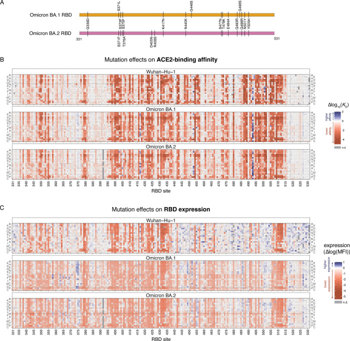
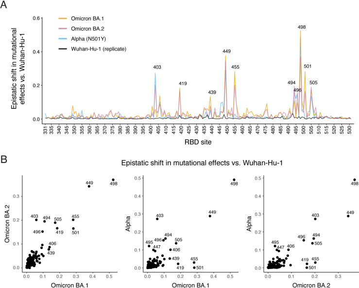
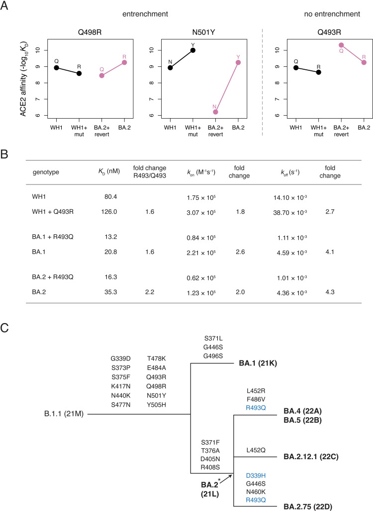
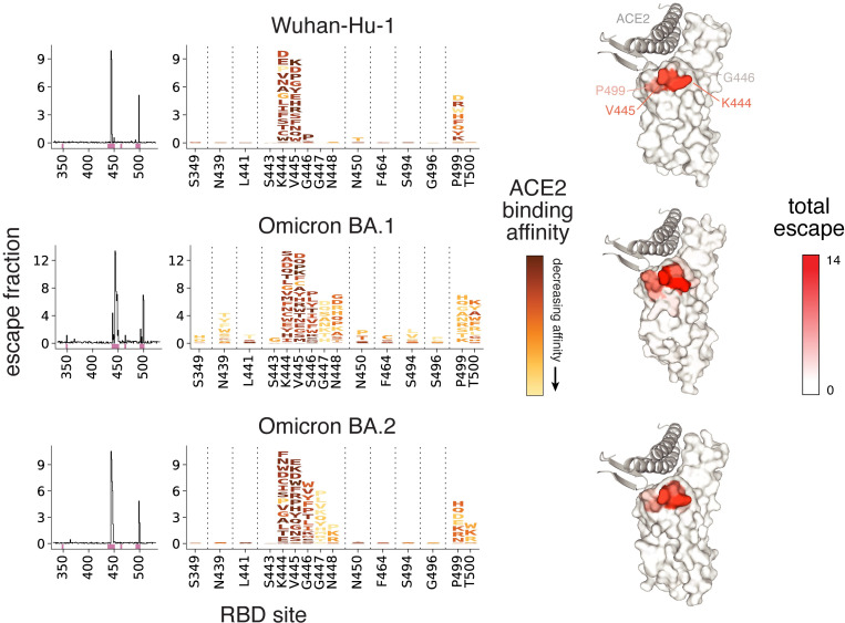
SARS-CoV-2 continues to acquire mutations in the spike receptor-binding domain (RBD) that impact ACE2 receptor binding, folding stability, and antibody recognition. Deep mutational scanning prospectively characterizes the impacts of mutations on these biochemical properties, enabling rapid assessment of new mutations seen during viral surveillance. However, the effects of mutations can change as the virus evolves, requiring updated deep mutational scans. We determined the impacts of all single amino acid mutations in the Omicron BA.1 and BA.2 RBDs on ACE2-binding affinity, RBD folding, and escape from binding by the LY-CoV1404 (bebtelovimab) monoclonal antibody. The effects of some mutations in Omicron RBDs differ from those measured in the ancestral Wuhan-Hu-1 background. These epistatic shifts largely resemble those previously seen in the Alpha variant due to the convergent epistatically modifying N501Y substitution. However, Omicron variants show additional lineage-specific shifts, including examples of the epistatic phenomenon of entrenchment that causes the Q498R and N501Y substitutions present in Omicron to be more favorable in that background than in earlier viral strains. In contrast, the Omicron substitution Q493R exhibits no sign of entrenchment, with the derived state, R493, being as unfavorable for ACE2 binding in Omicron RBDs as in Wuhan-Hu-1. Likely for this reason, the R493Q reversion has occurred in Omicron sub-variants including BA.4/BA.5 and BA.2.75, where the affinity buffer from R493Q reversion may potentiate concurrent antigenic change. Consistent with prior studies, we find that Omicron RBDs have reduced expression, and identify candidate stabilizing mutations that ameliorate this deficit. Last, our maps highlight a broadening of the sites of escape from LY-CoV1404 antibody binding in BA.1 and BA.2 compared to the ancestral Wuhan-Hu-1 background. These BA.1 and BA.2 deep mutational scanning datasets identify shifts in the RBD mutational landscape and inform ongoing efforts in viral surveillance.


Conflict of interest statement

I have read the journal’s policy and the authors of this manuscript have the following competing interests: J.D.B. has consulted for Moderna and Merck on viral evolution and epidemiology. T.N.S. and J.D.B. consult for Apriori Bio on deep mutational scanning. T.N.S., A.J.G., and J.D.B. receive a share of intellectual property revenue as inventors on Fred Hutchinson Cancer Center–optioned technology and patents related to deep mutational scanning of viral proteins and stabilization of SARS-CoV-2 RBDs.

Figures

Fig 1
Fig 1. Effects of mutations in Omicron BA.1 and BA.2 receptor-binding domains on ACE2-binding and RBD expression.
(A) Diagram of the RBD substitutions that distinguish Omicron BA.1 and BA.2 from Wuhan-Hu-1. We use Wuhan-Hu-1 spike numbering throughout. (B, C) Heatmaps illustrating the impacts of all single mutations on ACE2-binding affinity (B) and RBD surface expression (C), as determined from FACS-seq assays on yeast-displayed RBD mutant libraries. See S1–S3 Figs. for experimental details. Individual measurements are reported in S1 and S2 Data, and an interactive version of these heatmaps is available at https://jbloomlab.github.io/SARS-CoV-2-RBD_DMS_Omicron/RBD-heatmaps/.
Fig 2
Fig 2. Epistatic shifts in mutational effects on ACE2 binding.
(A) Epistatic shift in the effects of mutations on ACE2 binding at each RBD position compared to Wuhan-Hu-1. Interactive plot is available at https://jbloomlab.github.io/SARS-CoV-2-RBD_DMS_Omicron/epistatic-shifts/. Alpha and Wuhan-Hu-1 (replicate) datasets are from previously described deep mutational scanning [10]. Epistatic shifts of all variants are shown in S4 Fig. Epistatic shift represents the Jensen-Shannon divergence in the Boltzmann-weighted affinities for all amino acids at each site. See S5 Fig for patterns of mutation-level shifts at relevant sites. (B) Scatterplots of epistatic shifts versus Wuhan-Hu-1 at each RBD position between Omicron BA.1, BA.2, and Alpha backgrounds. Points that fall off of the 1:1 line indicate sites where mutational effects diverge from Wuhan-Hu-1 in one background more than the other.
Fig 3
Fig 3. Entrenchment (or lack of entrenchment) of Omicron substitutions.
(A) Patterns of entrenchment (or lack thereof) of Omicron substitutions Q498R, N501Y, and Q493R. Each plot shows the effect of the labeled mutation (“mut”) as measured in Wuhan-Hu-1 (WH1, black) or its reversion (“revert”) in Omicron BA.2 (pink). A difference in the slope of the lines connecting the ancestral (left) to derived (right) state affinities illustrates epistasis. Entrenchment is evident in mutations where the slope of the pink line is greater (more positive or less negative) than the slope of the black line and arises from favorable epistasis between the entrenched substitution and one or more other co-occurring substitutions. See S6 Fig for entrenchment patterns at all substituted Omicron BA.1 and BA.2 positions. (B) Measurement of binding kinetics of Q493R mutants via biolayer interferometry (BLI). Kinetic parameters and fold-change of R493 compared to Q493 value are given. See S7 Fig for raw BLI sensorgram traces and parameters from a second biological replicate. (C) Schematic cladogram illustrating the relationship of major Omicron sub-variants and history of RBD substitutions. Secondary substitutions and reversions of basal Omicron substitutions are marked in blue. Asterisk indicates that the representation of BA.2 being a direct ancestor of all shown descendant lineages is approximate/uncertain.
Fig 4
Fig 4. Complete maps of RBD mutations that escape LY-CoV1404 binding.
Escape from LY-CoV1404 binding in the Wuhan-Hu-1 (top), Omicron BA.1 (middle), and Omicron BA.2 (bottom) backgrounds. The lineplots at left show the total escape of mutations at each site in the RBD. Sites of strong escape indicated by pink bars are shown at the mutation level in logoplots at center. Mutations are colored by their effects on ACE2 binding (scaled according to effects in each background). At right, antibody escape in each background is mapped to the RBD structure bound to ACE2 (key motifs in gray) from PDB 6M0J (Wuhan-Hu-1), 7WPB (Omicron BA.1), and 7XB0 (Omicron BA.2), respectively. “Escape fraction” represents the fraction of cells with that mutation that are sampled in an antibody-escape bin representing 10-fold decreased binding (S8A Fig). See S3 Data for underlying measurement values.

References

    1. Starr TN, Greaney AJ, Hilton SK, Ellis D, Crawford KHD, Dingens AS, et al. Deep Mutational Scanning of SARS-CoV-2 Receptor Binding Domain Reveals Constraints on Folding and ACE2 Binding. Cell. 2020;182: 1295–1310.e20. doi: 10.1016/j.cell.2020.08.012 - DOI - PMC - PubMed
    1. Greaney AJ, Starr TN, Gilchuk P, Zost SJ, Binshtein E, Loes AN, et al. Complete Mapping of Mutations to the SARS-CoV-2 Spike Receptor-Binding Domain that Escape Antibody Recognition. Cell Host Microbe. 2021;29: 44–57.e9. doi: 10.1016/j.chom.2020.11.007 - DOI - PMC - PubMed
    1. Starr TN, Greaney AJ, Addetia A, Hannon WW, Choudhary MC, Dingens AS, et al. Prospective mapping of viral mutations that escape antibodies used to treat COVID-19. Science. 2021;371: 850–854. doi: 10.1126/science.abf9302 - DOI - PMC - PubMed
    1. Chan KK, Tan TJC, Narayanan KK, Procko E. An engineered decoy receptor for SARS-CoV-2 broadly binds protein S sequence variants. Sci Adv. 2021;7. doi: 10.1126/sciadv.abf1738 - DOI - PMC - PubMed
    1. Cao Y, Wang J, Jian F, Xiao T, Song W, Yisimayi A, et al. Omicron escapes the majority of existing SARS-CoV-2 neutralizing antibodies. Nature. 2022. doi: 10.1038/s41586-021-04385-3 - DOI - PMC - PubMed

Publication types

Substances

Supplementary concepts