. 2023 Feb 1;22(2):264-273.
doi: 10.1158/1535-7163.MCT-22-0365.

Retinoblastoma Expression and Targeting by CDK4/6 Inhibitors in Small Cell Lung Cancer

Affiliations

Retinoblastoma Expression and Targeting by CDK4/6 Inhibitors in Small Cell Lung Cancer

Gary Wildey et al. Mol Cancer Ther. .

Abstract

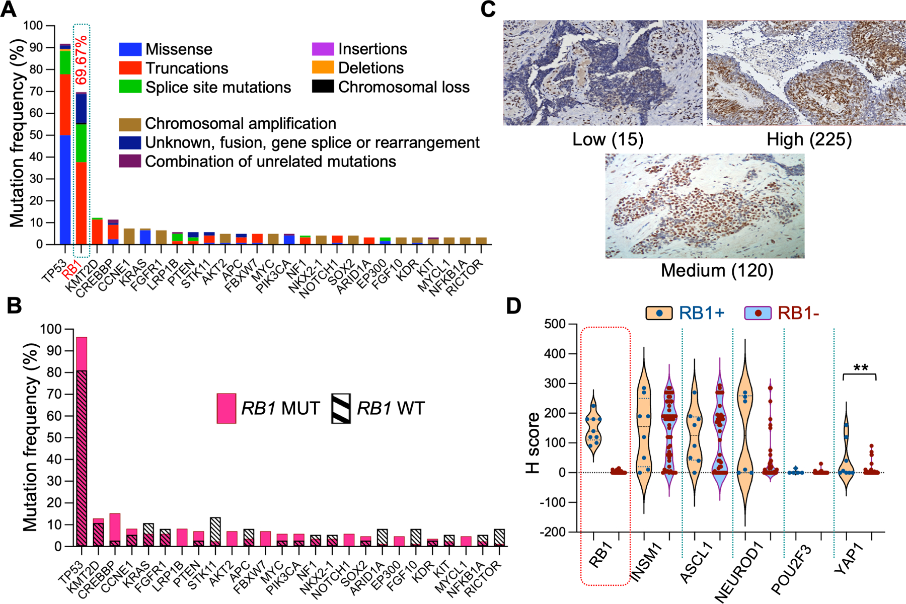
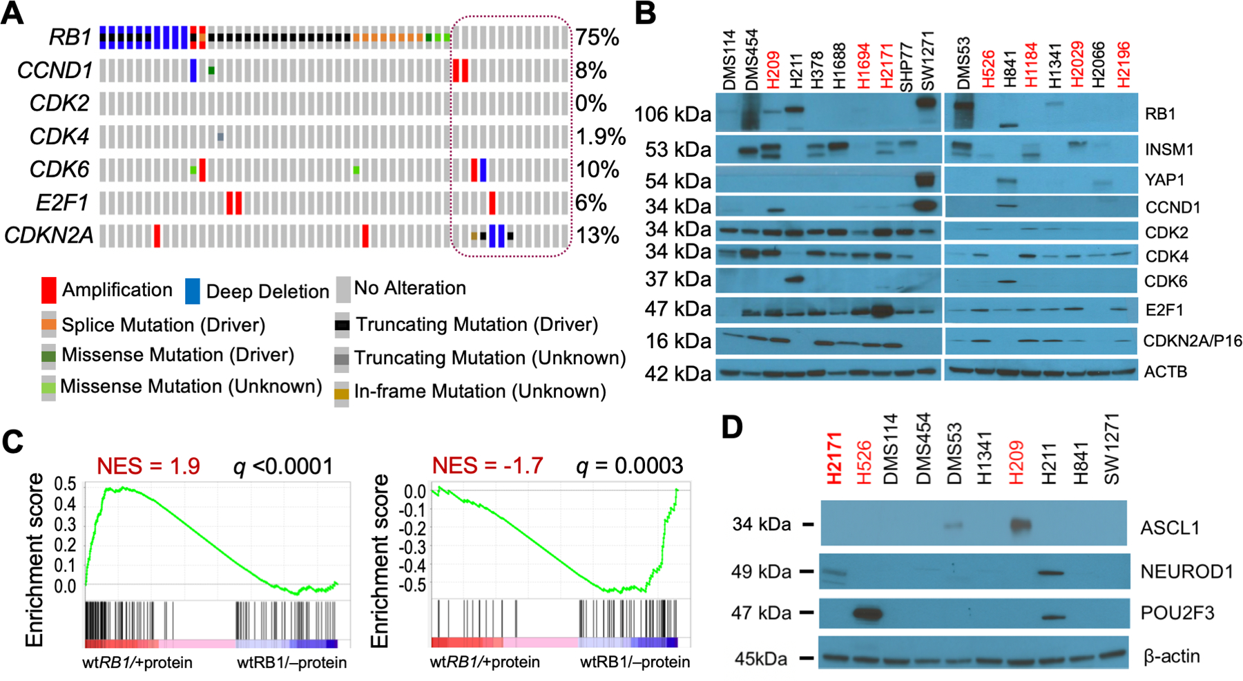
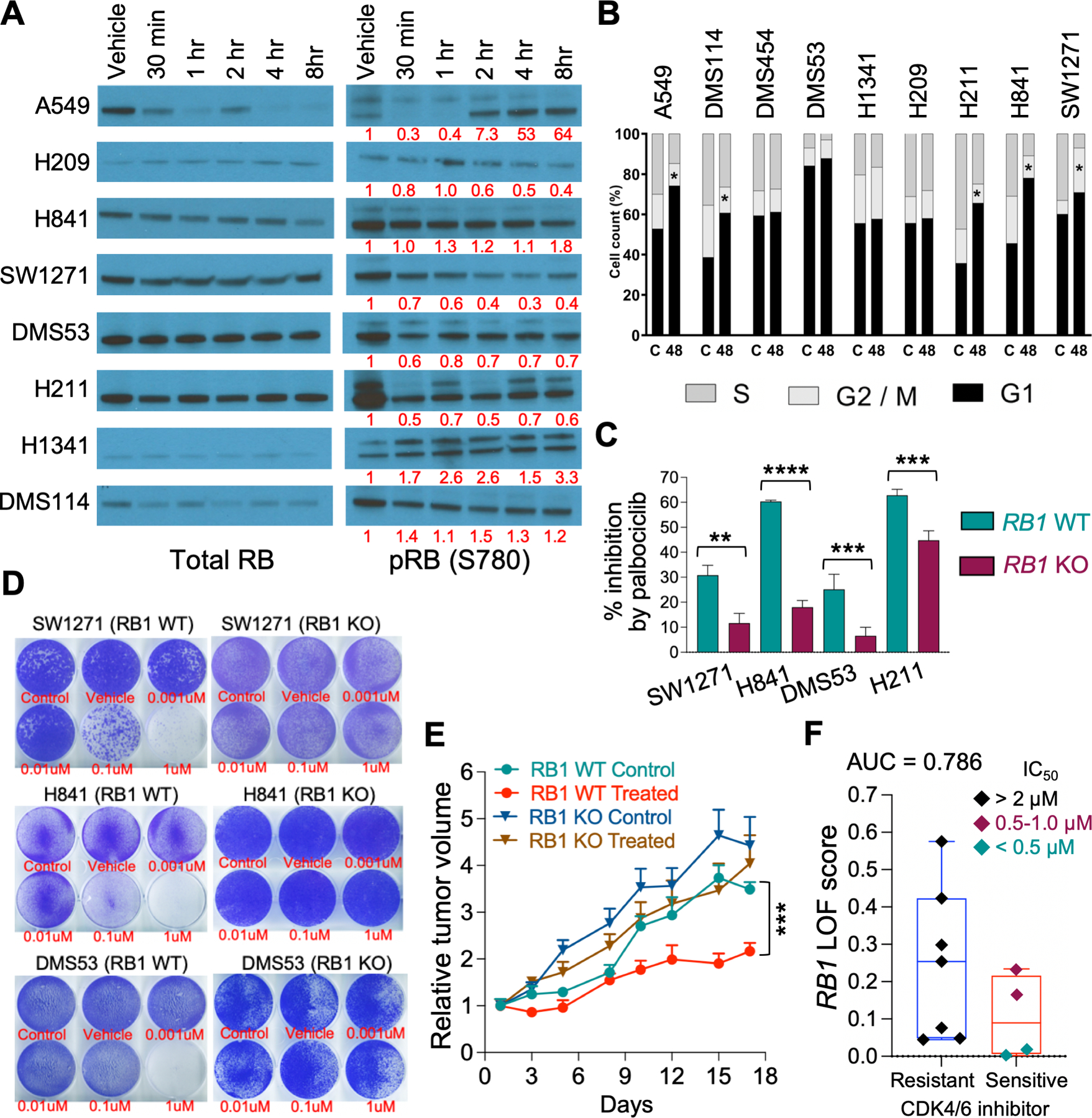
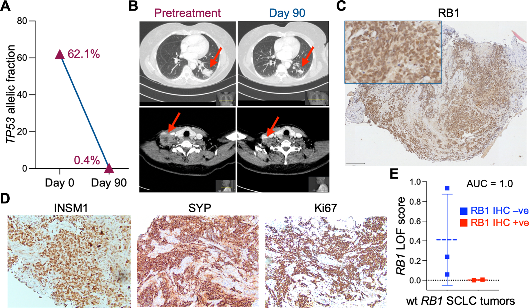
The canonical model of "small cell lung cancer" (SCLC) depicts tumors arising from dual inactivation of TP53 and RB1. However, many genomic studies have persistently identified tumors with no RB1 mutations. Here, we examined RB1 protein expression and function in SCLC. RB1 expression was examined by IHC analysis of 62 human SCLC tumors. These studies showed that ∼14% of SCLC tumors expressed abundant RB1 protein, which is associated with neuroendocrine gene expression and is enriched in YAP1 expression, but no other lineage proteins that stratify SCLC. SCLC cells and xenograft tumors with RB1 protein expression were sensitive to growth inhibition by the CDK4/6 inhibitor palbociclib, and this inhibition was shown to be dependent on RB1 expression by CRISPR knockout. Furthermore, a patient with biopsy-validated wild-type RB1 SCLC who received the CDK4/6 inhibitor abemaciclib demonstrated a dramatic decrease in mutant TP53 ctDNA allelic fraction from 62.1% to 0.4% and decreased tumor mass on CT scans. Importantly, IHC of the diagnostic biopsy specimen showed RB1 positivity. Finally, we identified a transcriptomics-based RB1 loss-of-function signature that discriminates between SCLC cells with or without RB1 protein expression and validated it in the patient who was responsive to abemaciclib, suggesting its potential use to predict CDK4/6 inhibitor response in patients with SCLC. Our study demonstrates that RB1 protein is an actionable target in a subgroup of SCLC, a cancer that exhibits no currently targetable mutations.


Conflict of interest statement

Conflict-of-interest statement: “The authors declare that no conflicts of interest exist.”

Figures

Figure 1.
Figure 1.. RB1 mutation and expression in SCLC tumors.
A. Gene mutation profile in SCLC tumors in the N=120 genomic cohort. Only genes that had ≥ 4 mutations are shown. Variants of unknown significance (VUS) were not considered. B. Same as in panel A except that the percentage of gene mutations between wt RB1 (magenta) and mutant RB1 (cross hatched) SCLC tumors is compared. TP53 (0.004) and CREBBP (0.045) exhibited a significantly lower % mutation rate in wt RB1; whereas STK11 (0.015), ARID1A (0.048), FGF10 (0.048), and RICTOR (0.048) exhibited a significantly higher mutation rate in wt RB1 (P values for individual genes in parentheses). Only genes that had ≥ 4 mutations were considered for analysis. C. Representative images of tumor RB1 staining (showing H scores). D. Violin plots of complete IHC profiles for the entire RB+ and RB- cohorts. Statistical differences in H score distribution between cohorts was only achieved for YAP1 and not for ASCL1 and NEUROD1. POU2F3 was not calculated.
Figure 2.
Figure 2.. RB1 mutation and expression in SCLC cell lines.
A. Oncoprint of mutations in select E2F pathway genes in SCLC cells. CCLE data (2019) for all 52 SCLC cells was visualized in cBioPortal using Oncoprint. The cells annotated as wt RB1 are circled. The genes queried were restricted to those examined by western blotting in Figure 2B. All the wt RB1 cells shown in Figure 2B are represented in the circle (column 1=DMS53, 2=DMS114, 3=DMS454, 4=H211, 5=H841, 6=H1092, 7=H1339, 8=H1341, 9=H2066, 10=H2286, 11=SBC5, 12=SHP77, 13=SW1271, L to R). B. Western blot analyses of wt RB1 (black text) and mutant RB1 (red text) cell lysates for expression of the proteins listed at right. Molecular size of bands is shown on the left. β-actin was used as a loading control. The specific mutations in the mutant RB1 cells are: H209 = missense p.C706F, H1694 = missense p.Q444L, H2171 = nonsense p.S567*, H526 = nonsense p.E31*, H1184 = nonsense p.E19*, H2029 = splice site, H2196 = splice site p.L738fs. C. GSEA analyses of wt RB1 SCLC cells with RB1 protein expression compared to wt RB1 SCLC cells lacking RB1 expression. GSEA analysis was performed using 186 RB1 LOF signature genes, including 118 downregulated and 68 upregulated genes. Downregulated genes due to RB1 loss are enriched in RB1 protein expressing cells (left panel), while upregulated genes due to RB1 loss are enriched in cells without RB1 protein (right panel). Cells used in wt RB1/+protein group: DMS114, DMS53, H1341, H211, H841, SW1271; and wt RB1/–protein group: H378, H1688, SHP77, H2066. D. Same as panel B.
Figure 3.
Figure 3.. RB1 is functional in wt RB1 SCLC cell lines.
A. Western blot analyses of total RB1 and pSer780-RB1 expression in wt RB1 cell lysates after incubation with 1.0 µM palbociclib for the indicated times. β-actin was used as a loading control but is not shown. The bands for SCLC total RB1 were the correct molecular size and generally mirrored the β-actin loading control. Bands for RB1 were quantified by IMAGE J and the numbers under the pRB1 bands represent the pRB1/total RB1 ratio normalized to the vehicle control. B. FACS analyses of growth inhibition by 1.0 µM palbociclib after 48 hours. Results show the average of triplicates for the percentage of cells in each of the three cell cycles. C. Plot of the percent cell growth inhibition by 1.0 μM palbociclib in vector control (WT) and matching RB1 CRISPR knockout (KO) cells measured by real-time imaging after five (SW1271, H841, H211) or ten (DMS53) days. D. Images of growth inhibition by various concentrations of palbociclib, shown below wells in red text, in vector control (C) and matching RB1 CRISPR knockout (KO) WT RB1 cells, as determined by colony formation assays over five (SW1271 and H841) or ten (DMS53) days. E. Growth inhibition by 100 mg/kg palbociclib in xenograft tumors of H841 vector control (Control) and matching RB1 CRISPR knockout (KO) wt RB1 cells. Statistical differences were determined at Day 17. F. RB1 LOF signature scores between palbociclib resistant and sensitive cells. SCLC cell lines were treated with PD0332991 (palbociclib) and IC50 values calculated using linear regression analysis (18). Box plot showing RB1 LOF scores between palbociclib resistant (IC50 >2 μM: H1048, H1092, H1694, H209, H2171, H446, and SHP77) and sensitive (IC50 <1 μM: DMS114, H211, H841, and SW1271) groups. Area under the ROC curve analysis (AUC = 0.786) showed the accuracy of RB1 LOF classifier.
Figure 4.
Figure 4.. Response of a wt RB1 SCLC patient to abemaciclib treatment.
A. The allelic fraction of mutant TP53 in ctDNA was measured before (day 0) and during (day 92) abemaciclib treatment. B. CT scans from different axial planes of patient 2 taken before (Day 0) and during (Day 90) treatment with abemaciclib. Arrows indicate tumor in lung (top panels) and neck (bottom panels). C. RB1 IHC staining of a pretreatment biopsy of patient 2 tumor. Scale bar = 200 μM. D. Additional IHC stains of a second pretreatment biopsy of patient 2 tumor. E. RB1 LOF signature scores of wt RB1 SCLC patients with RB1 positive (+ive) IHC compared to wt RB1 patients with RB1 negative (-ve) IHC. Area under the ROC curve analysis (AUC = 1.0) showed high accuracy of the RB1 LOF classifier. Analysis of patient 2 data is included. Patient 1 had no RNA-seq data available. The four additional wt RB1 patients analyzed were not enrolled in the clinical trial.

References

    1. Majeed U, et al. Targeted therapy in advanced non-small cell lung cancer: current advances and future trends. J Hematol Oncol 2021,14:108. - PMC - PubMed
    1. Chen Z, et al. Non-small-cell lung cancers: a heterogeneous set of diseases. Nat Rev Can 2014,14:535–546. - PMC - PubMed
    1. Thomas A, et al. Refining the treatment of NSCLC according to histological and molecular subtypes. Nat Rev Clin Oncol 2015,12:511–526. - PubMed
    1. Tan W-L, et al. Novel therapeutic targets on the horizon for lung cancer. Lancet Oncol 2016,17:e347–362. - PubMed
    1. Rudin CM, et al. Comprehensive genomic analysis identifies SOX2 as a frequently amplified gene in small-cell lung cancer. Nat Gen 2012,44:1111–1116. - PMC - PubMed

Publication types

MeSH terms