. 2023 Jan 4;43(1):56-67.
doi: 10.1523/JNEUROSCI.1181-22.2022. Epub 2022 Nov 18.

Preferential Modulatory Action of 5-HT2A Receptors on the Dynamic Regulation of Basal Ganglia Circuits

Affiliations

Preferential Modulatory Action of 5-HT2A Receptors on the Dynamic Regulation of Basal Ganglia Circuits

Lise Guilhemsang et al. J Neurosci. .

Erratum in

Abstract

In rodents, cortical information is transferred to the substantia nigra pars reticulata (SNr) through motor and medial prefrontal (mPF) basal ganglia (BG) circuits implicated in motor and cognitive/motivational behaviors, respectively. The serotonergic 5-HT2A receptors are located in both of these neuronal networks, displaying topographical differences with a high expression in the associative/limbic territories, and a very low expression in the subthalamic nucleus. This study investigated whether the stimulation of 5-HT2A receptors could have a specific signature on the dynamic regulation of BG circuits, preferentially modulating the mPF information processing through trans-striatal pathways. We performed in vivo single-unit extracellular recordings to assess the effect of the 5-HT2A agonist TCB-2 on the spontaneous and cortically evoked activity of lateral and medial SNr neurons in male rats (involved in motor and mPF circuits, respectively). TCB-2 (50-200 µg/kg, i.v.) increased the basal firing rate and enhanced the cortically evoked inhibitory response of medial SNr neurons (transmission through the direct striato-nigral pathway). A prior administration of the preferential 5-HT2A receptor antagonist MDL11939 (200 µg/kg, i.v.) did not modify any electrophysiological parameter, but occluded TCB-2-induced effects. In animals treated with the 5-HT synthesis inhibitor pCPA (4-chloro-dl-phenylalanine methyl ester hydrochloride), TCB-2 failed to induce the above-mentioned effects, thus suggesting the contribution of endogenous 5-HT. However, the mobilization of 5-HT induced by the acute administration of fluoxetine (10 mg/kg, i.p.) did not mimic the effects triggered by TCB-2. Overall, these data suggest that 5-HT2A receptors have a preferential modulatory action on the dynamic regulation of BG circuitry.SIGNIFICANCE STATEMENT Motor and medial prefrontal (mPF) basal ganglia (BG) circuits play an important role in integrative brain functions like movement control or cognitive/motivational behavior, respectively. Although these neuronal networks express 5-HT2A receptors, the expression is higher in associative/limbic structures than in the motor ones. We show a topographical-dependent dissociation in the effects triggered by the 5HT2A agonist TCB-2, which specifically increases the medial substantia nigra pars reticulata neuron activity and has a preferential action on mPF information processing through the striato-nigral direct pathway. These are very likely to be 5-HT2A receptor-mediated effects that require mobilization of the endogenous 5-HT system. These findings provide evidence about the specific signature of 5-HT2A receptors on the dynamic regulation of BG circuits.

Keywords: 5-HT2A receptor; TCB-2; basal ganglia circuits; fluoxetine; pCPA; serotonin.


Figures

Figure 1.
Figure 1.
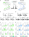
Schematic illustration of the electrophysiological recordings and firing pattern analysis of neurons from the lateral or medial subdivisions of the substantia nigra pars reticulata. A, Left, Representation of the in vivo experimental setup for single-unit extracellular recordings of lateral and medial SNr neurons with simultaneous electrical stimulation (1 mA, 1 Hz, 0.6 ms) of the MC or mPFC in anesthetized rats. Middle, Magnified recording trace of an SNr neuron (top; scale bar, 5 s), and a trace showing the bursts detected by the Poisson surprise algorithm (bottom). From this, we could calculate the number of bursts and burst recurrences in the analyzed period as well as the duration, number of spikes, and frequency for each of these bursts. Right, Characteristic narrow spike from an SNr neuron (calibration: 0.5 V/10 ms), which indicates the interspike interval (ISI) that was used to assess the firing pattern and regularity of these neurons, with the Poisson surprise algorithm and the ISI coefficient of variation, respectively. B, Representative coronal brain slices showing the placement of the stimulation and recording electrodes. Left, When studying the motor circuits, the stimulation electrode was placed in the MC (M1) and the recording site was in the lateral SNr, as shown by the deposition of pontamine sky blue. Right, When studying the mPF circuits, the stimulation electrode was placed in the mPFC (Cg1) and the recording site was in the medial SNr, as shown by the deposition of pontamine sky blue. Arrowheads indicate the place where these electrodes were placed.
Figure 2.
Figure 2.
Effect of systemic administration of cumulative doses of the 5-HT2A receptor agonist TCB-2, and its blockade by the preferential 5-HT2A receptor antagonist MDL11939, on electrophysiological parameters of lateral and medial substantia nigra pars reticulata neurons. A–F, Histograms illustrating the effect of cumulative doses of TCB-2 (50–200 µg/kg, i.v.) on the mean firing rate, and the mean coefficient of variation and firing pattern of SNr neurons [percentage of bursting (color) vs nonbursting (gray)] from the motor (AC, left) and from the mPF (DF, left) circuits recorded in anesthetized rats 24 h after the last vehicle injection. Administration of TCB-2 increased the basal firing rate of medial SNr neurons. Administration of MDL11939 (200 µg/kg, i.v.) did not show any effect by itself in the mean firing rate, and mean coefficient of variation and firing pattern of SNr neurons [percentage of bursting (color) vs nonbursting (gray)] from the motor (AC, right) and the mPF (DF, right) circuits recorded in control anesthetized rats. However, this dose of MDL11939 was able to block the effects observed after TCB-2 administration in the firing rate of medial SNr neurons. Each bar represents the mean ± SEM of n neurons. Each dot represents a single neuron. *p < 0.05, ***p < 0.001 versus basal (Bonferroni's post hoc test).
Figure 3.
Figure 3.
Effect of systemic administration of cumulative doses of the 5-HT2A receptor agonist TCB-2, and its blockade by the preferential 5-HT2A receptor antagonist MDL11939, on cortically evoked responses in lateral and medial substantia nigra pars reticulata neurons. A, Schematic anatomic diagram illustrating the motor (left) and mPF (right) cortico-BG circuits within the rat brain. Neurons from the MC or mPFC project to the lateral or medial STN, respectively. STN neurons then reach the SNr, constituting the hyperdirect pathway [dotted line (1)]. Additionally, MC and mPF cortical neurons send projections to the dorsolateral (DL) or dorsomedial (DM) striatum, respectively. From the striatum, there are two main projections: the direct pathway to the lateral or medial part of the SNr [dashed line (2)] and the indirect pathway connecting the DL/DM striatum to the lateral/medial globus pallidus (GP), the lateral/medial STN, and finally the lateral/medial SNr [solid line (3)]. Middle, PSTHs showing the characteristic triphasic response evoked in the SNr after MC or mPFC stimulation, consisting of EE [hyperdirect pathway (1)], I [direct pathway (2)], and LE [indirect pathway (3)], and the definition of these responses. The dashed line represents the threshold dividing excitatory and inhibitory responses; those above the threshold were considered excitatory. Duration indicates how long a response occurred, latency represents the time period between the occurrence of stimulation and response, and the amplitude indicates the magnitude of the excitations. B, Representative PSTHs illustrating the effect of the administration of the 5HT2A agonist TCB-2 (100 µg/kg, i.v.) on the cortically evoked activity of SNr neurons from the motor circuits recorded in vehicle-treated anesthetized rats (top), and its blockade by previous administration of the preferential 5-HT2A receptor antagonist MDL11939 (200 µg/kg, i.v.; bottom). C, Histograms illustrating the effect of the administration of TCB-2 (100 µg/kg, i.v.) in vehicle-treated anesthetized rats (left) on the different components (i.e., EE, I, and LE) of the cortically evoked activity of lateral SNr neurons, and the different electrophysiological parameters analyzed (i.e., latency, duration, and amplitude). Administration of TCB-2 (100 µg/kg, i.v.) caused a reduction in the duration of the LE. Previous administration of MDL11939 (200 µg/kg, i.v.) blocked the TCB-2-induced effects on the duration of the LE. MDL11939 caused no effect by itself on the cortically evoked activity of these neurons (right). D, Representative peristimulus time histograms illustrating the effect of the administration of TCB-2 (100 µg/kg, i.v.) on the cortically evoked activity of medial SNr neurons recorded in vehicle-treated anesthetized rats (top), and its blockade by previous administration of MDL11939 (200 µg/kg, i.v.; bottom). E, Histograms illustrating the effect of the administration of TCB-2 (100 µg/kg, i.v.) in vehicle-treated anesthetized rats (left) on the different components (i.e., EE, I, and LE) of the cortically evoked activity of medial SNr neurons, and the different electrophysiological parameters analyzed (i.e., latency, duration, and amplitude). Administration of TCB-2 (100 µg/kg, i.v.) caused an increase in the duration of the I and reduced the duration of the LE. Previous administration of MDL11939 (200 µg/kg, i.v.) blocked the TCB-2-induced effects on the duration of the I and LE. MDL11939 caused no effect by itself on the cortically evoked activity of these neurons (right). Each bar represents the mean ± SEM of n neurons. Each dot represents a single neuron. *p < 0.05, versus basal (Bonferroni's post hoc test).
Figure 4.
Figure 4.
Effects of the systemic administration of cumulative doses of the nonselective 5-HT receptor antagonist methiothepin on the spontaneous and cortically evoked activity of lateral and medial substantia nigra pars reticulata neurons. A, B, Mean firing rate, coefficient of variation, firing pattern [percentage of bursting (color) vs nonbursting (gray)], and cortically evoked activity of lateral (A) and medial (B) SNr neurons recorded in anesthetized control animals. Cumulative doses of the nonselective 5-HT receptor antagonist methiothepin (25–100 µg/kg, i.v.) induced no changes in either of the analyzed spontaneous electrophysiological parameters or in those regarding cortically evoked responses observed in lateral and medial SNr neurons. Each bar represents the mean ± SEM of n neurons. Each dot represents a single neuron.
Figure 5.
Figure 5.
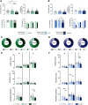
Effects of the administration of the 5-HT synthesis inhibitor pCPA (300 mg/kg/d, i.p., 3 d) or the selective 5-HT reuptake inhibitor fluoxetine (10 mg/kg, i.p.) on the spontaneous and cortically evoked electrophysiological activity of lateral and medial substantia nigra pars reticulata neurons from anesthetized rats. A, B, Histograms illustrating the effect on the mean firing rate, mean coefficient of variation, firing pattern [percentage of bursting (color) vs nonbursting (gray)] and on the different patterns of response displayed by neurons of the administration of vehicle or pCPA (300 mg/kg/d, i.p., 3 d) in lateral (A) and medial (B) SNr neurons recorded 24 h after the last injection, and of the acute dose of fluoxetine (10 mg/kg, i.p.) during the 6 h after its administration. pCPA administration increased the mean firing rate of lateral SNr neurons, while fluoxetine decreased their firing rate when compared with vehicle-treated rats. In the case of medial SNr neurons, their firing pattern changed after fluoxetine administration. The percentage of neurons displaying an EE, an I, or an LE is shown in A and B (bottom, right). Fluoxetine administration caused a decrease in the number of medial SNr neurons displaying an EE. After cortical stimulation, single SNr neurons would display different patterns of response consisting of different combinations of EE and/or I and/or LE. C, D, Fluoxetine was able to alter these patterns of response, producing a decrease in the number of lateral SNr neurons displaying LE (C), and in the number of medial SNr neurons displaying triphasic responses (EE + I + LE; D). E, F, Histograms illustrating the effect of the treatment with vehicle or pCPA (300 mg/kg/d, i.p., 3 d), and fluoxetine (10 mg/kg, i.p.) on the electrophysiological parameters analyzed from cortically evoked responses in lateral (E) and medial (F) SNr neurons. In lateral SNr neurons, fluoxetine was able to increase the duration of the EE and decrease the amplitude of the LE. Fluoxetine was also able to decrease the latency of the LE and the amplitude of the EE in neurons recorded in the medial SNr. In these neurons, pCPA increased the duration of the I. Each bar represents the mean ± SEM of n neurons. Each dot represents a single neuron. *p < 0.05, **p < 0.01 versus vehicle; &p < 0.05, &&p < 0.01, &&&p < 0.001 versus pCPA (firing rate, latency, duration and amplitude, Bonferroni's post hoc test; neurons exhibiting burst firing pattern, and patterns of response, χ2 test).
Figure 6.
Figure 6.
Effect of the systemic administration of cumulative doses of the 5-HT2A receptor agonist TCB-2 on the spontaneous activity and cortically evoked responses of lateral and medial substantia nigra pars reticulata neurons from pCPA-treated anesthetized rats. A, B, Histograms illustrating the effect on the mean firing rate, the mean coefficient of variation, and the firing pattern [percentage of bursting (color) vs nonbursting (gray)] of the administration of TCB-2 (50–200 µg/kg, i.v.) in lateral (A) and medial (B) SNr neurons recorded in pCPA-treated rats (300 mg/kg/d, i.p., 3 d) 24 h after the last injection. In neurons from the medial SNr, the administration of TCB-2 at the lowest dose (50 µg/kg, i.v.) was able to reduce the percentage of bursting neurons with no effect at higher doses (100–200 µg/kg, i.v.). C, D, Histograms illustrating the effect on the electrophysiological parameters analyzed from cortically evoked responses in lateral (C) and medial (D) SNr neurons of the administration of TCB-2 (50–200 µg/kg, i.v.) in pCPA-treated rats (300 mg/kg/d, i.p., 3 d) 24 h after the last injection. In pCPA-treated rats, TCB-2 (100 µg/kg, i.v.) was unable to alter cortico-nigral information transmission through any of the pathways that constitute the motor and mPF circuits. Each bar represents the mean ± SEM of n neurons. Each dot represents a single neuron. &p < 0.05 versus basal (neurons exhibiting burst firing pattern, χ2 test).

Similar articles

References

    1. Aghajanian GK, Foote WE, Sheard MH (1970) Action of psychotogenic drugs on single midbrain raphe neurons. J Pharmacol Exp Ther 171:178–187. - PubMed
    1. Alexander GE, DeLong MR, Strick PL (1986) Parallel organization of functionally segregated circuits linking basal ganglia and cortex. Annu Rev Neurosci 9:357–381. 10.1146/annurev.ne.09.030186.002041 - DOI - PubMed
    1. Aliane V, Pérez S, Nieoullon A, Deniau JM, Kemel ML (2009) Cocaine-induced stereotypy is linked to an imbalance between the medial prefrontal and sensorimotor circuits of the basal ganglia. Eur J Neurosci 30:1269–1279. 10.1111/j.1460-9568.2009.06907.x - DOI - PubMed
    1. Antonazzo M, Gutierrez-Ceballos A, Bustinza I, Ugedo L, Morera-Herreras T (2019) Cannabinoids differentially modulate cortical information transmission through the sensorimotor or medial prefrontal basal ganglia circuits. Br J Pharmacol 176:1156–1169. 10.1111/bph.14613 - DOI - PMC - PubMed
    1. Antonazzo M, Gomez-Urquijo SM, Ugedo L, Morera-Herreras T (2021) Dopaminergic denervation impairs cortical motor and associative/limbic information processing through the basal ganglia and its modulation by the CB1 receptor. Neurobiol Dis 148:105214. 10.1016/j.nbd.2020.105214 - DOI - PubMed

Publication types

LinkOut - more resources