

Assessment of the measurement methods in midshaft clavicle fracture
- PMID: 36401258
- PMCID: PMC9673337
- DOI: 10.1186/s12891-022-05961-y
Assessment of the measurement methods in midshaft clavicle fracture
Abstract
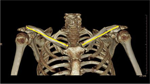
Background: Clavicle fractures account for approximately 5% of all fractures in adults and 75% of clavicle fractures occur in the midshaft. Shortening greater than two centimeters is an indicative of surgical treatment. Radiographic exams are often used to diagnose and evaluate clavicle fractures but computed tomography (CT) scan is currently considered the best method to assess these deformities and shortening.
Goal: 1- To investigate whether different methods of performing the radiographic exam interfere on the measurement of the fractured clavicle length. 2- Compare the clavicle length measurements obtained by the different radiographic exam methods with the CT scan measurements, used as a reference.
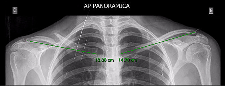
Materials and methods: Twenty-five patients with acute (< 3 weeks) midshaft clavicle fracture were evaluated. Patients underwent six radiographic images: PA Thorax (standing and lying), AP Thorax (standing and lying) and at 10° cephalic tilt (standing and lying), and the computed tomography was used as reference.
Results: The mean length (cm) obtained were: 14,930 on CT scan, 14,860 on PA Thorax Standing, 14,955 on PA Thorax Lying, 14,896 on AP Thorax Standing, 14,960 AP Thorax Lying, 15,098 on 10° cephalic tilt Standing and 15,001 on 10° cephalic tilt Lying, (p > 0,05).
Conclusion: 1- There is no significant statistical difference in the clavicle fracture length measurement among the variety of radiographic exam performances. 2- The method that comes closest to computed tomography results is the PA thorax incidence, with the patient in the lying position.
Keywords: Bone fractures; Clavicle; Methods; Radiography.
© 2022. The Author(s).
Conflict of interest statement
The authors declare that they have no conflict of interest.
Figures
References
-
- Neer C. Nonunion of the clavicle. JAMA. 1960;172(March 1960):96–99. - PubMed
MeSH terms
LinkOut - more resources
Full Text Sources
Medical
