

Use of a solid-state nanopore for profiling the transferrin receptor protein and distinguishing between transferrin receptor and its ligand protein
- PMID: 36401829
- PMCID: PMC9839655
- DOI: 10.1002/elps.202200147
Use of a solid-state nanopore for profiling the transferrin receptor protein and distinguishing between transferrin receptor and its ligand protein
Abstract
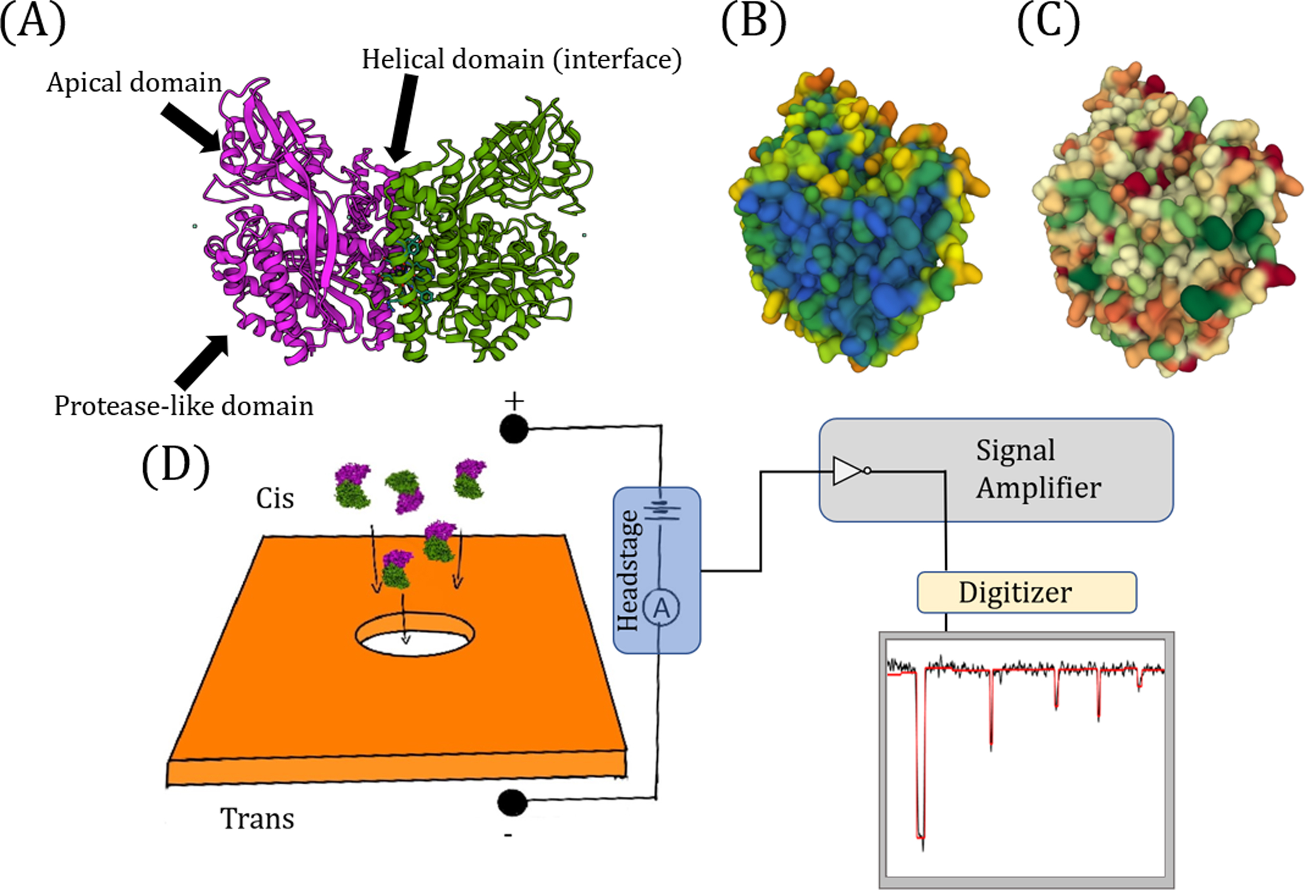
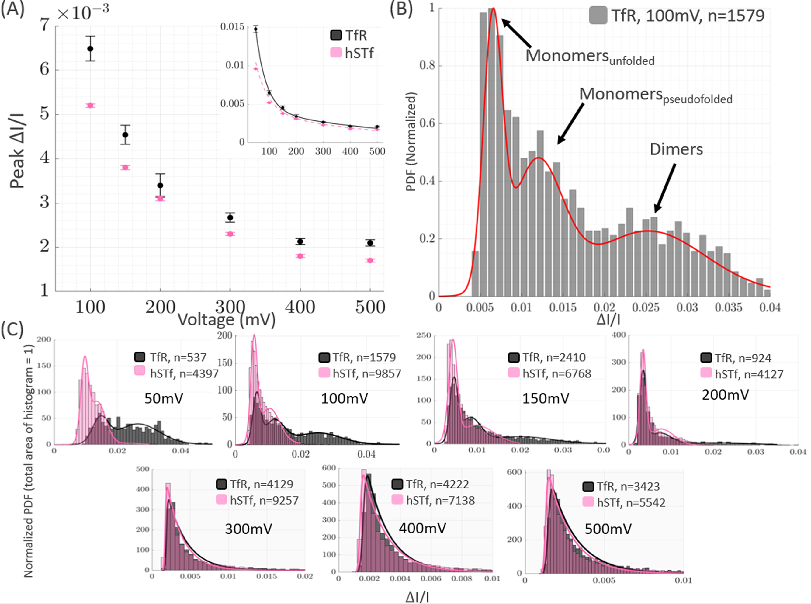
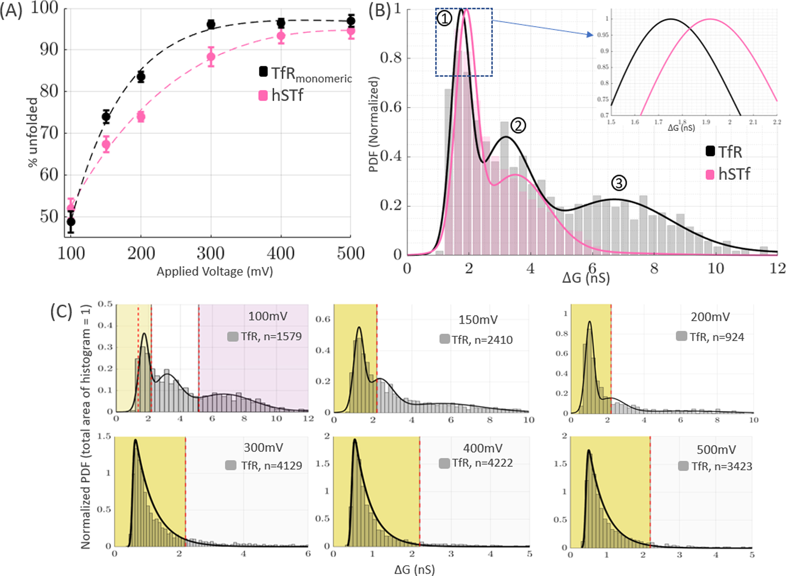
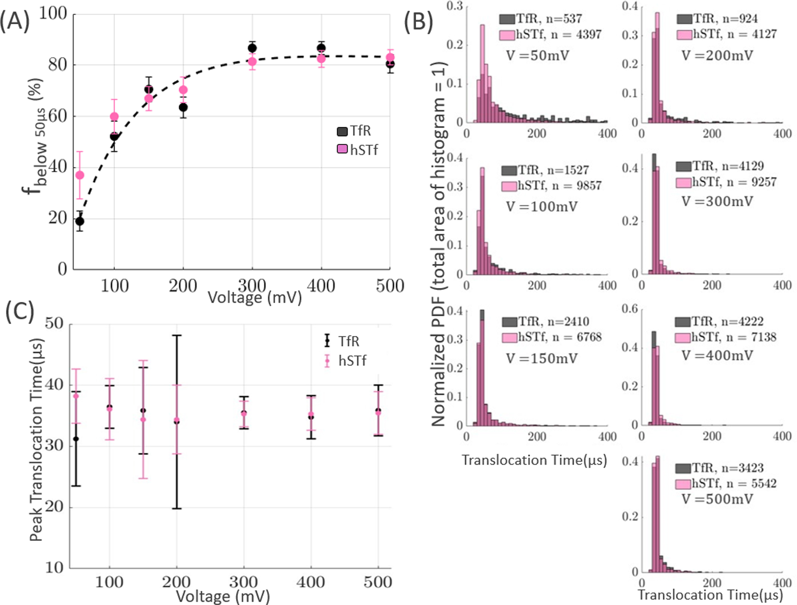
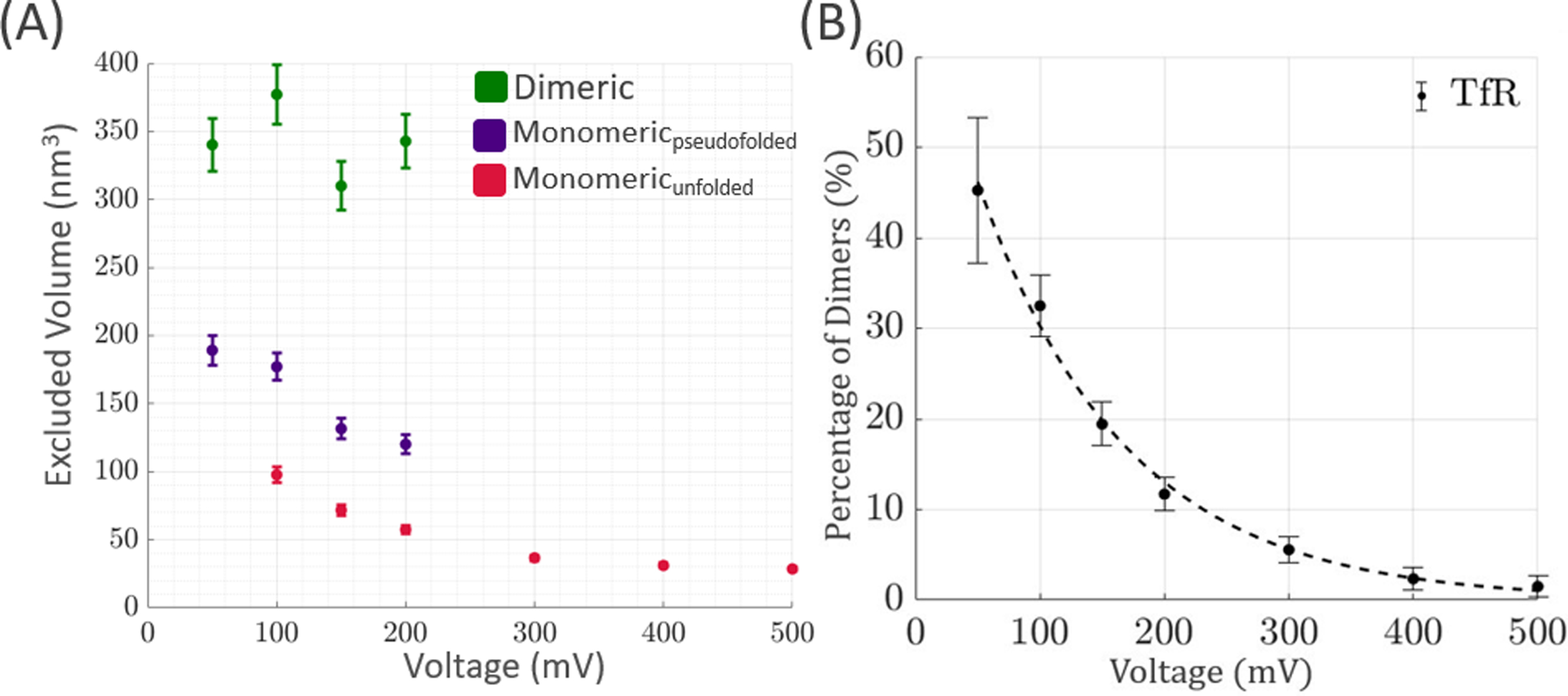
A nanopore device is capable of providing single-molecule level information of an analyte as they translocate through the sensing aperture-a nanometer-sized through-hole-under the influence of an applied electric field. In this study, a silicon nitride (Six Ny )-based nanopore was used to characterize the human serum transferrin receptor protein (TfR) under various applied voltages. The presence of dimeric forms of TfR was found to decrease exponentially as the applied electric field increased. Further analysis of monomeric TfR also revealed that its unfolding behaviors were positively dependent on the applied voltage. Furthermore, a comparison between the data of monomeric TfR and its ligand protein, human serum transferrin (hSTf), showed that these two protein populations, despite their nearly identical molecular weights, could be distinguished from each other by means of a solid-state nanopore (SSN). Lastly, the excluded volumes of TfR were experimentally determined at each voltage and were found to be within error of their theoretical values. The results herein demonstrate the successful application of an SSN for accurately classifying monomeric and dimeric molecules while the two populations coexist in a heterogeneous mixture.
Keywords: dielectric breakdown; human serum transferrin; nanopore; protein unfolding; transferrin receptor protein.
© 2022 Wiley-VCH GmbH.
Conflict of interest statement
Conflict of interest
The authors have declared no conflict of interest.
Figures
Similar articles
-
Molecular-Level Profiling of Human Serum Transferrin Protein through Assessment of Nanopore-Based Electrical and Chemical Responsiveness.ACS Nano. 2019 Apr 23;13(4):4246-4254. doi: 10.1021/acsnano.8b09293. Epub 2019 Mar 18. ACS Nano. 2019. PMID: 30844233
-
Fabrication of hexagonal boron nitride based 2D nanopore sensor for the assessment of electro-chemical responsiveness of human serum transferrin protein.Electrophoresis. 2020 Apr;41(7-8):630-637. doi: 10.1002/elps.201900336. Epub 2019 Nov 25. Electrophoresis. 2020. PMID: 31709550
-
Characterization of protein unfolding with solid-state nanopores.Protein Pept Lett. 2014 Mar;21(3):256-65. doi: 10.2174/09298665113209990077. Protein Pept Lett. 2014. PMID: 24370259 Free PMC article. Review.
-
Beyond nanopore sizing: improving solid-state single-molecule sensing performance, lifetime, and analyte scope for omics by targeting surface chemistry during fabrication.Nanotechnology. 2020 Aug 14;31(33):335707. doi: 10.1088/1361-6528/ab8f4d. Epub 2020 May 1. Nanotechnology. 2020. PMID: 32357346
-
Structure and dynamics of drug carriers and their interaction with cellular receptors: focus on serum transferrin.Adv Drug Deliv Rev. 2013 Jul;65(8):1012-9. doi: 10.1016/j.addr.2012.11.001. Epub 2012 Nov 23. Adv Drug Deliv Rev. 2013. PMID: 23183585 Free PMC article. Review.
Cited by
-
Exploring single-molecule interactions: heparin and FGF-1 proteins through solid-state nanopores.Nanoscale. 2024 May 2;16(17):8352-8360. doi: 10.1039/d4nr00274a. Nanoscale. 2024. PMID: 38563277 Free PMC article.
-
Probing nanopore surface chemistry through real-time measurements of nanopore conductance response to pH changes.Rev Sci Instrum. 2023 Oct 1;94(10):104101. doi: 10.1063/5.0155611. Rev Sci Instrum. 2023. PMID: 37812049 Free PMC article.
References
-
- Prabhu AS, Jubery TZN, Freedman KJ, Mulero R, Dutta P, Kim MJ. Chemically modified solid state nanopores for high throughput nanoparticle separation. J Phys Condens Matter. 2010;22(45):454107. - PubMed
-
- Han A, Creus M, Schürmann G, Linder V, Ward TR, De Rooij NF, et al. Label-free detection of single protein molecules and protein− protein interactions using synthetic nanopores. Analytical chemistry. 2008;80(12):4651–8. - PubMed
Publication types
MeSH terms
Substances
Grants and funding
LinkOut - more resources
Full Text Sources
