. 2023 Jan:118:108360.
doi: 10.1016/j.jmgm.2022.108360. Epub 2022 Oct 22.

Atomistic insights into the binding of SARS-CoV-2 spike receptor binding domain with the human ACE2 receptor: The importance of residue 493

Affiliations

Atomistic insights into the binding of SARS-CoV-2 spike receptor binding domain with the human ACE2 receptor: The importance of residue 493

Camryn Carter et al. J Mol Graph Model. 2023 Jan.

Abstract

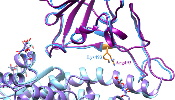
SARS-CoV-2 is a coronavirus that has created a global pandemic. The virus contains a spike protein which has been shown to bind to the ACE2 receptor on the surface of human cells. Vaccines have been developed that recognize elements of the SARS-CoV-2 spike protein and they have been successful in preventing infection. Recently, the Omicron variant of the SARS-CoV-2 virus was reported and quickly became a variant of concern due to its transmissibility. This variant contained an unusually large number (32) of point mutations, of which 15 of those mutations are in the receptor binding domain of the spike protein. While several computational and experimental investigations comparing the binding of the Omicron and wild type RBD to the human ACE2 receptor have been conducted, many of these report contradictory findings. In order to assess the differential binding ability, we conducted 2 μs of classical molecular dynamics (cMD) simulation to estimate the binding affinities and behaviors. Based upon MM-GBSA binding affinity, per-residue energy decomposition analysis, center of mass distance measurements, ensemble clustering, pairwise residue decomposition and hydrogen bonding analysis, our results suggest that a single point mutation is responsible for the enhanced binding of the Omicron mutant relative to the WT. While the 15-point mutations in the receptor binding domain contribute positively and negatively to the affinity of the spike protein for the human ACE2 receptor, it is the point mutation Q493R that confers enhanced binding while the Q493K mutation results in similar binding. The MM-GBSA binding estimations over a 2 μs trajectory, suggest that the wild type binds to ACE2 with a value of -29.69 kcal/mol while the Q493K and Q493R Omicron mutants bind with energy values of -26.67 and -34.56 kcal/mol, respectively. These values are significantly different, given the error estimates associated with the MM-GBSA method. In general, while some mutations increase binding, more mutations diminish binding, leading to an overall similar picture of binding for Q493K and enhanced binding for Q493R.

Keywords: COVID-19; Human ACE2 receptor; MM-GBSA; Molecular dynamics; Omicron; Receptor binding domain; Residue mutations; SARS-CoV-2; Spike protein.


Conflict of interest statement

Declaration of competing interest The authors declare that they have no known competing financial interests or personal relationships that could have appeared to influence the work reported in this paper.

Figures

Image 1
Graphical abstract
Fig. 1
Fig. 1
Omicron SARS-CoV-2 RBD - hACE2 interaction. Omicron SARS-CoV-2 RBD is displayed in blue and hACE2 is displayed in a lighter blue. In green are the Omicron SARS-CoV-2 RBD residue mutations: G339D, S371L, S373P, S375F, K417 N, N440K, G446S, S477 N, T478K, E484A, Q493K, G496S, Q498R, N501Y, and Y505H. The binding site for the RBD - hACE2 interaction and the residues K417 N, G446S, S477 N, T478K, E484A, Q493K, G496S, N501Y, and Y505H are shown in the inset.
Fig. 2
Fig. 2
Comparison of the 6LZG and 7WSA binding sites. The 6LZG structure is in blue with the RBD depicted in dark blue and the hACE2 receptor depicted in light blue. The 7WSA structure is in purple with the RBD depicted in dark purple and the hACE2 receptor depicted in light purple. The Omicron mutation of the residue 493 is highlighted in orange on both structures. On the 6LZG WT structure residue 493 is a lysine and on the 7WSA Omicron structure this residue is an arginine. Both mutated residues have similar lengths and placement of the side chain conformers.
Fig. 3
Fig. 3
Wild Type and Omicron SARS-CoV-2 RBD and hACE2 Root Mean Square Fluctuation Graphs. [A] RMSF graph of the WT and Omicron RBD concatenated 1 μs trajectories. Notably, differences in RMSF in RBD residues 358–376 and 384–390 are highlighted in the black box. [B] The Omicron Q493K RBD with the WT RBD superimposed. The most prevalent average structures of both models are shown. The Omicron Q493K RBD is displayed in blue and hACE2 in lighter blue, while the WT RBD and hACE2 are displayed in the same colors with a transparency effect applied. Amino acids highlighted in green are mutated residues that are notably close to the residues highlighted in red. In orange are WT RBD residues 384–390.
Fig. 4
Fig. 4
WT and Omicron Per-Residue Decomposition of SARS-CoV-2 RBD Mutated Residues. This bar graph demonstrates the per-residue decomposition energies for the 15 WT and Omicron (Q493K and Q493R) mutated RBD residues. The energies depicted are WT (blue), Omicron Q493K (orange) and Omicron Q493R (green). Standard deviations are reported in black.
Fig. 5
Fig. 5
Omicron Q493K SARS-CoV-2 RBD - hACE2 interactions. Highlighted are residues that most significantly affect binding based upon hydrogen bonding occurrence and pairwise residue decomposition changes in the WT and Omicron variant. Shown in red are mutated residues that display less favorable or less significant interaction. Shown in green are the mutated residues that became more significant for RBD - hACE2 interactions. See Fig. S11 for a complete visualization of all residues that play a significant role in WT and Omicron binding to hACE2.
Fig. 6
Fig. 6
Comparison of the WT and Omicron Q493K Cluster Families. [A] Representative structures from WT and Omicron Q493K clustering using backbone-atom RMSD. The 4 different WT cluster families are represented in shades of blue and the two different Omicron cluster families are depicted in shades of red. Individual images of representative structures from each cluster family are shown in Figs. S12 and S13. [B] Representative structures from WT and Omicron Q493K clustering using pairwise distance-based clustering. The WT cluster family is displayed in green and the two different Omicron cluster families are depicted in shades of orange. Individual images of the representative structures from each cluster family are shown in Figs. S15 and S16.

Similar articles

Cited by

References

    1. Berlin D.A., Gulick R.M., Martinez F.J. Severe covid-19. New England Journal of Medicine. 2020;383:2451–2460. - PubMed
    1. Pascarella G., Strumia A., Piliego C., Bruno F., Del Buono R., Costa F., et al. COVID-19 diagnosis and management: a comprehensive review. J Intern Med. 2020;288:192–206. - PMC - PubMed
    1. Wang C., Horby P.W., Hayden F.G., Gao G.F. A novel coronavirus outbreak of global health concern. The Lancet. 2020;395:470–473. - PMC - PubMed
    1. Harvey W.T., Carabelli A.M., Jackson B., Gupta R.K., Thomson E.C., Harrison E.M., et al. SARS-CoV-2 variants, spike mutations and immune escape. Nature Reviews Microbiology. 2021;19:409–424. - PMC - PubMed
    1. Lauring A.S., Hodcroft E.B. Genetic variants of SARS-CoV-2—what do they mean? JAMA. 2021;325:529–531. - PubMed

Publication types

Substances

Supplementary concepts