. 2023 Mar 1;227(5):651-662.
doi: 10.1093/infdis/jiac451.

Immunogenicity of an Additional mRNA-1273 SARS-CoV-2 Vaccination in People With HIV With Hyporesponse After Primary Vaccination

Affiliations

Immunogenicity of an Additional mRNA-1273 SARS-CoV-2 Vaccination in People With HIV With Hyporesponse After Primary Vaccination

Marlou J Jongkees et al. J Infect Dis. .

Abstract

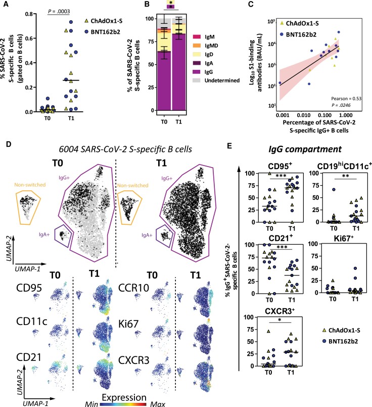
Background: The COVIH study is a prospective coronavirus disease 2019 (COVID-19) vaccination study in 1154 people with HIV (PWH), of whom 14% showed reduced antibody levels after primary vaccination. We evaluated whether an additional vaccination boosts immune responses in these hyporesponders.

Methods: The primary end point was the increase in antibodies 28 days after additional mRNA-1273 vaccination. Secondary end points included neutralizing antibodies, S-specific T-cell and B-cell responses, and reactogenicity.

Results: Of the 66 participants, 40 previously received 2 doses ChAdOx1-S, 22 received 2 doses BNT162b2, and 4 received a single dose Ad26.COV2.S. The median age was 63 years (interquartile range [IQR], 60-66), 86% were male, and median CD4+ T-cell count was 650/μL (IQR, 423-941). The mean S1-specific antibody level increased from 35 binding antibody units (BAU)/mL (95% confidence interval [CI], 24-46) to 4317 BAU/mL (95% CI, 3275-5360) (P < .0001). Of all participants, 97% showed an adequate response and the 45 antibody-negative participants all seroconverted. A significant increase in the proportion of PWH with ancestral S-specific CD4+ T cells (P = .04) and S-specific B cells (P = .02) was observed.

Conclusions: An additional mRNA-1273 vaccination induced a robust serological response in 97% of PWH with a hyporesponse after primary vaccination. Clinical Trials Registration. EUCTR2021-001054-57-N.

Keywords: COVID-19; HIV; SARS-CoV-2 vaccines; additional dose; nonresponder.


Conflict of interest statement

Potential conflicts of interest. S. J. received grants from the Dutch research council (NWO), European Union Horizon 2020, and the Bill and Melinda Gates Foundation. W. F. W. B. declares reimbursement to institution for participation of patient in trial by GSK. R. D. d. V. is listed as inventor of the fusion inhibitory lipopeptide [SARSHRC-PEG4]2-chol on a provisional patent application. K. C. E. S. received honorariums for advisory boards from Gilead and ViiV. C. R. has received research grants from ViiV, Gilead, ZonMw, AIDSfonds, Erasmus MC, and Health∼Holland; and honorariums for advisory boards from Gilead and ViiV. B. J. A. R. declares research grants from Gilead and MSD; and honorary for advisory boards for AstraZeneca, Roche, Gilead, and F2G. K. B. received research and educational grants from ViiV and Gilead; and consulting fees for advisory boards for ViiV, Gilead, MSD, and AstraZeneca. A. R. received grants from the Bill and Melinda Gates foundation and the Leids Universitair Fonds. All other authors report no potential conflicts of interest. All authors have submitted the ICMJE Form for Disclosure of Potential Conflicts of Interest. Conflicts that the editors consider relevant to the content of the manuscript have been disclosed.

Figures

Figure 1.
Figure 1.
SARS-CoV-2 S1-specific binding antibody levels in PWH after additional mRNA-1273 vaccination. Levels of S1-specific binding antibodies measured 28 days after the additional mRNA-1273 vaccination in all 66 PWH (squares), in 22 PWH after primary vaccination with BNT162b2 (circles), in 40 PWH after primary vaccination with ChadOx1-S (triangles), and in 4 PWH after primary vaccination with Ad26.COV2.S (diamonds). The thick horizontal bar shows the mean S1-specific binding antibody level, also indicated above the graph, with error bars showing the standard error of the mean. The horizontal lines show the lower limit of detection of the performed test (4.81 BAU/mL), the positivity cutoff (33.8 BAU/mL), and the hyporesponse cutoff (300 BAU/mL). Comparisons of time points were performed by paired t test. Abbreviations: BAU, binding antibody unit; LLoD, lower limit of detection; PWH, people with human immunodeficiency virus; S, spike; SARS-CoV-2, severe acute respiratory syndrome coronavirus 2; T0, before additional vaccination; T1, 28 days after additional vaccination.
Figure 2.
Figure 2.
Neutralizing antibodies to SARS-CoV-2 in subgroup participants (n = 40) after additional mRNA-1273 vaccination. A, PRNT50 titer measured 28 days after the additional mRNA-1273 vaccination against the ancestral SARS-CoV-2 (D614G) and Omicron (BA.1) variant after primary vaccination with ChAdOx1-S (triangles) and BNT162b2 (circles). The thick horizontal bar shows the mean neutralizing antibody titer, also indicated above the graph, with error bars showing the standard error of the mean. LLoD is 10. Comparisons between the 2 different primary vaccination groups were performed using unpaired t test. B, Correlation between the S1-specific binding antibody levels and neutralizing antibody levels targeting the ancestral SARS-CoV-2 by linear regression analysis on transformed data; R = 0.66, P < .0001. C, Correlation between the S1-specific binding antibody levels and neutralizing antibody levels targeting the Omicron BA.1 variant by linear regression analysis on transformed data; R = 0.45, P < .0001. Adequate responder cutoff is 300 BAU/mL (dotted line). Abbreviations: BAU, binding antibody unit; LLoD, lower limit of detection; PRNT50, 50% plaque reduction neutralization test; S, spike; SARS-CoV-2, severe acute respiratory syndrome coronavirus.
Figure 3.
Figure 3.
Frequency of SARS-CoV-2 S-specific T cells in subgroup participants (n = 40) before and 28 days after additional mRNA-1273 vaccination. CD4+ (CD4+CD40L+CD137+) and CD8+ (CD8+CD69+CD137+) T-cell responses to ancestral spike (Wuhan-Hu1) and Omicron spike (BA.1) measured by the AIM assay before additional vaccination (T0) compared to 28 days after additional vaccination (T1). Participants who received ChAdOx1-S as primary vaccination are shown as triangles, participants who received BNT162b2 as primary vaccination are shown as circles. The horizontal line shows the median, also indicated above the graph. The total numbers of participants with detectable S-specific T cells are indicated below the graphs. Comparisons of time points were performed by unpaired t test. Abbreviations: AIM, activation induced marker assay; S, spike; SARS-CoV-2, severe acute respiratory syndrome coronavirus; T0, before additional vaccination; T1, 28 days after additional vaccination.
Figure 4.
Figure 4.
Frequency and phenotype of SARS-CoV-2 S-specific B cells in subgroup participants (n = 18) before and 28 days after an additional mRNA-1273 vaccination. A, Percentages of S-specific B cells are shown as frequencies from total B cells per individual. Each individual is colored according to the primary vaccination regimen (triangles, ChAdOx1-s, n = 8; circles, BNT162b2, n = 10). The horizontal line shows the median. B, Isotype usage of S-specific B cells are shown as stacked bars at T0 and T1. C, Correlation plot between S1-specific binding antibody levels and IgG+ S-specific B cells. Pearson correlation analysis on nontransformed data is depicted and linear regression results shown as a black line with red shaded 95% confidence intervals. D, UMAP for all 6004 S-specific B cells (grey) to cluster cells based on 24 different markers. S-specific B cells are overlaid based on time point (black) on top of all cells (grey). Normalized expression of 6 selected markers is shown below the overlaid UMAP. Expression plots for all markers are in Supplementary Figure 7. E, Manually gated B-cell subsets are shown within the IgG+ B-cell compartment at each time point. A, B, and E, Statistical analyses, Wilcoxon matched pairs tests were performed to compare T0 versus T1. *P < .05, **P < .01, ***P < .001. Individuals and median values (A and E) and means with standard error of means (B) are shown. Abbreviations: IgG, immunoglobulin G; S, spike; SARS-CoV-2, severe acute respiratory syndrome coronavirus; T0, before additional vaccination; T1, 28 days after additional vaccination; UMAP, uniform manifold approximation and projection.

References

    1. van den Berg R, van Hoogstraten I, van Agtmael M. Non-responsiveness to hepatitis B vaccination in HIV seropositive patients; possible causes and solutions. AIDS Rev 2009; 11:157–64. - PubMed
    1. Pallikkuth S, De Armas LR, Pahwa R, et al. . Impact of aging and HIV infection on serologic response to seasonal influenza vaccination. Aids 2018; 32:1085–94. - PMC - PubMed
    1. Madhi SA, Koen AL, Izu A, et al. . Safety and immunogenicity of the ChAdOx1 nCoV-19 (AZD1222) vaccine against SARS-CoV-2 in people living with and without HIV in South Africa: an interim analysis of a randomised, double-blind, placebo-controlled, phase 1B/2A trial. Lancet HIV 2021; 8:e568–e80. - PMC - PubMed
    1. Frater J, Ewer KJ, Ogbe A, et al. . Safety and immunogenicity of the ChAdOx1 nCoV-19 (AZD1222) vaccine against SARS-CoV-2 in HIV infection: a single-arm substudy of a phase 2/3 clinical trial. Lancet HIV 2021; 8:e474–e85. - PMC - PubMed
    1. Levy I, Wieder-Finesod A, Litchevsky V, et al. . Immunogenicity and safety of the BNT162b2 mRNA COVID-19 vaccine in people living with HIV-1. Clin Microbiol Infect 2021; 27:1851–5. - PMC - PubMed

Publication types