. 2022 Nov 19;12(1):487.
doi: 10.1038/s41398-022-02252-x.

Constitutive 5-HT2C receptor knock-out facilitates fear extinction through altered activity of a dorsal raphe-bed nucleus of the stria terminalis pathway

Affiliations

Constitutive 5-HT2C receptor knock-out facilitates fear extinction through altered activity of a dorsal raphe-bed nucleus of the stria terminalis pathway

Sandra T Süß et al. Transl Psychiatry. .

Abstract

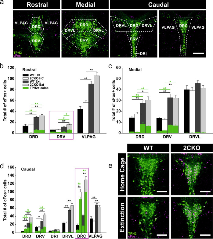
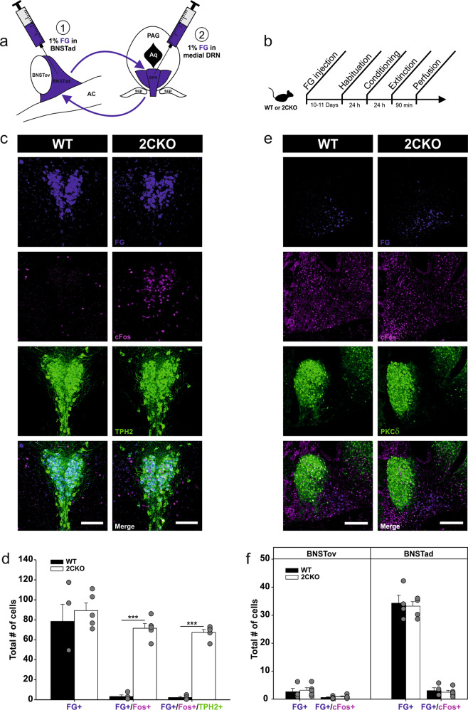
Serotonin 2C receptors (5-HT2CRs) are widely distributed throughout the brain and are strongly implicated in the pathophysiology of anxiety disorders such as post-traumatic stress disorder (PTSD). Although in recent years, a considerable amount of evidence supports 5-HT2CRs facilitating effect on anxiety behavior, the involvement in learned fear responses and fear extinction is rather unexplored. Here, we used a 5-HT2CR knock-out mouse line (2CKO) to gain new insights into the involvement of 5-HT2CRs in the neuronal fear circuitry. Using a cued fear conditioning paradigm, our results revealed that global loss of 5-HT2CRs exclusively accelerates fear extinction, without affecting fear acquisition and fear expression. To investigate the neuronal substrates underlying the extinction enhancing effect, we mapped the immediate-early gene product cFos, a marker for neuronal activity, in the dorsal raphe nucleus (DRN), amygdala and bed nucleus of the stria terminalis (BNST). Surprisingly, besides extinction-associated changes, our results revealed alterations in neuronal activity even under basal home cage conditions in specific subregions of the DRN and the BNST in 2CKO mice. Neuronal activity in the dorsal BNST was shifted in an extinction-supporting direction due to 5-HT2CR knock-out. Finally, the assessment of DRN-BNST connectivity using antero- and retrograde tracing techniques uncovered a discrete serotonergic pathway projecting from the most caudal subregion of the DRN (DRC) to the anterodorsal portion of the BNST (BNSTad). This serotonergic DRC-BNSTad pathway showed increased neuronal activity in 2CKO mice. Thus, our results provide new insights for the fear extinction network by revealing a specific serotonergic DRC-BNSTad pathway underlying a 5-HT2CR-sensitive mechanism with high significance in the treatment of PTSD.


Conflict of interest statement

The authors declare no competing interests.

Figures

Fig. 1
Fig. 1. Constitutive 5-HT2CR knock-out facilitates fear extinction.
a Schematic representation of the experimental paradigm. b Freezing behavior during conditioning (day 2) and extinction (day 3) session in respect to conditioned stimulus (CS) time bins and baseline (Bl) period. Circles outlined in green reflect the fear retrieval test (averaged freezing to the first two CS of the extinction session). 2CKO mice showed less CS-freezing during extinction: Mann–Whitney Rank Sum test (WT vs. 2CKO); bin2(CS): P = 0.023, bin3(CS): P = 0.001, bin4–bin7(CS): P ≤ 0.001. c Freezing behavior during conditioning (day 2) and extinction (day 3) session in respect to inter-trial interval (ITI) time bins and baseline (Bl) period. 2CKO mice showed less ITI-freezing during extinction: Mann–Whitney Rank Sum test (WT vs. 2CKO); bin3(ITI): P = 0.001, bin4(ITI): P = 0.009, bin5–6(ITI): P ≤ 0.001. d Comparison of baseline (Bl) and post-stimulus time (PST) freezing during conditioning (day 2) and extinction (day 3) session. Conditioning procedure increased freezing in both genotypes: Wilcoxon Signed Rank test (Bl vs. PST); WT: P ≤ 0.001, 2CKO: P ≤ 0.001. WT mice showed higher freezing in extinction PST: Wilcoxon Signed Rank test (Bl vs. PST); WT: P ≤ 0.001. Mann–Whitney Rank Sum test (WT vs. 2CKO); PST: P = 0.011. e Maximum movement velocity as unconditioned stimulus (US, foot shock) reactivity. Movement velocity was compared between baseline (BL) and CS + US time, whereby US was presented during the last second of each 30 s CS interval of the conditioning session (day 2). US presentation increased movement velocity in both genotypes: Wilcoxon Signed Rank test (Bl vs. PST); WT: P ≤0.001, 2CKO: P ≤ 0.001. 2CKO mice showed a higher US reactivity: Mann–Whitney Rank Sum test (WT vs. 2CKO); CS + US1-5: P = 0.021. f Total distance moved during habituation (day 1) was similar in both genotypes. On freezing time curves (b and c), each bin reflects two to three averaged time intervals depicted underneath the x-axis. For all graphs (bf), WT mice (n = 29), 2CKO mice (n = 30, for total distance moved in f n = 29). Data are shown as means ± SEM. *P < 0.05, **P < 0.01, ***P < 0.001.
Fig. 2
Fig. 2. Neuronal activity in the DRN is altered in 2CKO mice.
a Representative images of the DRN levels analyzed. The pattern of TPH2+ 5-HT cells (green) was used to define the respective level. Boundaries are outlined by dashed lines. DRD dorsal raphe nucleus, dorsal part; DRV dorsal raphe nucleus, ventral part; DRI dorsal raphe nucleus, interfascicular part; DRC dorsal raphe nucleus, caudal part; DRVL dorsal raphe nucleus, ventrolateral part; VLPAG ventrolateral periaqueductal gray. Scale bar = 250 µm. b cFos quantification in the rostral DRN. Significant effects DRD: Kruskal–Wallis one-way ANOVA on ranks (cFos+): P ≤ 0.001; pairwise Dunn’s test: WT HC vs. WT Ext: P ≤ 0.01, 2CKO HC vs. 2CKO Ext: P ≤ 0.01. Kruskal–Wallis one-way ANOVA on ranks (TPH2+/cFos+): P ≤ 0.001; pairwise Dunn’s test: WT HC vs. WT Ext: P ≤ 0.01, 2CKO HC vs. 2CKO Ext: P ≤ 0.01. Significant effects DRV: Kruskal–Wallis one-way ANOVA on ranks (cFos+): P ≤ 0.001; pairwise Dunn’s test: 2CKO HC vs. 2CKO Ext: P ≤ 0.01. Kruskal–Wallis one-way ANOVA on ranks (TPH2+/cFos+): P = 0.004; pairwise Dunn’s test: WT HC vs. WT Ext: P ≤ 0.05, WT Ext vs. 2CKO Ext: P ≤ 0.05. Significant effects VLPAG: Kruskal–Wallis one-way ANOVA on ranks (cFos+): P ≤ 0.001; pairwise Dunn’s test: WT HC vs. WT Ext: P ≤ 0.01, 2CKO HC vs. 2CKO Ext: P ≤ 0.01. c cFos quantification in the medial DRN. Significant effects DRD: Kruskal–Wallis one-way ANOVA on ranks (cFos+): P ≤ 0.001; pairwise Dunn’s test: WT HC vs. WT Ext: P ≤ 0.01, 2CKO HC vs. 2CKO Ext: P ≤ 0.05. Kruskal–Wallis one-way ANOVA on ranks (TPH2+/cFos+): P ≤ 0.001; pairwise Dunn’s test: WT HC vs. WT Ext: P ≤ 0.01, 2CKO HC vs. 2CKO Ext: P ≤ 0.01. Significant effects DRV: Kruskal–Wallis one-way ANOVA on ranks (cFos+): P ≤ 0.001; pairwise Dunn’s test: WT HC vs. WT Ext: P ≤ 0.01, 2CKO HC vs. 2CKO Ext: P ≤ 0.01. Kruskal–Wallis one-way ANOVA on ranks (TPH2+/cFos+): P ≤ 0.001; pairwise Dunn’s test: WT HC vs. WT Ext: P ≤ 0.01, 2CKO HC vs. 2CKO Ext: P ≤ 0.05. d cFos quantification in the caudal DRN. Significant effects DRD: Kruskal–Wallis one-way ANOVA on ranks (cFos+): P ≤ 0.001; pairwise Dunn’s test: WT HC vs. WT Ext: P ≤ 0.01, 2CKO HC vs. 2CKO Ext: P ≤ 0.01. Kruskal–Wallis one-way ANOVA on ranks (TPH2+/cFos+): P ≤ 0.001; pairwise Dunn’s test: WT HC vs. WT Ext: P ≤ 0.01, 2CKO HC vs. 2CKO Ext: P ≤ 0.05. Significant effects DRV: Kruskal–Wallis one-way ANOVA on ranks (cFos+): P ≤ 0.001; pairwise Dunn’s test: WT HC vs. WT Ext: P ≤ 0.05, 2CKO HC vs. 2CKO Ext: P ≤ 0.01. Kruskal–Wallis one-way ANOVA on ranks (TPH2+/cFos+): P ≤ 0.001; pairwise Dunn’s test: 2CKO HC vs. 2CKO Ext: P ≤ 0.01. Significant effects DRVL: Kruskal–Wallis one-way ANOVA on ranks (cFos+): P ≤ 0.001; pairwise Dunn’s test: WT HC vs. WT Ext: P ≤ 0.01, 2CKO HC vs. 2CKO Ext: P ≤ 0.01. Significant effects DRC: Kruskal–Wallis one-way ANOVA on ranks (cFos+): P ≤ 0.001; pairwise Dunn’s test: WT HC vs. 2CKO HC: P ≤ 0.01, WT Ext vs. 2CKO Ext: P ≤ 0.01. Kruskal–Wallis one-way ANOVA on ranks (TPH2+/cFos+): P ≤ 0.001; pairwise Dunn’s test: WT HC vs. 2CKO HC: P ≤ 0.01, WT Ext vs. 2CKO Ext: P ≤ 0.01. Significant effects VLPAG: Kruskal–Wallis one-way ANOVA on ranks (cFos+): P ≤ 0.001; pairwise Dunn’s test: WT HC vs. WT Ext: P ≤ 0.01, 2CKO HC vs. 2CKO Ext: P ≤ 0.01. e Representative immuno-stained DRC sections. cFos+ cells (magenta) showed a high colocalization with TPH2+ 5-HT cells (green) in 2CKO animals under home cage and extinction conditions. Scale bars = 100 µm. Boxes in magenta highlight subregions with significant genotype effects. For all graphs (bd), HC: WT mice (n = 10), 2CKO mice (n = 11); Ext: WT mice (n = 13), 2CKO mice (n = 13, for caudal in d n = 12). Data are shown as means ± SEM. *P < 0.05, **P < 0.01.
Fig. 3
Fig. 3. Altered neuronal activity in the dorsal BNST supports faster fear extinction in 2CKO mice.
a Schematic illustration of the dorsal BNST subregions analyzed. BNSTov bed nucleus of the stria terminalis, oval nucleus; BNSTad bed nucleus of the stria terminalis, anteriodorsal part; LV lateral ventricle; AC anterior commissure. b cFos quantification in the BNSTov and BNSTad. In the BNSTov, 2CKO mice showed reduced cFos levels under home cage conditions and extinction treatment reduced cFos in WT mice: Two-way ANOVA (treatment): F(1,19) = 4.973, P = 0.038; pairwise Holm–Sidak test: WT HC vs. 2CKO HC: P = 0.036, WT HC vs. WT Ext: P = 0.011. In the BNSTad, 2CKO mice showed increased cFos levels under home cage and extinction conditions and extinction treatment increased cFos in both genotypes: Two-way ANOVA (genotype): F(1,19) = 20.736, P ≤ 0.001; Two-way ANOVA (treatment): F(1,19) = 114.923, P ≤ 0.001; pairwise Holm–Sidak test: WT HC vs. 2CKO HC: P = 0.034, WT Ext vs. 2CKO Ext: P ≤ 0.001, WT HC vs. WT Ext: P ≤ 0.001, KO HC vs. KO Ext: P ≤ 0.001. HC: WT mice (n = 5), 2CKO mice (n = 6); Ext: WT mice (n = 5), 2CKO mice (n = 7). Data are shown as means ± SEM. *P < 0.05, ***P < 0.001. c Representative immuno-stained BNST sections. PKCδ (green), used as a marker for the BNSTov subregion, in combination with cFos (magenta). Scale bars = 200 µm.
Fig. 4
Fig. 4. The DRN and the dorsal BNST are reciprocally connected.
a AAV-based labeling strategy of anterograde terminals. AAV1.CAG.FLEX.tdTomato virus was injected either into the DRC of ePet1-Cre mice (1) to label 5-HT terminals innervating the BNST, or into the BNSTad subregion of Gad2-Ires-Cre mice (2) to label GABAergic terminals innervating the DRN. b DRC injection site in an ePet1-Cre mouse indicates selective tdTomato expression (magenta) in TPH2 + 5-HT cells (green). Scale bar = 200 µm. c BNST target site in an ePet1-Cre mouse showing tdTomato+ 5-HT terminals (magenta) arising from the DRC. PKCδ (green) was used as a marker for the BNSTov subregion. Scale bar = 200 µm. d Overview of the dorsal BNST in an ePet1-Cre mouse reflecting the bilateral innervation of the BNSTad with tdTomato+ 5-HT terminals (magenta) arising from the DRC. PKCδ (green) was used as a marker for the BNSTov subregion. Boxed region is separately shown in c. Scale bar = 300 µm. e Quantification of tdTomato fluorescence intensity in the dorsal BNST. tdTomato+ 5-HT terminals innervate primarily the BNSTad subregion. Two-tailed Student’s paired t-test (BNSTov vs. BNSTad): P = 0.007. Injected mice (n = 3). Data are shown as means ± SEM. **P < 0.01. f Overview of bilateral BNSTad injection sites in a Gad2-Ires-Cre mouse. tdTomato+ GABAergic cells (magenta). PKCδ (green) was used as a marker for the BNSTov subregion. Scale bar = 300 µm. g Quantification of tdTomato fluorescence intensity in the DRN. tdTomato+ GABAergic terminals arising from the BNSTad innervate all rostrocauldal DRN levels. Rostral and medial level show the strongest innervation. One-way RM ANOVA (fluorescence): F(3,15) = 34.018, P ≤ 0,001; pairwise Holm–Sidak test: rostral vs. caudal anterior: P ≤ 0.001, rostral vs. caudal posterior: P ≤ 0.001, medial vs. caudal anterior: P ≤ 0.001, medial vs. caudal posterior: P ≤ 0.001. Injected mice (n = 6). Data are shown as means ± SEM. ***P < 0.001. h DRN target site in a Gad2-Ires-Cre mouse showing tdTomato+ GABAergic terminals (magenta) arising from the BNSTad. The pattern of TPH2 + 5-HT cells (green) was used to define the respective level. Boundaries are outlined by dashed lines. DRD dorsal raphe nucleus, dorsal part; DRV dorsal raphe nucleus, ventral part; DRI dorsal raphe nucleus, interfascicular part; DRC dorsal raphe nucleus, caudal part; DRVL dorsal raphe nucleus, ventrolateral part; VLPAG ventrolateral periaqueductal gray. Scale bar = 250 µm.
Fig. 5
Fig. 5. A BNSTad-DRN pathway is not involved in faster fear extinction in 2CKO mice.
a Fluorogold (FG)-based retrograde labeling strategy in 2CKO and WT mice. FG was injected either into the BNSTad (1) for retrograde labeling of DRN cells, or into the medial DRN (2), for retrograde labeling of BNSTad cells. b Experimental workflow. c Representative immuno-stained DRC sections of mice injected into the BNSTad. Retrogradely labeled FG cells (blue) are highly colocalized with cFos (magenta) and TPH2 (green) in the 2CKO mouse. Scale bar = 100 µm. d Evaluation of retrogradely labeled FG cells in the DRN revealed a higher number of FG+/cFos+ and (FG+/cFos+/TPH2+) in 2CKO mice. Two-tailed Student’s t test (WT vs. 2CKO); FG+/cFos+: P ≤ 0.001, FG+/cFos+/TPH2+: P ≤ 0.001. WT mice (n = 4), 2CKO mice (n = 5). e Representative immuno-stained dorsal BNST sections of mice injected into the medial DRN. Extinction-induced cFos (magenta) is not associated with FG-labeled neurons (blue) projecting to the DRN. PKCδ (green) was used as a marker for the BNSTov subregion. Scale bar = 200 µm. f Evaluation of retrogradely labeled FG cells in the dorsal BNST revealed no genotype differences. WT mice (n = 4), 2CKO mice (n = 5). Data are shown as means ± SEM. ***P < 0.001.

Similar articles

Cited by

References

    1. Homberg JR. Serotonergic modulation of conditioned fear. Scientifica. 2012;2012:821549. doi: 10.6064/2012/821549. - DOI - PMC - PubMed
    1. Chagraoui A, Thibaut F, Skiba M, Thuillez C, Bourin M. 5-HT2C receptors in psychiatric disorders: a review. Prog Neuro Psychopharmacol Biol psychiatry. 2016;66:120–35. doi: 10.1016/j.pnpbp.2015.12.006. - DOI - PubMed
    1. Millan MJ, Marin P, Bockaert J, La Mannoury Cour C. Signaling at G-protein-coupled serotonin receptors: recent advances and future research directions. Trends Pharmacol Sci. 2008;29:454–64. doi: 10.1016/j.tips.2008.06.007. - DOI - PubMed
    1. Jensen NH, Cremers TI, Sotty F. Therapeutic potential of 5-HT2C receptor ligands. TheScientificWorldJournal. 2010;10:1870–85. doi: 10.1100/tsw.2010.180. - DOI - PMC - PubMed
    1. Kent JM, Coplan JD, Gorman JM. Clinical utility of the selective serotonin reuptake inhibitors in the spectrum of anxiety. Biol Psychiatry. 1998;44:812–24. doi: 10.1016/S0006-3223(98)00210-8. - DOI - PubMed

Publication types

Substances