. 2022 Nov 19;13(1):7109.
doi: 10.1038/s41467-022-34765-w.

How Carvedilol activates β2-adrenoceptors

Affiliations

How Carvedilol activates β2-adrenoceptors

Tobias Benkel et al. Nat Commun. .

Abstract

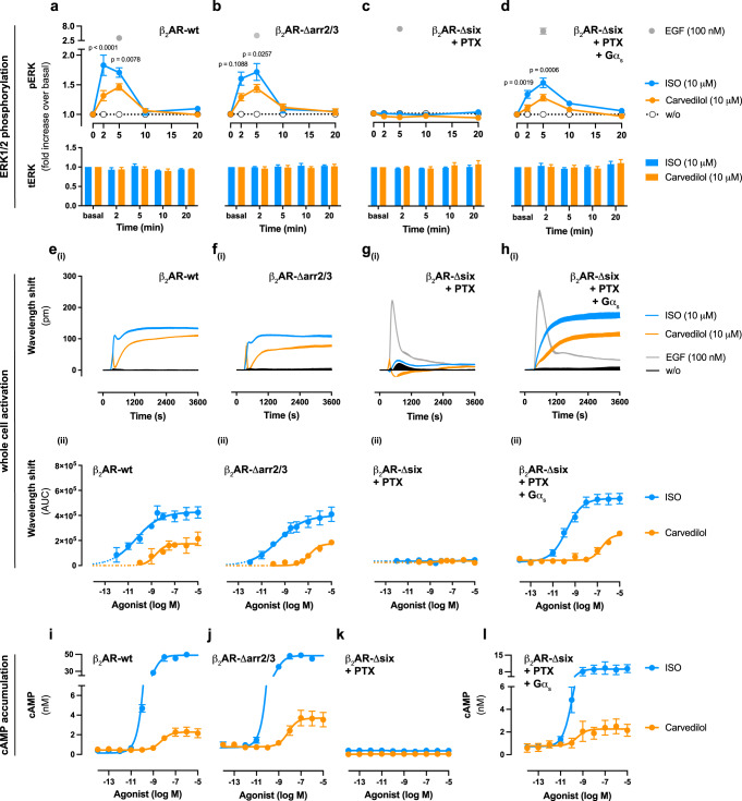
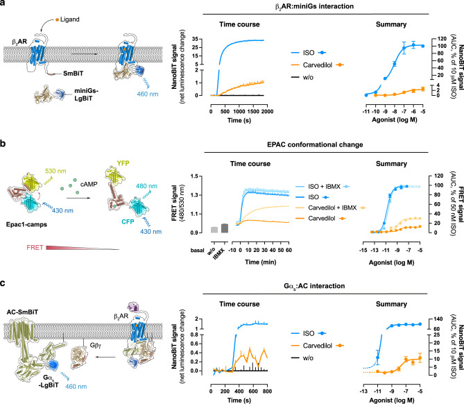
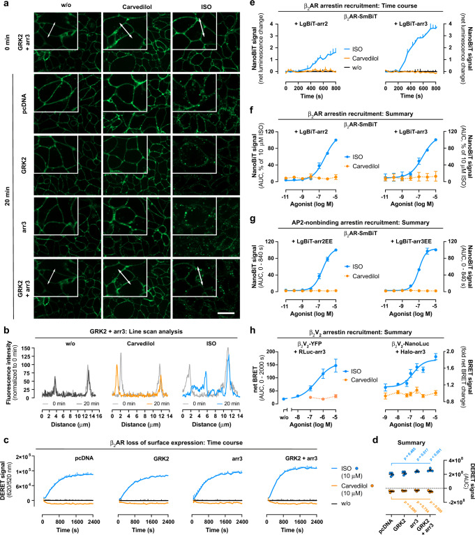
Carvedilol is among the most effective β-blockers for improving survival after myocardial infarction. Yet the mechanisms by which carvedilol achieves this superior clinical profile are still unclear. Beyond blockade of β1-adrenoceptors, arrestin-biased signalling via β2-adrenoceptors is a molecular mechanism proposed to explain the survival benefits. Here, we offer an alternative mechanism to rationalize carvedilol's cellular signalling. Using primary and immortalized cells genome-edited by CRISPR/Cas9 to lack either G proteins or arrestins; and combining biological, biochemical, and signalling assays with molecular dynamics simulations, we demonstrate that G proteins drive all detectable carvedilol signalling through β2ARs. Because a clear understanding of how drugs act is imperative to data interpretation in basic and clinical research, to the stratification of clinical trials or to the monitoring of drug effects on the target pathway, the mechanistic insight gained here provides a foundation for the rational development of signalling prototypes that target the β-adrenoceptor system.


Conflict of interest statement

S.S. is the founder and scientific advisor of 7TM Antibodies GmbH, Jena, Germany. The remaining authors declare no competing interests.

Figures

Fig. 1
Fig. 1. Carvedilol initiation of signalling downstream of β2AR requires Gs proteins.
ERK1/2 phosphorylation, cell morphological changes and cAMP accumulation in wild-type (wt) and genome-edited HEK293 cells, stably expressing SNAP-tagged β2ARs, upon stimulation with either carvedilol or Isoproterenol (ISO). ad Temporal pattern of ERK1/2 phosphorylation and total ERK1/2 in a wild-type, b Δarr2/3, c, d Δsix cells pre-treated with PTX in the absence (c) and presence (d) of re-expressed heterotrimeric G protein subunit Gαs after stimulation with the indicated ligands. eh Representative optical recordings of cell morphological changes (eihi) and corresponding concentration-response curves (eiihii) upon stimulation with carvedilol, ISO or epidermal growth factor (EGF) as viability control. il Quantification of carvedilol- or ISO-induced cAMP accumulation in i wild-type, j Δarr2/3 and k, l Δsix cells pre-treated with PTX in the absence (k) and after re-expression of Gαs (l). Kinetic traces in eihi of a representative experiment (from 3–9 independent experiments) are shown as means + SD, measured in triplicate. Summarized data are depicted as means +/± SEM (a, b: n  =  6; c, d: n  =  9; eiigii: n = 3; hii: n = 9; i–k: n = 3–5 per ligand; l: n = 3–4 per ligand). ad: two-way ANOVA followed by Šídák’s post hoc test. pm picometre, w/o without. Source data are provided as a Source Data file.
Fig. 2
Fig. 2. Recruitment and activation of Gs by carvedilol-occupied β2ARs.
a Schematic of the NanoBiT complementation assay between ligand-activated β2AR C-terminally tagged with the small fragment (SmBiT) of Nanoluciferase (NanoLuc®) and miniGs N-terminally fused to the large fragment of NanoLuc® (LgBiT). Ligand application to the transfected cells induces proximity between the labelled proteins and thus, complementation of a functional NanoLuc® enzyme. Shown are representative kinetic traces of NanoBiT complementation in HEK293 wt cells transiently transfected with β2AR-SmBiT and LgBiT-miniGs after treatment with either 10 µM carvedilol or 10 µM ISO as well as concentration-response curves derived therefrom. b Assay principle underlying detection of cAMP as a FRET decrease with the cytosolic Epac1-camps sensor. Data shown are representative FRET recordings in SNAP-β2AR-HEK293 wt cells after addition of either carvedilol (10 µM) or ISO (50 nM) in the presence or absence of the pan-PDE inhibitor IBMX (500 µM) and corresponding concentration-response curves. FRET recordings of cAMP dynamics were performed in the subsaturating sensor range for carvedilol but at sensor saturation for ISO. c Cartoon of NanoBiT-based detection of interactions between Gαs-LgBiT and AC5-SmBiT, accompanied by representative NanoBiT time courses after treatment of cells with either 10 µM carvedilol or 10 µM ISO and corresponding concentration-effect relationships. Kinetic traces of a representative experiment (from 3–4 independent experiments) are shown as means + SD of 2–4 technical replicates. Summarized data are presented as means ± SEM (a: n = 3; b: n = 4; c: n = 3). w/o without. Source data are provided as a Source Data file.
Fig. 3
Fig. 3. Carvedilol elevates cAMP and spontaneous beating in primary cardiomyocytes.
a Assay design for cAMP detection in adult mouse ventricular myocytes (AMVM) using the plasma membrane-bound Epac1-camps sensor (pmEpac1-camps). b Representative ligand-mediated kinetic pmEpac1-camps recordings in AMVM single cells without and after pre-treatment with the β2AR selective blocker ICI-118,551 (ICI, 1 μM) or the β1AR selective blocker CGP-20712A (CGP, 1 μM). c Summarized ligand-mediated cAMP response of AMVM single cells treated with ISO (n = 20) or carvedilol without (n = 7) or with ICI (n = 10) or CGP (n = 10) relative to the effect achieved with pan-PDE inhibitor IBMX (100 μM) in combination with AC activator Fsk (10 μM). d Representative impedance signals from spontaneously beating neonatal mouse ventricular cardiomyocytes before (w/o) and after application of 300 nM ISO (blue, left) or 300 nM Carvedilol (orange, right). e, f Effect of ISO in control conditions (w/o, n = 5) or in the presence of 10 μM ICI (n = 6) or 10 μM CGP (n = 4) on absolute beating frequency. g, h Effect of carvedilol in control conditions (w/o, n = 11), in the presence of CGP (n = 6) or in the presence of both CGP and ICI (10 μM each, n = 11) on absolute beating frequency. Kinetic recordings of single-cell FRET microscopy in b and beating frequency analyses in d, e, g are from single cells and cell populations, respectively, obtained from three different animal preparations. Only cardiomyocytes with spontaneous beating ≤60 bpm were considered for the impedance time course analysis in eh. Data in c, eh are presented as means ± SEM. e, g: two-tailed paired Student’s t-test; c, f, h: one-way ANOVA with Tukey’s post hoc test. Source data are provided as a Source Data file.
Fig. 4
Fig. 4. β2ARs do not internalize after carvedilol addition.
a Structured-illumination micrographs (representative of three independent experiments) of HEK293 wt cells stably expressing SNAP-tagged β2ARs treated with buffer (w/o), 10 µM carvedilol or 10 µM ISO for 20 min using focus control. b Line scan analysis along a trajectory, indicated by double-faced arrows in a, of buffer-, carvedilol- or ISO-mediated changes in fluorescence intensity. c Schematic illustrating the DERET assay principle displaying how the terbium donor allows resonance energy transfer (RET) to occur with fluorescein acceptor molecules. Agonist-induced receptor internalization decreases the RET to the cell impermeable fluorescein acceptor and is visible as upward deflected trace over time. d Representative kinetic recording of ligand-induced DERET in wt SNAP-β2AR-HEK293, treated either with 10 µM carvedilol or 10 µM ISO, and concentration-response curves derived from three independent experiments. e CCP trapping analysis showing representative overlays of β2AR trajectories and the CCP mask. The coloured traces indicate trajectories of ligand-stimulated β2ARs relative to clathrin-coated structures, imaged with TIRF microscopy in CHO cells. Trajectory portions are coloured according to whether they are detected as free (grey), trapped in CCPs (yellow) or confined outside CCPs (green). f, g Proportion of trapped or freely diffusing receptors per cell (each dot is averaged data from one cell) (f), as well as the empirical probability density estimated for all or only the freely diffusing trajectory portions of the generalized diffusion coefficient D (g). d, f: One-way ANOVA with Kruskal–Wallis post hoc test. Summarized data is shown as mean ± SEM of three independent experiments. Scale bars are 20 µm for a and 1 µm for e, respectively. Source data are provided as a Source Data file.
Fig. 5
Fig. 5. Carvedilol-liganded β2AR is a poor target for arrestin binding.
a Structured-illumination micrographs (representative of three independent experiments) of SNAP-β2AR-HEK293 wt cells transiently transfected with either GRK2, arr3 or both prior to and 20 min after addition of solvent control (w/o), 10 µM carvedilol or 10 µM ISO. b Line scans along a trajectory, indicated by double-faced arrows, corresponding to the panels in a showing fluorescence intensity distribution of cells transfected with GRK2 and arr3 after treatment with solvent control, carvedilol and ISO. c, d Representative DERET measurements in SNAP-β2AR-HEK293 wt cells transiently transfected with pcDNA (n = 4), GRK2 (n = 4), arr3 (n = 3) or both (n = 4), treated either with 10 µM carvedilol or 10 µM ISO (c) and corresponding quantification (d). e, f Representative NanoBiT-based interaction between β2AR-SmBiT and LgBiT-arr2/3 after addition of 10 µM carvedilol or 10 µM ISO (e) and corresponding concentration-effect quantifications (n = 3, f). g NanoBiT-based complementation assays in HEK293 wt cells transiently transfected with β2AR-SmBiT and LgBiT-arr2EE or LgBiT-arr3EE (n = 4). h BRET-based interaction in HEK293 wt cells between pairs of β2V2 C-terminally fused to EYFP (β2V2-EYFP) and Renilla luciferase fused to arrestin-3 (RLuc-arr3; n = 3), as well as NanoLuc®-labelled β2V22V2-NanoLuc®) and Halo-tagged arrestin-3 (Halo-arr3; n = 9), after addition of ISO or carvedilol. Kinetic traces are shown as mean + SD of one representative experiment, measured at least in duplicate. Summarized data is depicted as mean ± SEM. d: two-way ANOVA with Tukey’s post hoc test. Scale bar is 20 µm. Source data are provided as a Source Data file.
Fig. 6
Fig. 6. Ligand-specific β2AR phosphorylation and molecular dynamics simulations fully explain carvedilol’s signalling profile.
a NanoBiT-based enzyme complementation between the Gs-mimetic Nb80-LgBiT and β2AR-SmBiT upon treatment with carvedilol or ISO. b Metadynamics simulations of activation states of the β2AR. Free energy surfaces (FES), revealing energetically favourable conformations at their respective minima, of unliganded apo-, carvedilol- and ISO-bound β2AR plotted against the A100 activation index, a measure of receptor activation. The vertical dashed lines indicate the value of A100 for active (at A100 of 34.4) and inactive (at A100 of −24.1) conformations taken from crystal structures, respectively, and are provided for comparison. c Representative frames of selected ICL2 clusters from minima of apo- (second cluster, 20.7% of the frames), carvedilol- (second cluster, 32.6% of the frames) and ISO-bound (first cluster, 93% of the frames). The first clusters of apo simulations consist of 63.7% of the frames, and 59.7% for carvedilol-bound, respectively. Conformations in both clusters contain mostly alpha-helical ICL2. Thus, whereas in almost all ISO-bound conformations ICL2 is alpha-helical, apo- and carvedilol-bound simulations contain a significant fraction of conformations in which ICL2 is unstructured. d, e NanoBRET-based quantification of proximity between β2AR-NanoLuc® and phosphorylation-independent Halo-arr3-R170E or Halo-arr3-F388A in HEK293 wt cells (d) and the quadruple GRK2/3/5/6 KO HEK293 cell line (aka ΔQ-GRK in ref. 88) (e) after treatment with carvedilol or ISO. f Snake plot spanning the entire sequence of the β2AR including the intracellular loop 3 (ICL3) and the receptor C-terminus. The PKA phosphorylation site S261 in the ICL3 is highlighted in dark magenta, and the C-terminal GRK phosphorylation sites S355, S356, T360 and S364 are marked in light green. g Characterization of β2AR phosphorylation with phosphosite-specific antibodies against pS261, pS355/356 and pT360/364 in HEK293 wt cells stably expressing 3xHA-tagged β2ARs after treatment with 10 µM carvedilol or 10 µM ISO in the absence or presence of overexpressed GRK2 or GRK6. Summarized data are shown as means ± SEM (a: n  =  5; d, e: n  =  9). a: one-way ANOVA with Dunnett´s multiple comparisons post hoc test. The blot in g is representative of at least three independent experiments with similar results. Source data are provided as a Source Data file.

Comment in

  • How carvedilol does not activate β2-adrenoceptors.
    Lefkowitz RJ, Rockman HA, Shim PJ, Liu S, Ahn S, Pani B, Rajagopal S, Shenoy SK, Bouvier M, Benovic JL, Liggett SB, Ruffolo RR, Bristow MR, Packer M. Lefkowitz RJ, et al. Nat Commun. 2023 Nov 30;14(1):7866. doi: 10.1038/s41467-023-42848-5. Nat Commun. 2023. PMID: 38036531 Free PMC article. No abstract available.

References

    1. Nature Medicine Editorial. Mechanism matters. Nat. Med.16, 347 (2010). - PubMed
    1. Davis RL. Mechanism of action and target identification: a matter of timing in drug discovery. iScience. 2020;23:101487. doi: 10.1016/j.isci.2020.101487. - DOI - PMC - PubMed
    1. McDonagh TA, et al. 2021 ESC Guidelines for the diagnosis and treatment of acute and chronic heart failure. Eur. Heart J. 2021;42:3599–3726. doi: 10.1093/eurheartj/ehab368. - DOI - PubMed
    1. Mancia G, et al. 2013 ESH/ESC guidelines for the management of arterial hypertension: the Task Force for the Management of Arterial Hypertension of the European Society of Hypertension (ESH) and of the European Society of Cardiology (ESC) Eur. Heart J. 2013;34:2159–2219. doi: 10.1093/eurheartj/eht151. - DOI - PubMed
    1. Martinez-Milla J, Raposeiras-Roubin S, Pascual-Figal DA, Ibanez B. Role of beta-blockers in cardiovascular disease in 2019. Rev. Esp. Cardiol. (Engl. Ed.) 2019;72:844–852. - PubMed

Publication types

MeSH terms