. 2022 Nov 19;13(1):7114.
doi: 10.1038/s41467-022-34870-w.

PEAR1 regulates expansion of activated fibroblasts and deposition of extracellular matrix in pulmonary fibrosis

Affiliations

PEAR1 regulates expansion of activated fibroblasts and deposition of extracellular matrix in pulmonary fibrosis

Yan Geng et al. Nat Commun. .

Erratum in

Abstract

Pulmonary fibrosis is a chronic interstitial lung disease that causes irreversible and progressive lung scarring and respiratory failure. Activation of fibroblasts plays a central role in the progression of pulmonary fibrosis. Here we show that platelet endothelial aggregation receptor 1 (PEAR1) in fibroblasts may serve as a target for pulmonary fibrosis therapy. Pear1 deficiency in aged mice spontaneously causes alveolar collagens accumulation. Mesenchyme-specific Pear1 deficiency aggravates bleomycin-induced pulmonary fibrosis, confirming that PEAR1 potentially modulates pulmonary fibrosis progression via regulation of mesenchymal cell function. Moreover, single cell and bulk tissue RNA-seq analysis of pulmonary fibroblast reveals the expansion of Activated-fibroblast cluster and enrichment of marker genes in extracellular matrix development in Pear1-/- fibrotic lungs. We further show that PEAR1 associates with Protein Phosphatase 1 to suppress fibrotic factors-induced intracellular signalling and fibroblast activation. Intratracheal aerosolization of monoclonal antibodies activating PEAR1 greatly ameliorates pulmonary fibrosis in both WT and Pear1-humanized mice, significantly improving their survival rate.


Conflict of interest statement

The authors have submitted a patent application (application numbers PCT/CN2021/073547, CN202011212639.8) based on the results reported in this study. And the authors declare no other competing interests.

Figures

Fig. 1
Fig. 1. Pear1 deficiency exacerbated PF by direct regulation of mesenchymal cells function.
a Forced vital capacity (FVC) was measured for evaluating lung function of 12-month-old WT mice and Pear1−/− mice (n = 10 mice in WT group; n = 9 mice in Pear1−/− group). WT, wild type. b Masson staining of lung tissue in 12-month-old WT mice and Pear1−/− mice. The collagen area (green) was calculated (n = 10 mice per group) (scale bars, 50 μm). c FVC of WT and Pear1−/− mice was measured on day 21 after bleo administration (n = 9 mice in WT group; n = 5 mice in Pear1−/− group). d Survival curves of WT and Pear1−/− mice induced by 1.5 µg/g or 2 µg/g bleo through endotracheal atomization (n = 10 mice per group). e Representative images of masson staining on lung sections from WT mice and Pear1−/− mice on day 21 after bleo administration. The collagen area (green) was calculated (n = 10 mice per group) (scale bars, 50 μm). f The proportion of leukocytes (CD45+), endothelial cells (CD31+), epithelial cells (CD326+) and Pdgfra+ mesenchymal cells (MSC, CD45CD31CD326) were analyzed in the lung tissue of Pear1−/− mice with or without treatment of bleo (n = 5 mice in PBS group; n = 8 mice in bleo group). g PEAR1+ cell proportion in Pdgfra+ mesenchymal cells (Pdgfra+CD45CD31CD326) from WT mice lung treated with or without bleo was investigated by flow cytometry. h FVC was measured of Pear1f/f and Col1a2-CreERPear1f/f mice on day 21 after bleo administration (n = 9 mice in Pear1f/f group; n = 5 mice in Col1a2-CreERPear1f/f group). i Survival curves of Pear1f/f and Col1a2-CreERPear1f/f mice induced by 1.5 µg/g bleo through endotracheal atomization (n = 15 mice in Pear1f/f group; n = 17 mice in Col1a2-CreERPear1f/f group). j Representative images of masson staining on lung sections from Pear1f/f and Col1a2-CreERPear1f/f mice on day 21 after bleo administration. The collagen area (green) was calculated (n = 9 mice in Pear1f/f group; n = 5 mice in Col1a2-CreERPear1f/f group) (scale bars, 50 μm). For ac, e, h, j, two-tailed t test was used. For f, one-way ANOVA was used. Data are presented as mean ± SD. Source data are provided as a Source data file.
Fig. 2
Fig. 2. Pear1 deficiency expanded activated fibroblasts in bleo model.
ac All of the 17,934 mesenchymal cells across four experiment conditions were integrated for umap clustering (a), cell type annotation and cluster-tree dendrogram (b), and fraction of each cell type in each experimental condition (c). d GSEA analysis of activated fibroblasts signature of cluster 7 compared with the rest clusters. e Dotplot of markers for the selected cluster. f, g Top 10 GO biological process (f) and KEGG pathways (g) enriched with Cluster 7 markers in Pear1−/− bleo mice. h Differentially expressed genes in Cluster 7 between Pear1−/− bleo and WT bleo. For d, nominal P value for the statistical significance of the enrichment score is adjusted using Benjamini–Hochberg method for multiple gene sets and hypothesis testing. For f, g, one-sided Fisher’s exact test was used.
Fig. 3
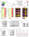
Fig. 3. PEAR1 associated with PP1 to suppress fibrotic factor induced fibroblast activation.
Pdgfra+ cells were selected for bulk RNA-seq assay. a Overlapping pattern of the differentially expressed genes (DEGs) in three comparisons identified 155 core DEGs. b Principle component analysis (PCA) of replicated samples in each condition. Squares represent the centroid of corresponding sample group. c Top 10 GO biological process enriched with upregulated genes in Pear1−/− Bleo compared to WT Bleo. d Clustering of the leading-edge EMT genes in GSEA across 12 samples in four experiment conditions. Colored squares on bottom left represent experiment conditions. Colored bar on bottom right represents the scaled expression levels. e Leading-edge EMT genes in GSEA were predominantly expressed in cluster 7 (activated fibroblasts) cells of scRNA-seq data. Color bar on bottom left represents the scaled expression levels. Color dot on bottom left represents Pdgfra+ cell clusters in scRNA-seq data: 1-Col14a1 Matrix Fibroblasts, 2-Col14a1/Col13a1 Matrix Fibroblasts, 5-Col13a1 Matrix Fibroblasts, 7-Activated Fibroblasts, 8-Col14a1 Matrix Fibroblasts (Mmp3/Cyp1b1 high). f The phosphorylation levels of Smad2/3, AKT, ERK1/2, P38, and JNK1/2 were evaluated in WT and Pear1−/− fibroblasts stimulated by TGFβ, FGF, or PDGF for 30 min, respectively. GAPDH was used as a loading control. The experiments were repeated three times and the results were similar. g, h The binding of PEAR1 and PP1 were detected by immunoprecipitation in cultured human and mouse pulmonary fibroblasts. The experiments were repeated three times and the results were similar. i The phosphorylation levels of, AKT, P38, ERK1/2, and JNK1/2 were evaluated in fibroblasts incubated with 1 nM, 10 nM, or 100 nM PP1 inhibitor for 15 min, respectively. GAPDH was used as a loading control. The experiments were repeated three times and the results were similar. j The relative mRNA expression levels of ECM genes in cultured pulmonary fibroblasts incubated with PP1 inhibitor or DMSO as a negative control (n = 3 biologically independent samples in each group). For c, one-sided Fisher’s exact test was used. For j, two-tailed t test was used. Data are presented as mean ± SD. Source data are provided as a Source data file.
Fig. 4
Fig. 4. Monoclonal antibody targeted human PEAR1 for PF therapy.
a The mRNA expression levels of extracellular matrix genes in human pulmonary fibroblasts (HFL1) treated with 1 µg/mL anti-human PEAR1 monoclonal antibodies (LF2) by qPCR (n = 3 biologically independent samples in each group). LF2 is a humanized antibody with an ADCC/ADCP-weak human IgG4 Fc with S228P mutation. b Survival curves of humanized Pear1 mice induced by 1.5 µg/g (body weight) bleo treated with 300 mg/kg pirfenidone daily by gavage (PFD), 0.5 mg/kg (LF2L), 1 mg/kg (LF2H) LF2 through the trachea once a week. Saline was used as control (Ctrl) (n = 11 mice in Ctrl group; n = 16 mice in PFD group; n = 12 mice in LF2L group and n = 10 mice in LF2H group). c FVC was measured of Ctrl group mice, PFD group mice, LF2L group mice, and LF2H group mice (n = 3 mice in Ctrl group; n = 11 mice in PFD group; n = 7 mice in LF2L group; n = 9 mice in LF2H group). d Representative images of masson staining on lung sections from Ctrl group mice, PFD group mice, LF2L group mice, and LF2H group mice. The collagen area (green) was calculated and statistics were performed (n = 11 mice in Ctrl group; n = 16 mice in PFD group; n = 12 mice in LF2L group; n = 9 mice in LF2H group) (scale bars, 50 μm). e, f Representative images of immunofluorescence staining on lung sections from Ctrl group mice, PFD group mice, LF2L group mice, and LF2H group mice for Pdgfra (red), collagen IV (green), and DAPI (blue). The fluorescence positive area was calculated and statistics were performed (n = 11 mice in saline control group; n = 15 mice in PFD group; n = 8 mice in LF2L group; n = 8 mice in LF2H group) (scale bars, 75 μm). g A schematic diagram of the mechanism of PEAR1 in regulation of pulmonary fibrosis. ECM, extracellular matrix; MAb, monoclonal antibody. For c, d, f, one-way ANOVA was used. Data are presented as mean ± SD. Source data are provided as a Source data file.

Similar articles

Cited by

References

    1. Sgalla G, Kulkarni T, Antin-Ozerkis D, Thannickal VJ, Richeldi L. Update in pulmonary fibrosis 2018. Am. J. Respir. Crit. Care Med. 2019;200:292–300. doi: 10.1164/rccm.201903-0542UP. - DOI - PubMed
    1. Rockey DC, Bell PD, Hill JA. Fibrosis—a common pathway to organ injury and failure. N. Engl. J. Med. 2015;372:1138–1149. doi: 10.1056/NEJMra1300575. - DOI - PubMed
    1. Lederer DJ, Martinez FJ. Idiopathic pulmonary fibrosis. N. Engl. J. Med. 2018;378:1811–1823. doi: 10.1056/NEJMra1705751. - DOI - PubMed
    1. Martinez FJ, et al. Idiopathic pulmonary fibrosis. Nat. Rev. Dis. Prim. 2017;3:17074. doi: 10.1038/nrdp.2017.74. - DOI - PubMed
    1. Wolters PJ, Collard HR, Jones KD. Pathogenesis of idiopathic pulmonary fibrosis. Annu. Rev. Pathol. 2014;9:157–179. doi: 10.1146/annurev-pathol-012513-104706. - DOI - PMC - PubMed

Publication types