

Melt electrowriting of PLA, PCL, and composite PLA/PCL scaffolds for tissue engineering application
- PMID: 36402790
- PMCID: PMC9675866
- DOI: 10.1038/s41598-022-24275-6
Melt electrowriting of PLA, PCL, and composite PLA/PCL scaffolds for tissue engineering application
Abstract
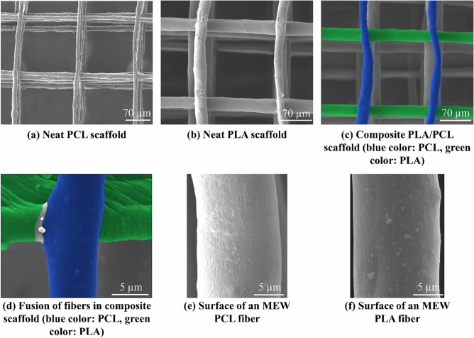
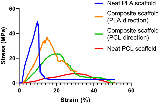
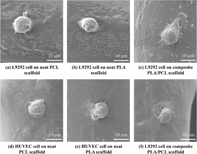
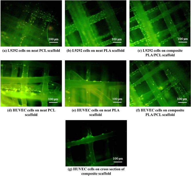
Fabrication of well-ordered and bio-mimetic scaffolds is one of the most important research lines in tissue engineering. Different techniques have been utilized to achieve this goal, however, each method has its own disadvantages. Recently, melt electrowriting (MEW) as a technique for fabrication of well-organized scaffolds has attracted the researchers' attention due to simultaneous use of principles of additive manufacturing and electrohydrodynamic phenomena. In previous research studies, polycaprolactone (PCL) has been mostly used in MEW process. PCL is a biocompatible polymer with characteristics that make it easy to fabricate well-arranged structures using MEW device. However, the mechanical properties of PCL are not favorable for applications like bone tissue engineering. Furthermore, it is of vital importance to demonstrate the capability of MEW technique for processing a broad range of polymers. To address aforementioned problems, in this study, three ten-layered box-structured well-ordered scaffolds, including neat PLA, neat PCL, and PLA/PCL composite are fabricated using an MEW device. Printing of the composite PLA/PCL scaffold using the MEW device is conducted in this study for the first time. The MEW device used in this study is a commercial fused deposition modeling (FDM) 3D printer which with some changes in its setup and configuration becomes prepared for being used as an MEW device. Since in most of previous studies, a setup has been designed and built for MEW process, the use of the FDM device can be considered as one of the novelties of this research. The printing parameters are adjusted in a way that scaffolds with nearly equal pore sizes in the range of 140 µm to 150 µm are fabricated. However, PCL fibers are mostly narrower (diameters in the range of 5 µm to 15 µm) than PLA fibers with diameters between 15 and 25 µm. Unlike the MEW process of PCL, accurate positioning of PLA fibers is difficult which can be due to higher viscosity of PLA melt compared to PCL melt. The printed composite PLA/PCL scaffold possesses a well-ordered box structure with improved mechanical properties and cell-scaffold interactions compared to both neat PLA and PCL scaffolds. Besides, the composite scaffold exhibits a higher swelling ratio than the neat PCL scaffold which can be related to the presence of less hydrophobic PLA fibers. This scaffold demonstrates an anisotropic behavior during uniaxial tensile test in which its Young's modulus, ultimate tensile stress, and strain to failure all depend on the direction of the applied tensile force. This anisotropy makes the composite PLA/PCL scaffold an exciting candidate for applications in heart tissue engineering. The results of in-vitro cell viability test using L929 mouse murine fibroblast and human umbilical vein endothelial (HUVEC) cells demonstrate that all of the printed scaffolds are biocompatible. In particular, the composite scaffold presents the highest cell viability value among the fabricated scaffolds. All in all, the composite PLA/PCL scaffold shows that it can be a promising substitution for neat PCL scaffold used in previous MEW studies.
© 2022. The Author(s).
Conflict of interest statement
The authors declare no competing interests.
Figures


Similar articles
-
Facile manufacturing of fused-deposition modeled composite scaffolds for tissue engineering-an embedding model with plasticity for incorporation of additives.Biomed Mater. 2020 Dec 17;16(1):015028. doi: 10.1088/1748-605X/abc1b0. Biomed Mater. 2020. PMID: 33331292
-
Melt electrowriting of a biocompatible photo-crosslinkable poly(D,L-lactic acid)/poly(ε-caprolactone)-based material with tunable mechanical and functionalization properties.J Biomed Mater Res A. 2023 Jun;111(6):851-862. doi: 10.1002/jbm.a.37536. Epub 2023 Mar 23. J Biomed Mater Res A. 2023. PMID: 36951312
-
Preparation and characterization of PLA/PCL/HA composite scaffolds using indirect 3D printing for bone tissue engineering.Mater Sci Eng C Mater Biol Appl. 2019 Nov;104:109960. doi: 10.1016/j.msec.2019.109960. Epub 2019 Jul 6. Mater Sci Eng C Mater Biol Appl. 2019. PMID: 31500051
-
Polymers for Melt Electrowriting.Adv Healthc Mater. 2021 Jan;10(1):e2001232. doi: 10.1002/adhm.202001232. Epub 2020 Sep 17. Adv Healthc Mater. 2021. PMID: 32940962 Free PMC article. Review.
-
Unveiling the potential of melt electrowriting in regenerative dental medicine.Acta Biomater. 2023 Jan 15;156:88-109. doi: 10.1016/j.actbio.2022.01.010. Epub 2022 Jan 10. Acta Biomater. 2023. PMID: 35026478 Free PMC article. Review.
Cited by
-
Characterization and In Vitro Evaluation of Porous Polymer-Blended Scaffolds Functionalized with Tricalcium Phosphate.J Funct Biomater. 2024 Feb 26;15(3):57. doi: 10.3390/jfb15030057. J Funct Biomater. 2024. PMID: 38535250 Free PMC article.
-
Biomaterials in Postoperative Adhesion Barriers and Uterine Tissue Engineering.Gels. 2025 Jun 9;11(6):441. doi: 10.3390/gels11060441. Gels. 2025. PMID: 40558740 Free PMC article. Review.
-
Non-Woven Fibrous Polylactic Acid/Hydroxyapatite Nanocomposites Obtained via Solution Blow Spinning: Morphology, Thermal and Mechanical Behavior.Nanomaterials (Basel). 2024 Jan 15;14(2):196. doi: 10.3390/nano14020196. Nanomaterials (Basel). 2024. PMID: 38251160 Free PMC article.
-
3D Printed Mesh Geometry Modulates Immune Response and Interface Biology in Mouse and Sheep Model: Implications for Pelvic Floor Surgery.Adv Sci (Weinh). 2025 Mar;12(11):e2405004. doi: 10.1002/advs.202405004. Epub 2024 Sep 19. Adv Sci (Weinh). 2025. PMID: 39297316 Free PMC article.
-
Detection of Limbal Stem Cells Adhered to Melt Electrospun Silk Fibroin and Gelatin-Modified Polylactic Acid Scaffolds.Polymers (Basel). 2023 Feb 3;15(3):777. doi: 10.3390/polym15030777. Polymers (Basel). 2023. PMID: 36772078 Free PMC article.
References
-
- Diomede F, Gugliandolo A, Cardelli P, Merciaro I, Ettorre V, Traini T, Bedini R, Scionti D, Bramanti A, Nanci A. Three-dimensional printed PLA scaffold and human gingival stem cell-derived extracellular vesicles: A new tool for bone defect repair. Stem Cell Res. Ther. 2018;9:1–21. doi: 10.1186/s13287-018-0850-0. - DOI - PMC - PubMed
MeSH terms
Substances
LinkOut - more resources
Full Text Sources
