. 2023 Feb;48(3):540-551.
doi: 10.1038/s41386-022-01496-x. Epub 2022 Nov 19.

Adolescent Δ-9-tetrahydrocannabinol exposure induces differential acute and long-term neuronal and molecular disturbances in dorsal vs. ventral hippocampal subregions

Affiliations

Adolescent Δ-9-tetrahydrocannabinol exposure induces differential acute and long-term neuronal and molecular disturbances in dorsal vs. ventral hippocampal subregions

Marta De Felice et al. Neuropsychopharmacology. 2023 Feb.

Abstract

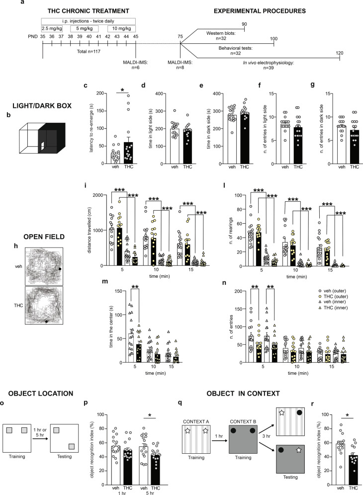
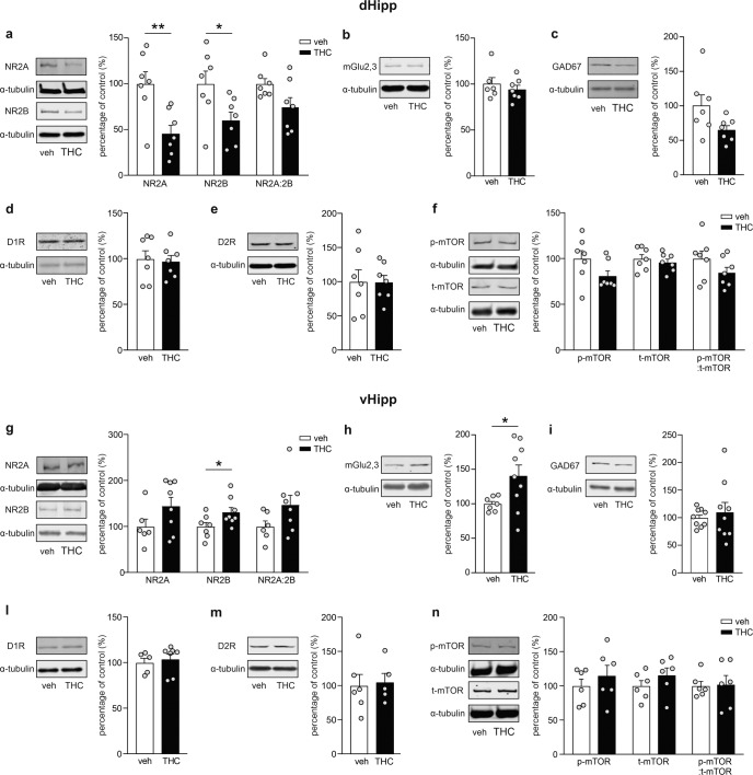
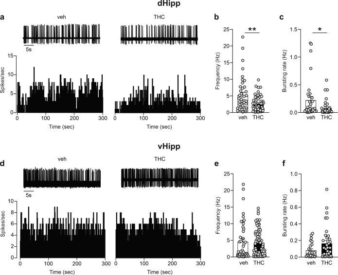
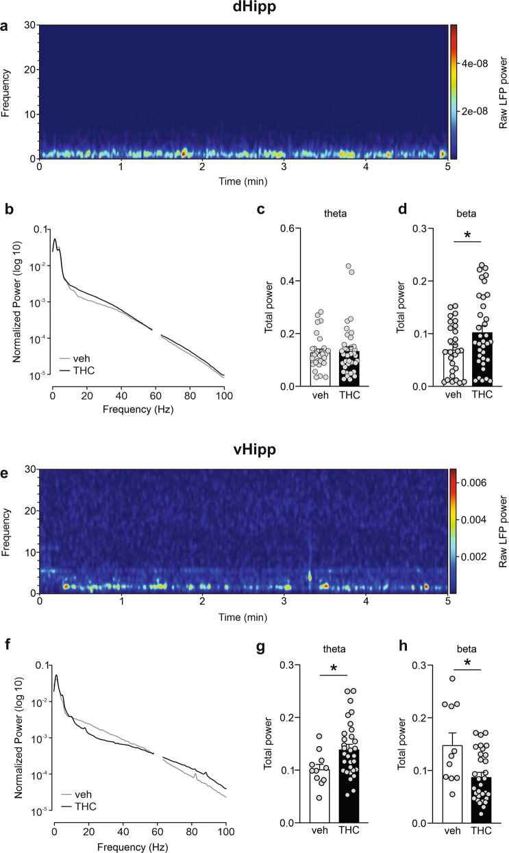
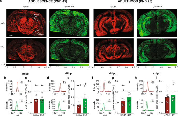
Chronic exposure to Δ-9-tetrahydrocannabinol (THC) during adolescence is associated with long-lasting cognitive impairments and enhanced susceptibility to anxiety and mood disorders. Previous evidence has revealed functional and anatomical dissociations between the posterior vs. anterior portions of the hippocampal formation, which are classified as the dorsal and ventral regions in rodents, respectively. Notably, the dorsal hippocampus is critical for cognitive and contextual processing, whereas the ventral region is critical for affective and emotional processing. While adolescent THC exposure can induce significant morphological disturbances and glutamatergic signaling abnormalities in the hippocampus, it is not currently understood how the dorsal vs. ventral hippocampal regions are affected by THC during neurodevelopment. In the present study, we used an integrative combination of behavioral, molecular, and neural assays in a neurodevelopmental rodent model of adolescent THC exposure. We report that adolescent THC exposure induces long-lasting memory deficits and anxiety like-behaviors concomitant with a wide range of differential molecular and neuronal abnormalities in dorsal vs. ventral hippocampal regions. In addition, using matrix-assisted laser desorption/ionization imaging mass spectrometry (MALDI-IMS), we show for the first time that adolescent THC exposure induces significant and enduring dysregulation of GABA and glutamate levels in dorsal vs. ventral hippocampus. Finally, adolescent THC exposure induced dissociable dysregulations of hippocampal glutamatergic signaling, characterized by differential glutamatergic receptor expression markers, profound alterations in pyramidal neuronal activity and associated oscillatory patterns in dorsal vs. ventral hippocampal subregions.


Conflict of interest statement

The authors declare no competing interests.

Figures

Fig. 1
Fig. 1. Long-lasting effects of adolescent THC exposure on anxiety and memory.
a Experimental timeline including the number of subjects used for each procedure. b Schematic representation of light–dark box apparatus for anxiety test. c Adolescent THC-treated rats re-emerged later from the dark environment than vehicle group (n = 16 for both groups), while the time spent in the light and dark environments (d, e) as well as the number of entries in both sides (f, g) were similar between vehicle- and THC-treated rats. h Representative open-field activity traces for vehicle- and THC-exposed rats. il Both groups (veh vs. THC: n = 16 vs. 14) exhibited larger locomotor activity and higher rearing behavior in the outer zone vs. the inner zone of the OF arena at any of the timepoints examined, however in the first 5 min of the test, THC-treated rats spent less time in the inner zone of the arena (m) and did less transitions between inner vs. outer zones (n) compared to their vehicle counterparts. o Schematic protocol representation for object location test. p THC-treated rats exhibited spatial memory impairments after 5-h delay but not after 1 h delay, when compared to their vehicle counterparts (n = 16 for both groups). q Schematic protocol representation for object in context test. r Adolescent THC exposure altered rats’ ability to recognize a familiar object in a novel context (veh vs. THC: n = 14 vs. 13). Mann–Whitney U test, Student’s t test, two-way or three-way ANOVAs, *P < 0.05, **P < 0.01, ***P < 0.001.
Fig. 2
Fig. 2. Long-term effects of THC treatment during adolescence on selected markers in dHipp and vHipp.
af, insets on the left side of the bar graphs, Representative western blots for NMDA2AR, NMDA2BR, mGluR2/3, GAD67, D1R, D2R and mTOR in dHipp. a, right, Densitometry analysis revealed a reduction in NMDA2AR and NMDA2BR levels induced by adolescent THC exposure, but not in their ratio of NMDA2A/NMDA2B (n = 7 for both groups). bf, right, No differences were observed between groups in mGluR2/3 (veh vs. THC: n = 6 vs. 7), GAD67 (n = 7 for both groups), D1R (n = 7 for both groups), D2R (n = 7 for both groups) and mTOR (n = 7 for both groups) expression. gn, insets on the left side of the bar graphs, Representative western blots for NMDA2AR, NMDA2BR, mGluR2/3, GAD67, D1R, D2R and mTOR in vHipp (left). g, h, right, Adolescent THC treatment induced a long-lasting increase in NMDA2BR (veh vs. THC: n = 7 vs. 8) and mGluR2/3 (veh vs. THC: n = 7 vs. 9) levels, but not in NMDA2AR or in the ratio of NMDA2A/NMDA2B (veh vs. THC: n = 6 vs. 8 for both). in, right, Chronic THC treatment did not affect GAD67 (n = 9 for both groups), D1R (veh vs. THC: n = 5 vs. 7), D2R (veh vs. THC: n = 6 vs. 5) and mTOR (n = 6 for both groups) expression. Mann–Whitney U test or Student’s t test, *P < 0.05, **P < 0.01.
Fig. 3
Fig. 3. Effects of adolescent THC exposure on dHipp and vHipp pyramidal spontaneous electrical activity.
a Traces and rate histograms of representative dHipp glutamatergic cells recorded from vehicle and THC group. b, c THC-treated rats showed a reduction in the firing rate (veh vs. THC: n = 41 cells/8 rats vs. 55 cells/13 rats) and bursting activity (veh vs. THC: n = 33 cells/8 rats vs. 35 cells/13 rats) in of pyramidal neurons in dHipp. d Traces and rate histograms of representative vHipp glutamatergic cells recorded from vehicle and THC group. e, f No differences were observed between groups in the firing rate (veh vs. THC: n = 45 cells/9 rats vs. 68 cells/9 rats) and bursting activity (veh vs. THC: n = 23 cells/9 rats vs. 34 cells/9 rats) of pyramidal neurons in vHipp. Mann–Whitney U test, *P < 0.05, **P < 0.01.
Fig. 4
Fig. 4. Effects of THC treatment during adolescence on spontaneous dHipp and vHipp theta (4–7 Hz) and beta (14–30 Hz) oscillations.
a Representative spectrogram of a 5-min recording in dHipp. b Average normalized LFP power spectra in dHipp of vehicle and THC groups. c, d THC exposure during adolescence did not affect the theta waves in dHipp, while it significantly enhanced the beta oscillations (veh vs. THC: n = 28 recording site/5 rats vs. 35 cells/9 rats). e Representative spectrogram of a 5-min recording in vHipp. f Average normalized LFP power spectra in vHipp of vehicle and THC groups. g, h The THC-treated group exhibited an increase in theta oscillations and a decrease in beta oscillation (veh vs. THC: n = 11 recording site/4 rats vs. 31 cells/4 rats). Student’s t test or Mann–Whitney U test, *P < 0.05.
Fig. 5
Fig. 5. Short- vs. long-term effects of adolescent THC exposure on neurotransmitters levels in dHipp and vHipp.
a MALDI-IMS images of representative vehicle and THC sections at PND 45 and 75. Representative ion-mass spectra in dHipp (b, f) and vHipp (d, h) of vehicle and THC groups at PND 45 (b, d) and 75 (f, h) for [GABA + K]+ at m/z 142 (top) and [glutamic acid + K]+ at m/z 186 (bottom). c MALDI-IMS relative quantification revealed a short-term reduction in GABA and glutamate levels (n = 6 for both groups and neurotransmitters) in dHipp of THC-exposed rat. e GABA levels were decreased in vHipp, while no differences were observed in glutamate (n = 6 for both groups and neurotransmitters). g MALDI-IMS relative quantification revealed a long-lasting reduction in GABA, but not glutamate levels (n = 8 for both groups and neurotransmitters), in dHipp of rats exposed to THC during adolescence. i No differences were observed in GABA and glutamate levels (n = 8 for both groups and neurotransmitters) in vHipp. One-sample t-test, *P < 0.05, **P < 0.01, ***P < 0.001.

Similar articles

Cited by

References

    1. Andréasson S, Allebeck P, Engström A, Rydberg U. Cannabis and schizophrenia. A longitudinal study of Swedish conscripts. Lancet. 1987;2:1483–6. doi: 10.1016/S0140-6736(87)92620-1. - DOI - PubMed
    1. Krebs MO, Kebir O, Jay TM. Exposure to cannabinoids can lead to persistent cognitive and psychiatric disorders. Eur J Pain. 2019;23:1225–33. - PubMed
    1. Murray RM, Englund A, Abi-Dargham A, Lewis DA, Di Forti M, Davies C, et al. Cannabis-associated psychosis: Neural substrate and clinical impact. Neuropharmacology. 2017;124:89–104. doi: 10.1016/j.neuropharm.2017.06.018. - DOI - PubMed
    1. Gobbi G, Atkin T, Zytynski T, Wang S, Askari S, Boruff J, et al. Association of cannabis use in adolescence and risk of depression, anxiety, and suicidality in young adulthood: a systematic review and meta-analysis. JAMA Psychiatry. 2019;76:426–34. doi: 10.1001/jamapsychiatry.2018.4500. - DOI - PMC - PubMed
    1. Mato S, Del Olmo E, Pazos A. Ontogenetic development of cannabinoid receptor expression and signal transduction functionality in the human brain. Eur J Neurosci. 2003;17:1747–54. doi: 10.1046/j.1460-9568.2003.02599.x. - DOI - PubMed

Publication types