

Chronic oligodendrocyte injury in central nervous system pathologies
- PMID: 36402839
- PMCID: PMC9675815
- DOI: 10.1038/s42003-022-04248-1
Chronic oligodendrocyte injury in central nervous system pathologies
Abstract
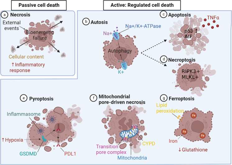
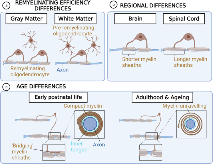
Myelin, the membrane surrounding neuronal axons, is critical for central nervous system (CNS) function. Injury to myelin-forming oligodendrocytes (OL) in chronic neurological diseases (e.g. multiple sclerosis) ranges from sublethal to lethal, leading to OL dysfunction and myelin pathology, and consequent deleterious impacts on axonal health that drive clinical impairments. This is regulated by intrinsic factors such as heterogeneity and age, and extrinsic cellular and molecular interactions. Here, we discuss the responses of OLs to injury, and perspectives for therapeutic targeting. We put forward that targeting mature OL health in neurological disease is a promising therapeutic strategy to support CNS function.
© 2022. The Author(s).
Conflict of interest statement
The authors declare no competing interests.
Figures

Similar articles
-
Oligodendrocyte regeneration: Its significance in myelin replacement and neuroprotection in multiple sclerosis.Neuropharmacology. 2016 Nov;110(Pt B):633-643. doi: 10.1016/j.neuropharm.2015.10.010. Epub 2015 Oct 22. Neuropharmacology. 2016. PMID: 26474658 Free PMC article. Review.
-
Extrinsic Factors Driving Oligodendrocyte Lineage Cell Progression in CNS Development and Injury.Neurochem Res. 2020 Mar;45(3):630-642. doi: 10.1007/s11064-020-02967-7. Epub 2020 Jan 29. Neurochem Res. 2020. PMID: 31997102 Free PMC article. Review.
-
R-Ras1 and R-Ras2 Are Essential for Oligodendrocyte Differentiation and Survival for Correct Myelination in the Central Nervous System.J Neurosci. 2018 May 30;38(22):5096-5110. doi: 10.1523/JNEUROSCI.3364-17.2018. Epub 2018 May 2. J Neurosci. 2018. PMID: 29720552 Free PMC article.
-
Myelin-based inhibitors of oligodendrocyte myelination: clues from axonal growth and regeneration.Neurosci Bull. 2013 Apr;29(2):177-88. doi: 10.1007/s12264-013-1319-x. Epub 2013 Mar 20. Neurosci Bull. 2013. PMID: 23516141 Free PMC article. Review.
-
Remyelination: the true regeneration of the central nervous system.J Comp Pathol. 2013 Aug-Oct;149(2-3):242-54. doi: 10.1016/j.jcpa.2013.05.004. Epub 2013 Jul 5. J Comp Pathol. 2013. PMID: 23831056 Review.
Cited by
-
Heterogeneity of mature oligodendrocytes in the central nervous system.Neural Regen Res. 2025 May 1;20(5):1336-1349. doi: 10.4103/NRR.NRR-D-24-00055. Epub 2024 Jun 26. Neural Regen Res. 2025. PMID: 38934385 Free PMC article.
-
Reflective imaging of myelin integrity in the human and mouse central nervous systems.Front Cell Neurosci. 2024 Jul 10;18:1408182. doi: 10.3389/fncel.2024.1408182. eCollection 2024. Front Cell Neurosci. 2024. PMID: 39049821 Free PMC article.
-
Insights on therapeutic potential of clemastine in neurological disorders.Front Mol Neurosci. 2023 Sep 28;16:1279985. doi: 10.3389/fnmol.2023.1279985. eCollection 2023. Front Mol Neurosci. 2023. PMID: 37840769 Free PMC article. Review.
-
Oligodendrocyte and Myelin Pathophysiology in Multiple Sclerosis.Adv Neurobiol. 2025;43:317-361. doi: 10.1007/978-3-031-87919-7_12. Adv Neurobiol. 2025. PMID: 40500503 Review.
-
Multisensory gamma stimulation mitigates the effects of demyelination induced by cuprizone in male mice.Nat Commun. 2024 Aug 8;15(1):6744. doi: 10.1038/s41467-024-51003-7. Nat Commun. 2024. PMID: 39112447 Free PMC article.
References
Publication types
MeSH terms
Grants and funding
LinkOut - more resources
Full Text Sources
Medical
