. 2022 Nov 19;13(1):7110.
doi: 10.1038/s41467-022-34882-6.

Structure of the human heparan sulfate polymerase complex EXT1-EXT2

Affiliations

Structure of the human heparan sulfate polymerase complex EXT1-EXT2

Francisco Leisico et al. Nat Commun. .

Abstract

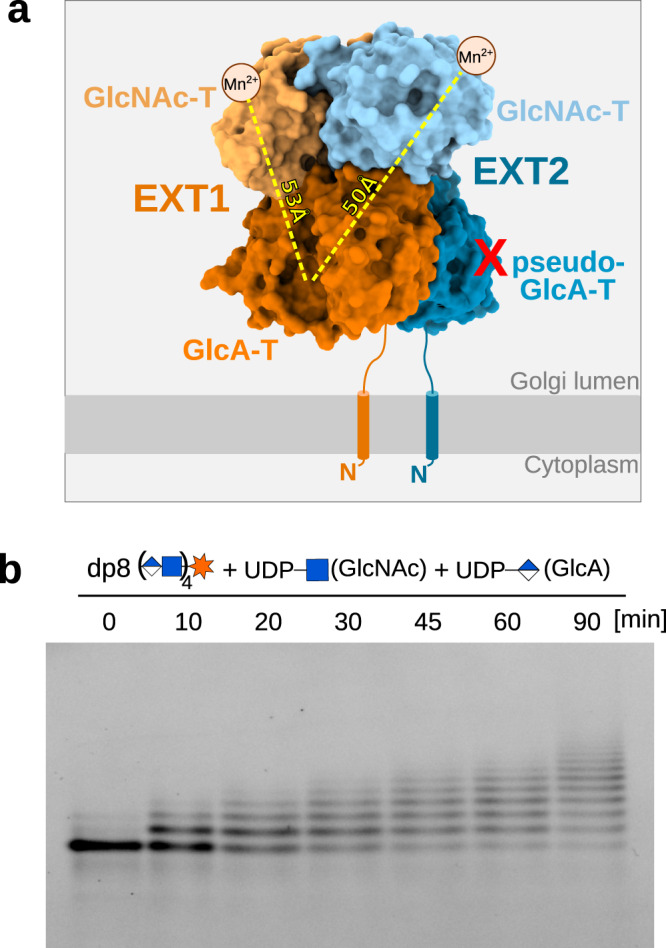
Heparan sulfates are complex polysaccharides that mediate the interaction with a broad range of protein ligands at the cell surface. A key step in heparan sulfate biosynthesis is catalyzed by the bi-functional glycosyltransferases EXT1 and EXT2, which generate the glycan backbone consisting of repeating N-acetylglucosamine and glucuronic acid units. The molecular mechanism of heparan sulfate chain polymerization remains, however, unknown. Here, we present the cryo-electron microscopy structure of human EXT1-EXT2, which reveals the formation of a tightly packed hetero-dimeric complex harboring four glycosyltransferase domains. A combination of in vitro and in cellulo mutational studies is used to dissect the functional role of the four catalytic sites. While EXT1 can catalyze both glycosyltransferase reactions, our results indicate that EXT2 might only have N-acetylglucosamine transferase activity. Our findings provide mechanistic insight into heparan sulfate chain elongation as a nonprocessive process and lay the foundation for future studies on EXT1-EXT2 function in health and disease.


Conflict of interest statement

The authors declare no competing interests.

Figures

Fig. 1
Fig. 1. Purification and functional characterization of the EXT1-EXT2 complex.
a Coomassie-stained SDS-PAGE analysis of purified EXT1-EXT2 complex, lacking the N-terminal membrane anchoring helix. Expected molecular sizes of EXT1 and EXT2 are about 85 kDa and 79 kDa, respectively. Source data is provided as a Source Data 1. b Mass photometry analysis of purified complex with a major peak at 160 kDa. Source data is provided as a Source Data 2. c The catalytic activity of EXT1-EXT2 was studied using an in vitro glycosylation assay. The specific transfer of N-acetylglucosamine (GlcNAc) and glucuronic acid (GlcA) molecules was followed using synthetic, fluorescently-labeled octa-saccharide (dp8) and nona-saccharide (dp9) substrate analogs. Source data is provided as a Source Data 3.
Fig. 2
Fig. 2. Structure of the human EXT1-EXT2 complex.
a EM map of the hetero-dimeric EXT1-EXT2 complex, with density covering EXT1 in orange and EXT2 in blue. Both proteins harbor a glucuronic acid transferase (GlcA-T) domain and a N-acetylglucosamine transferase (GlcNAc-T) domain colored in darker and lighter shades, respectively. Densities of N-glycans are colored in yellow. The N-terminal membrane anchoring helices, absent in our expression constructs, are drawn as cylinders. b Cartoon representation of the complex with coloring as in a and glycosylations shown in stick representation. Missing loops are indicated as dotted lines. c Rotated view showing the GlcA-T domains from the membrane plane.
Fig. 3
Fig. 3. The active sites of the EXT1-EXT2 complex.
a Cartoon representation of the EXT1 GlcA-T catalytic site shown in dark orange with important residues shown as sticks. b Catalytic site of the EXT2 pseudo-GlcA-T domain colored in dark blue and shown from the same view as in (a). Two anti-parallel beta-strands, colored in dark blue, intrude into the active site. c–e Cartoon representation of the GlcNAc-T active sites of EXT1 (light orange), EXTL2 (green), and EXT2 (light blue) with a UDP-GlcNAc donor substrate (yellow) in the crystal structure of EXTL2 (PDB-ID: 1ON6). Conserved residues involved in metal ion (shown as purple sphere) or UDP-GlcNAc binding are highlighted as sticks. f In vitro activity of EXT1-EXT2 in presence of different divalent metal ions. Fluorescently-labeled octa- (dp8) and nona- (dp9) oligosaccharide substrate analogs were used as acceptor substrates. Source data is provided as a Source Data 4. GlcNAc-T, N-Acetylglucosamine-transferase; GlcA-T, glucuronic acid-transferase.
Fig. 4
Fig. 4. In vitro and in cellulo mutational analysis.
a Table summarizing EXT1 and EXT2 mutants used in this study. b In vitro activity assay using fluorescently-labeled octa- (dp8) and nona- (dp9) oligosaccharide acceptor substrates and purified EXT1-EXT2 complexes harboring either EXT1 and EXT2 wild-type proteins (WT) or amino acid substitutions in one of the two proteins. Source data is provided as a Source Data 5. c Quantification of cell surface heparan sulfate levels by flow cytometry experiments in HeLa knock-out (KO) cell lines lacking either the EXT1 or EXT2 protein. The knock-out cell lines were not transfected or complemented with either WT EXT1 or EXT2 proteins or amino acid substituted versions thereof. Quantified heparan sulfate content from three independent experiments was normalized to the EXT expression level (see Supplementary Fig. 10) and blotted as percentage compared to wild-type HeLa cells (n = 3), shown as dots and average as bars. Error bars indicate SD. A one-sided paired student t-test was used to determine p-values: NS > 0.05, *<0.05 and **<0.01 or more precisely EXT1 WT vs D162N/D164N (0.0058), R280A (0.0559), R346A (0.0020) and D565N/D567N (0.0485) and EXT2 WT vs R266A (0.1313), D538N/D540N (0.0089) and N637A (0.0178). Source data is provided as a Source Data 6. d Mapping of EXT1 and EXT2 missense mutations in patients with hereditary multiple exostoses. EXT1-EXT2 structure is shown as cartoon representation and substituted residues are highlighted as yellow spheres. The four active sites are indicated as gray spheres. GlcNAc-T, N-Acetylglucosamine-transferase; GlcA-T, glucuronic acid-transferase.
Fig. 5
Fig. 5. Heparan sulfate chain elongation is nonprocessive.
a Surface representation of the EXT1-EXT2 complex. The metal-ion dependence of the GlcNAc-T reaction is indicated by small spheres with a Mn2+ label. A red cross illustrates that the pseudo-GlcA-T domain of EXT2 is catalytic inactive. Distances between the EXT1 GlcA-T active site and the GlcNAc-T active sites of EXT1 and EXT2 are marked by a yellow dotted line. b Time course experiment using an excess of fluorescently-labeled oligosaccharide (dp8) substrate and limiting amounts of purified EXT1-EXT2 complex. A ladder-like pattern suggests that GlcNAc and GlcA addition by the EXT1-EXT2 complex occur in a step-wise fashion. Source data is provided as a Source Data 7. GlcNAc-T, N-Acetylglucosamine-transferase; GlcA-T, glucuronic acid-transferase.

Similar articles

Cited by

References

    1. Karamanou K, Espinosa DCR, Fortuna-Costa A, Pavão MSG. Biological function of unique sulfated glycosaminoglycans in primitive chordates. Glycoconj. J. 2017;34:277–283. doi: 10.1007/s10719-016-9728-5. - DOI - PubMed
    1. Yamada S, Sugahara K, Özbek S. Evolution of glycosaminoglycans. Commun. Integr. Biol. 2011;4:150–158. doi: 10.4161/cib.4.2.14547. - DOI - PMC - PubMed
    1. Couchman JR, Pataki CA. An introduction to proteoglycans and their localization. J. Histochem. Cytochem. 2012;60:885–897. doi: 10.1369/0022155412464638. - DOI - PMC - PubMed
    1. Iozzo RV, Schaefer L. Proteoglycan form and function: A comprehensive nomenclature of proteoglycans. Matrix Biol. 2015;42:11–55. doi: 10.1016/j.matbio.2015.02.003. - DOI - PMC - PubMed
    1. Li J-P, Kusche-Gullberg M. Heparan sulfate: biosynthesis, structure, and function. Int. Rev. Cell Mol. Biol. 2016;325:215–273. doi: 10.1016/bs.ircmb.2016.02.009. - DOI - PubMed

Publication types

MeSH terms