. 2023 Jan;128(2):342-353.
doi: 10.1038/s41416-022-02031-x. Epub 2022 Nov 19.

Folate receptor alpha in ovarian cancer tissue and patient serum is associated with disease burden and treatment outcomes

Affiliations

Folate receptor alpha in ovarian cancer tissue and patient serum is associated with disease burden and treatment outcomes

Heather J Bax et al. Br J Cancer. 2023 Jan.

Abstract

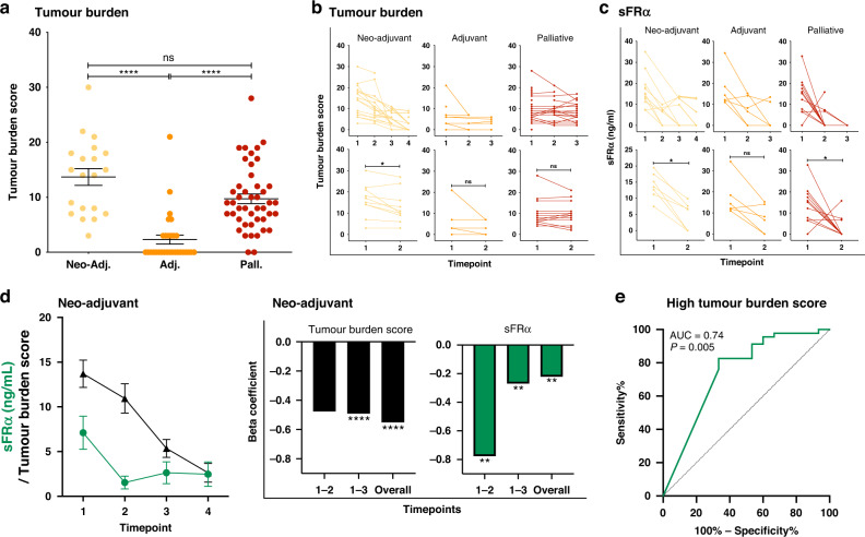
Background: Survival rates for ovarian cancer remain poor, and monitoring and prediction of therapeutic response may benefit from additional markers. Ovarian cancers frequently overexpress Folate Receptor alpha (FRα) and the soluble receptor (sFRα) is measurable in blood. Here we investigated sFRα as a potential biomarker.

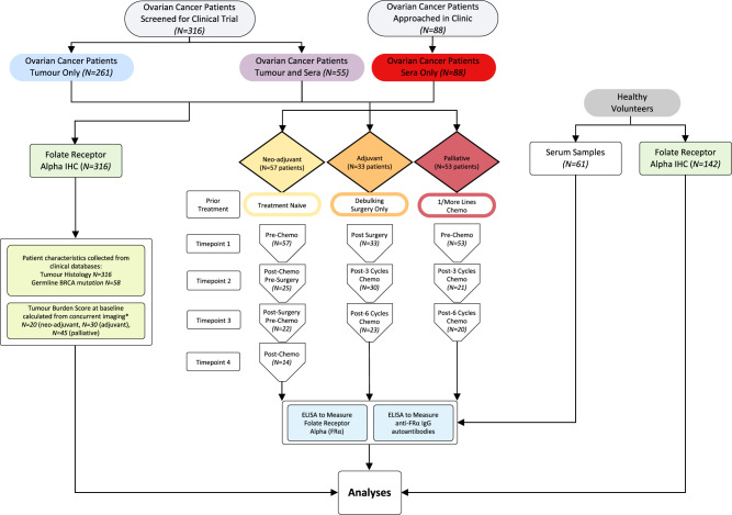
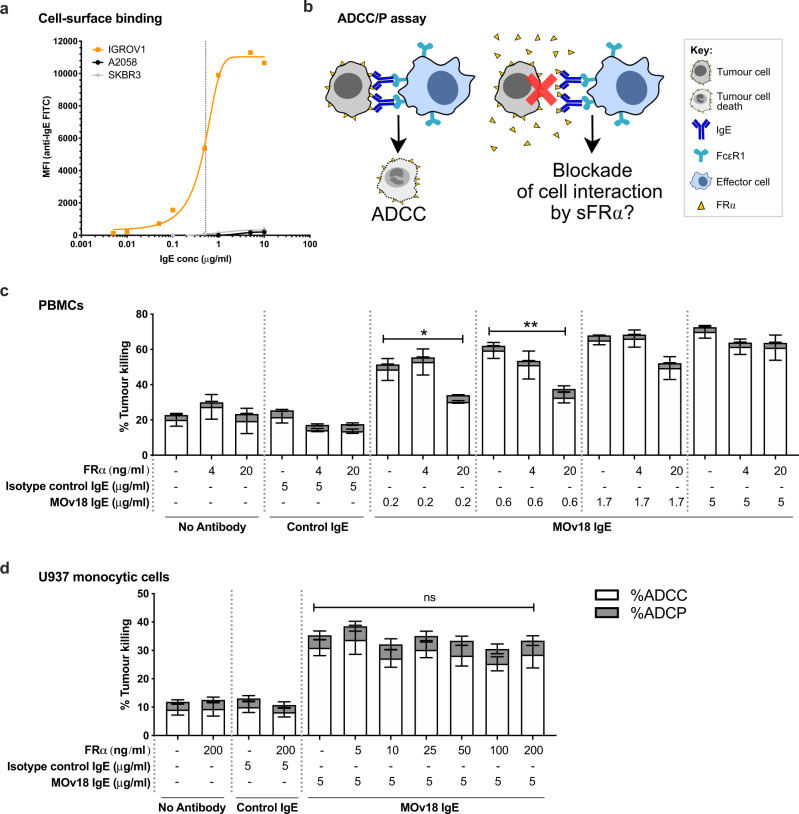
Methods: We evaluated sFRα longitudinally, before and during neo-adjuvant, adjuvant and palliative therapies, and tumour FRα expression status by immunohistrochemistry. The impact of free FRα on the efficacy of anti-FRα treatments was evaluated by an antibody-dependent cellular cytotoxicity assay.

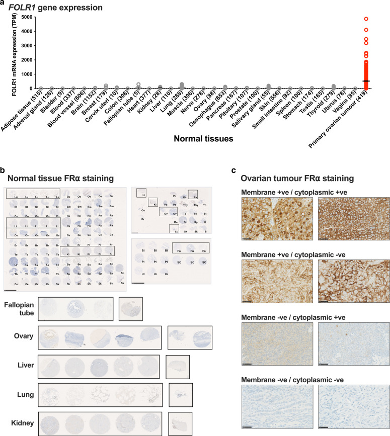
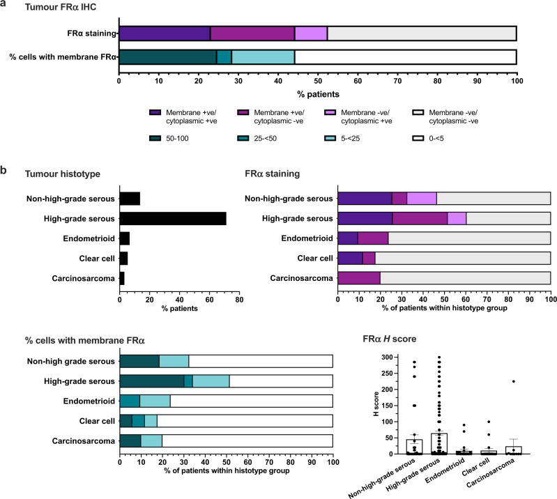
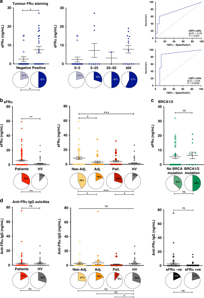
Results: Membrane and/or cytoplasmic FRα staining were observed in 52.7% tumours from 316 ovarian cancer patients with diverse histotypes. Circulating sFRα levels were significantly higher in patients, compared to healthy volunteers, specifically in patients sampled prior to neoadjuvant and palliative treatments. sFRα was associated with FRα cell membrane expression in the tumour. sFRα levels decreased alongside concurrent tumour burden in patients receiving standard therapies. High concentrations of sFRα partly reduced anti-FRα antibody tumour cell killing, an effect overcome by increased antibody doses.

Conclusions: sFRα may present a non-invasive marker for tumour FRα expression, with the potential for monitoring patient response to treatment. Larger, prospective studies should evaluate FRα for assessing disease burden and response to systemic treatments.


Conflict of interest statement

SNK and JS are founders and shareholders of Epsilogen Ltd. KFG is an employee of Epsilogen Ltd. HJB, MG, JL-A and LCGFP are employed through a fund provided by Epsilogen Ltd. SNK, JS, DHJ, HJB and KFG are inventors of patents on antibody technologies. All other authors have declared no conflict of interest.

Figures

Fig. 1
Fig. 1. Study design and sample workflow.
Serum samples from ovarian cancer patients were collected at timepoint 1 and up to 3 sequential treatment-related time points (e.g., for the treatment-naïve group: pre-chemo, post-chemo/pre-surgery, post-surgery/pre-chemo, and post-chemo). These were collected alongside serum samples from healthy volunteers. Serum samples were studied for sFRα and anti-FRα autoantibodies, and FRα protein expression in patient tumours and normal tissue microarrays was evaluated by immunohistochemistry. Patient characteristics, including tumour histotype, were collected from clinical databases (see Supplementary Table 1 for ovarian patients in the longitudinal study). Patient tumour burden scores were calculated from concurrent imaging (*see Supplementary Table 2 for a scoring system). N numbers are indicated in the figure.
Fig. 2
Fig. 2. Transcriptomic and immunohistochemical analyses of FRα expression in normal tissues and ovarian tumours.
a Low FOLR1 gene expression in normal tissues (grey) compared to primary ovarian tumours (red) (N numbers indicated in parentheses; The Human Protein Atlas online tool Xenabrowser online tool [35] (Xenabrowser.net). b No membrane or cytoplasmic FRα protein expression on a broad range of normal tissues (N = 142; scale bars = 2.5 mm). Key normal tissue types; fallopian tube (Fa), ovary (Ov), liver (Li), lung (Lu) and kidney (Ki) are highlighted with black boxes and zoomed images shown. c Representative images of ovarian tumour FRα staining (×40 magnification; scale bar = 50 μm). Bl bladder, Bo bone marrow, B brain, Br breast, Cb cerebrum; Ce cervix; Co colon; En endometrium, Fa fallopian tube, Il ileum, Ki kidney, Li liver, Lu lung, Ly lymph node, Mu muscle, striated, Oe oesophagus, Ov ovary, Pa pancreas, Pl placenta, Pr prostate, Re rectum, Sk skin, Sm small intestine, SC spinal cord, Sp spleen, St stomach, Te testis, Th thymus, Thy thyroid, Ur ureter.
Fig. 3
Fig. 3. Immunohistochemical analyses reveal a mixture of cell surface and cytoplasmic FRα protein expression in ovarian cancer tissues.
a Percentage of patients with tumours expressing FRα on the membrane, in the cytoplasm, both or neither, and with membrane staining on 0–<5, 5–<25, 25–<50 or ≥50% of tumour cells. b Top left: Overall percentage of patients with each tumour histotype; Top right: Percentage of patients with membrane and cytoplasmic positivity within each histotype subgroup; Bottom left: Percentage of patients within each histotype subgroup with different proportions of membrane positive tumours cells; Bottom right: Immunohistochemical H scores calculated in each histotype subgroup.
Fig. 4
Fig. 4. Soluble FRα but not anti-FRα autoantibodies were elevated in the circulation of ovarian cancer patients compared with healthy subjects.
sFRα and anti-FRα autoantibodies were measured in three patient cohorts (neo-adjuvant, adjuvant, palliative) at timepoint 1 (see Fig. 1). a sFRα levels were significantly-higher in patients with FRα tumour cell membrane expression, compared to patients with FRα-negative tumours (left) and with trend for higher levels in patients with tumours showing FRα cell membrane expression in a greater proportion of tumour cells (middle). In neo-adjuvant treatment-naïve patients, baseline sFRα concentration was predictive of FRα cell membrane expression in both ≥5% and ≥50% of tumour cells (right top and bottom, respectively). b Significantly-higher sFRα levels were measured in ovarian cancer patients compared to healthy volunteers, and between patient cohorts (proportion of samples with detectable sFRα indicated by filled pie chart sections below). c sFRα levels, or proportions of samples with detectable sFRα, were not associated with the patient’s germline BRCA1/2 mutational status. d Comparable levels of anti-FRα autoantibodies were detected in serum samples from ovarian cancer patients and healthy volunteers (left), across all patient cohorts (middle), and irrespective of detectable sFRα (right). N numbers in Fig. 1; *P ≤ 0.05, **P ≤ 0.01, ***P ≤ 0.001, ****P ≤ 0.0001. Statistical tests: t test, one-way ANOVA with Kruskal–Wallis multiple comparisons, receiver operating characteristic (ROC) curve analyses and Chi square test. Error bars represent the standard error of mean (SEM).
Fig. 5
Fig. 5. Serum markers and tumour burden score change during patient treatment.
a Patient tumour burden score at the start of treatment (calculated as described in Supplementary Table 2) was significantly greater in the neo-adjuvant and palliative ovarian cancer patient cohorts, compared to the adjuvant patient cohort. b, c Changes in tumour burden score (b) and levels of sFRα (c) were measured across treatment timepoints for all ovarian cancer patient cohorts. d (Left) Comparison of changes in serum sFRα and tumour burden score over time in patients receiving standard neo-adjuvant treatment regimen. (Right) Beta coefficients illustrate the variation in sFRα among the different time points, namely timepoints 1 and 2, timepoints 1 and 3, and overall across the earliest and latest timepoint for which data were collected. N numbers and treatment timepoints as indicated in Fig. 1. e Receiver operating characteristic (ROC) curve analysis evaluating the capacity sFRα to predict high values of tumour burden scores in the neo-adjuvant treatment-naive patient group. sFRα levels were predictive of high tumour burden scores (high defined as the upper quartile: ≥75th percentile of tumour burden scores). *P ≤ 0.05, **P ≤ 0.01, ****P ≤ 0.0001. Statistical tests: t test, one-way ANOVA with Kruskal–Wallis multiple comparisons, receiver operating characteristic (ROC) curve analyses and linear regression analysis. Error bars represent the standard error of mean (SEM).
Fig. 6
Fig. 6. Potential blockade of efficacy of a FRα-targeted therapeutic antibody candidate by soluble FRα.
a Binding of MOv18 IgE to FRα-expressing IGROV1 ovarian, but not to melanoma (A2058) or breast (SKBR3), cancer cells (EC50 = 0.53 µg/ml for binding to IGROV1 cells, indicated by vertical dotted line). b Schematic of potential blockade of MOv18 IgE anti-tumour function by sFRα. c Using PBMCs, the level of antibody-dependent cellular cytotoxicity (ADCC) mediated by the FRα-specific antibody MOv18 IgE was significantly reduced by a high concentration of FRα antigen (20 ng/ml) where MOv18 IgE was introduced at non-saturating concentrations of cancer cell-associated FRα (0.2 and 0.6 µg/ml; below or at the EC50 as shown in a). High concentrations of FRα antigen (20 ng/ml) did not block ADCC mediated by concentrations of MOv18 IgE above the EC50 (see a). d Similarly, with U937 monocytic cells, ADCC mediated by a saturating concentration of MOv18 IgE (5 µg/ml; see a) was not blocked by supraphysiological concentrations of FRα antigen (200 ng/ml). Antibody-dependent cellular phagocytosis (ADCP) was not mediated by MOv18 IgE. N = 6 and 4 independent experiments, respectively. *P ≤ 0.05, **P ≤ 0.01, ns not significantly different. Statistical test: one-way ANOVA with Kruskal–Wallis multiple comparisons. Error bars represent the standard error of mean (SEM).

References

    1. Menon U, Gentry-Maharaj A, Burnell M, Singh N, Ryan A, Karpinskyj C, et al. Ovarian cancer population screening and mortality after long-term follow-up in the UK Collaborative Trial of Ovarian Cancer Screening (UKCTOCS): a randomised controlled trial. Lancet. 2021;397:2182–93. - PMC - PubMed
    1. Gonzalez-Martin A, Pothuri B, Vergote I, DePont Christensen R, Graybill W, Mirza MR, et al. Niraparib in patients with newly diagnosed advanced ovarian cancer. N Engl J Med. 2019;381:2391–402. - PubMed
    1. Moore K, Colombo N, Scambia G, Kim BG, Oaknin A, Friedlander M, et al. Maintenance olaparib in patients with newly diagnosed advanced ovarian cancer. N Engl J Med. 2018;379:2495–505. - PubMed
    1. Perren TJ, Swart AM, Pfisterer J, Ledermann JA, Pujade-Lauraine E, Kristensen G, et al. A phase 3 trial of bevacizumab in ovarian cancer. N Engl J Med. 2011;365:2484–96. - PubMed
    1. Ray-Coquard I, Pautier P, Pignata S, Perol D, Gonzalez-Martin A, Berger R, et al. Olaparib plus bevacizumab as first-line maintenance in ovarian cancer. N Engl J Med. 2019;381:2416–28. - PubMed

Publication types