

Combining BN-PAGE and microscopy techniques to investigate pigment-protein complexes and plastid transitions in citrus fruit
- PMID: 36403000
- PMCID: PMC9675244
- DOI: 10.1186/s13007-022-00956-1
Combining BN-PAGE and microscopy techniques to investigate pigment-protein complexes and plastid transitions in citrus fruit
Abstract
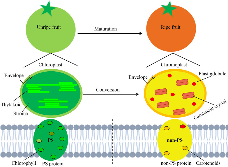
Background: Chlorophyll and carotenoids, the most widely distributed lipophilic pigments in plants, contribute to fruit coloration during development and ripening. These pigments are assembled with pigment-protein complexes localized at plastid membrane. Pigment-protein complexes are essential for multiple cellular processes, however, their identity and composition in fruit have yet to be characterized.
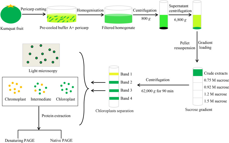
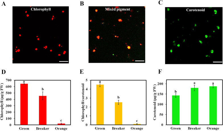
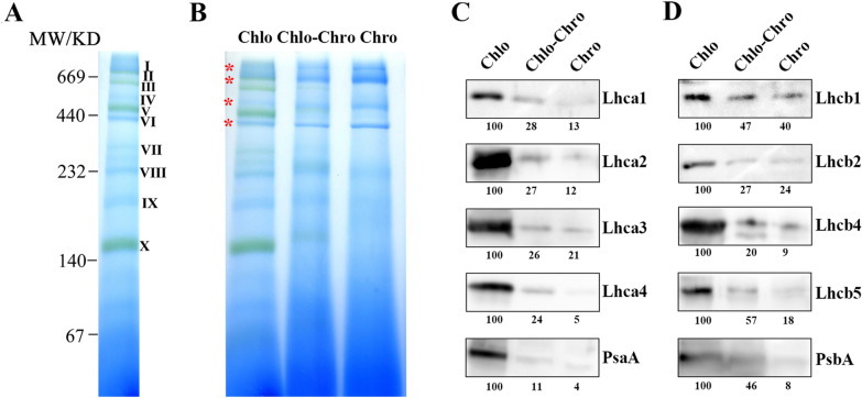
Results: By using BN-PAGE technique in combination with microscopy, we studied pigment-protein complexes and plastid transformation in the purified plastids from the exocarp of citrus fruit. The discontinuous sucrose gradient centrifugation was used to isolate total plastids from kumquat fruit, and the purity of isolated plastids was assessed by microscopy observation and western blot analysis. The isolated plastids at different coloring stages were subjected to pigment autofluorescence observation, western blot, two-dimensional electrophoresis analysis and BN-PAGE assessment. Our results demonstrated that (i) chloroplasts differentiate into chromoplasts during fruit coloring, and this differentiation is accompanied with a decrease in the chlorophyll/carotenoid ratio; (ii) BN-PAGE analysis reveals the profiles of macromolecular protein complexes among different types of plastids in citrus fruit; and (iii) the degradation rate of chlorophyll-protein complexes varies during the transition from chloroplasts to chromoplasts, with the stability generally following the order of LHCII > PS II core > LHC I > PS I core.
Conclusions: Our optimized methods for both plastid separation and BN-PAGE assessment provide an opportunity for developing a better understanding of pigment-protein complexes and plastid transitions in plant fruit. These attempts also have the potential for expanding our knowledge on the sub-cellular level synchronism of protein changes and pigment metabolism during the transition from chloroplasts to chromoplasts.
Keywords: BN-PAGE; Carotenoid; Chlorophyll; Citrus fruit; Pigment-protein complex; Plastid.
© 2022. The Author(s).
Conflict of interest statement
The authors declare that they have no competing interests.
Figures


Similar articles
-
Chloroplast to chromoplast transition in tomato fruit: spectral confocal microscopy analyses of carotenoids and chlorophylls in isolated plastids and time-lapse recording on intact live tissue.Ann Bot. 2011 Aug;108(2):291-7. doi: 10.1093/aob/mcr140. Ann Bot. 2011. PMID: 21788376 Free PMC article.
-
Microscopic Analyses of Fruit Cell Plastid Development in Loquat (Eriobotrya japonica) during Fruit Ripening.Molecules. 2019 Jan 27;24(3):448. doi: 10.3390/molecules24030448. Molecules. 2019. PMID: 30691226 Free PMC article.
-
Citrus chlorophyllase dynamics at ethylene-induced fruit color-break: a study of chlorophyllase expression, posttranslational processing kinetics, and in situ intracellular localization.Plant Physiol. 2008 Sep;148(1):108-18. doi: 10.1104/pp.108.124933. Epub 2008 Jul 16. Plant Physiol. 2008. PMID: 18633118 Free PMC article.
-
Carotenoid Biosynthesis and Plastid Development in Plants: The Role of Light.Int J Mol Sci. 2021 Jan 26;22(3):1184. doi: 10.3390/ijms22031184. Int J Mol Sci. 2021. PMID: 33530294 Free PMC article. Review.
-
Differentiation of chromoplasts and other plastids in plants.Plant Cell Rep. 2019 Jul;38(7):803-818. doi: 10.1007/s00299-019-02420-2. Epub 2019 May 11. Plant Cell Rep. 2019. PMID: 31079194 Free PMC article. Review.
Cited by
-
Research progress on differentiation and regulation of plant chromoplasts.Mol Biol Rep. 2024 Jul 13;51(1):810. doi: 10.1007/s11033-024-09753-6. Mol Biol Rep. 2024. PMID: 39001942 Review.
References
-
- Zeng Y, Du J, Wang L, Pan Z, Xu Q, Xiao S, Deng X. A comprehensive analysis of chromoplast differentiation reveals complex protein changes associated with plastoglobule biogenesis and remodeling of protein systems in sweet orange flesh. Plant Physiol. 2015;168(4):1648–1665. doi: 10.1104/pp.15.00645. - DOI - PMC - PubMed
-
- Iglesias DJ, Cercos M, Colmenero-Flores JM, Naranjo MA, Rios G, Carrera E, Ruiz-Rivero O, Lliso I, Morillon R, Tadeo FR, Talon M. Physiology of citrus fruiting. Brazilian J Plant Physiol. 2007;19(4):333–362. doi: 10.1590/s1677-04202007000400006. - DOI
-
- Sato N. Origin and evolution of plastids: Genomic view on the unification and diversity of plastids. In: Wise RR, Hoober JK, editors. Structure and function of plastids. Dordrecht: Springer; 2006. pp. 75–102.
Grants and funding
LinkOut - more resources
Full Text Sources
