. 2022 Nov 19;19(1):25.
doi: 10.1186/s12977-022-00611-6.

The envelope proteins from SARS-CoV-2 and SARS-CoV potently reduce the infectivity of human immunodeficiency virus type 1 (HIV-1)

Affiliations

The envelope proteins from SARS-CoV-2 and SARS-CoV potently reduce the infectivity of human immunodeficiency virus type 1 (HIV-1)

Wyatt Henke et al. Retrovirology. .

Abstract

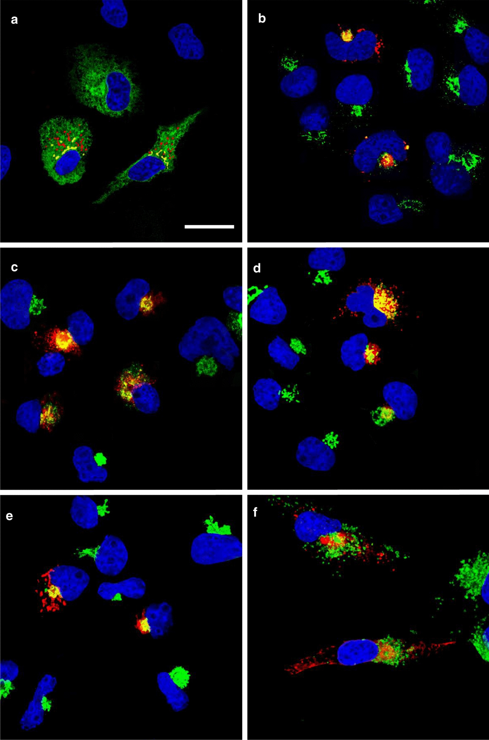
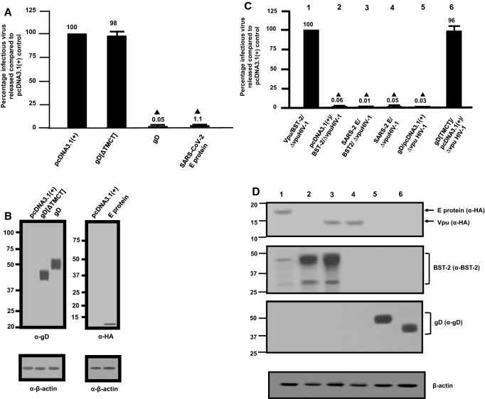
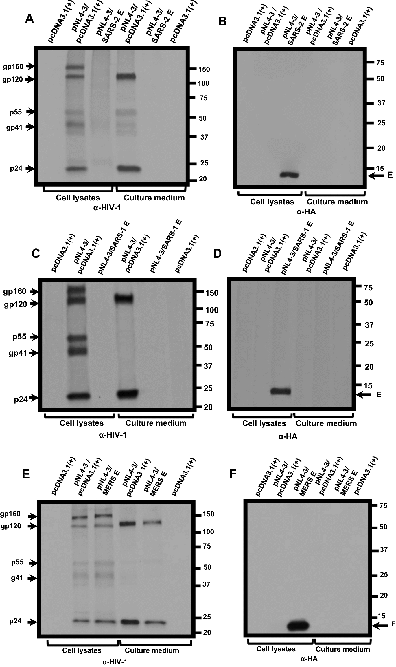
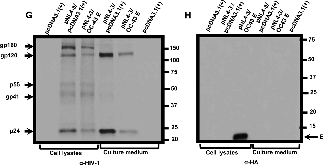
Background: Viroporins are virally encoded ion channels involved in virus assembly and release. Human immunodeficiency virus type 1 (HIV-1) and influenza A virus encode for viroporins. The human coronavirus SARS-CoV-2 encodes for at least two viroporins, a small 75 amino acid transmembrane protein known as the envelope (E) protein and a larger 275 amino acid protein known as Orf3a. Here, we compared the replication of HIV-1 in the presence of four different β-coronavirus E proteins.

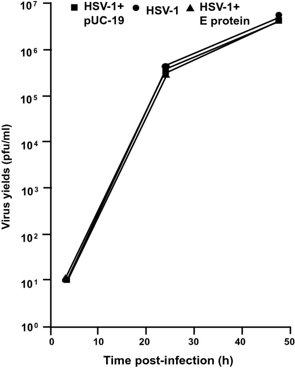
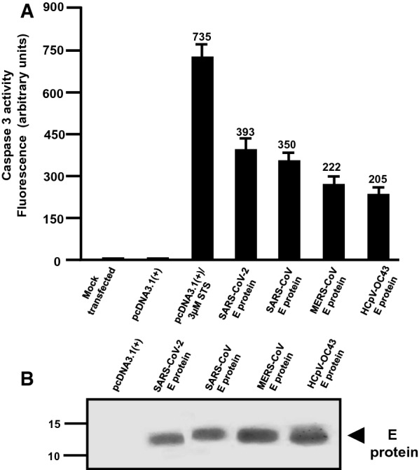
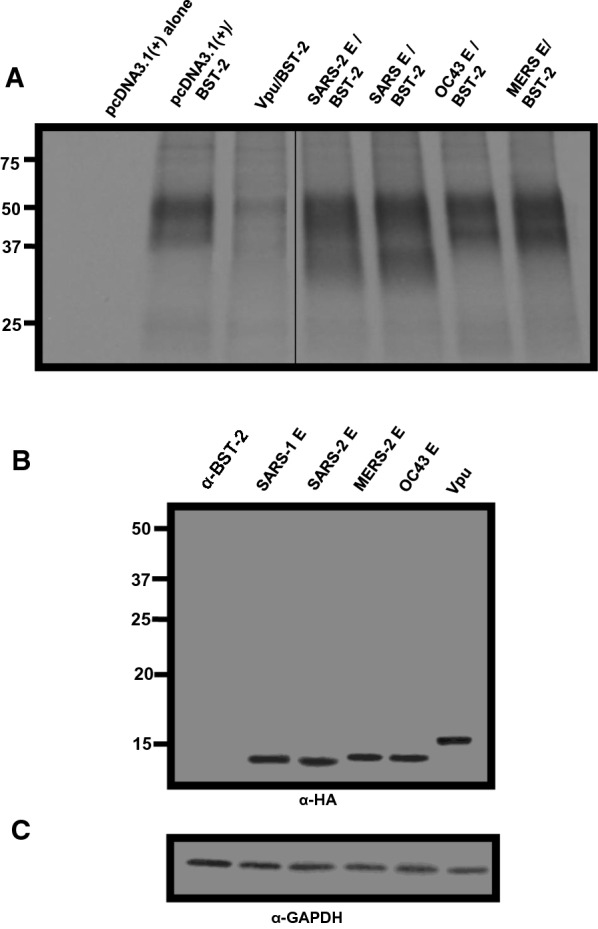
Results: We observed that the SARS-CoV-2 and SARS-CoV E proteins reduced the release of infectious HIV-1 yields by approximately 100-fold while MERS-CoV or HCoV-OC43 E proteins restricted HIV-1 infectivity to a lesser extent. Mechanistically, neither reverse transcription nor mRNA synthesis was involved in the restriction. We also show that all four E proteins caused phosphorylation of eIF2-α at similar levels and that lipidation of LC3-I could not account for the differences in restriction. However, the level of caspase 3 activity in transfected cells correlated with HIV-1 restriction in cells. Finally, we show that unlike the Vpu protein of HIV-1, the four E proteins did not significantly down-regulate bone marrow stromal cell antigen 2 (BST-2).

Conclusions: The results of this study indicate that while viroporins from homologous viruses can enhance virus release, we show that a viroporin from a heterologous virus can suppress HIV-1 protein synthesis and release of infectious virus.


Conflict of interest statement

The authors declare that they have no competing interests.

Figures

Fig. 1
Fig. 1
The expression of the SARS-CoV-2 E protein is restricted to intracellular compartments of the cell. COS-7 cells were transfected with the vector expressing the SARS-CoV-2 E-HA protein. At 24 h, cells were processed for immunostaining. The cells on coverslips were reacted with a mouse monoclonal antibody against HA-tag and with rabbit antibodies against ERGIC53, Golgin-97, or LAMP-1. The cells were washed and reacted with a secondary goat anti-rabbit antibody conjugated to AlexaFluor488 and with a chicken anti-mouse antibody conjugated to AlexaFluor594 for 1 h. Cells on coverslips were counterstained with DAPI and mounted on glass slides with a glycerol-containing mounting medium. To examine the intracellular localization of the E protein with the RER, cis-medial Golgi, or trans Golgi network (TGN), COS-7 cells were co-transfected with the plasmid expressing the E-HA protein, and vectors expressing ERMoxGFP, mNeonGreen-Giantin, or TGN38-GFP. Cells were prepared as above and immunostained with an anti-HA antibody and mounted as above. Coverslips were viewed with a Leica TCS SP8 Confocal Microscope with a 100X objective and a 2X digital zoom using the Leica Application Suite X (LASX) as previously described. A 405 nm filter was used to visualize the DAPI, a 594 nm filter to visualize HA staining, and a 488 nm filter to visualize the cellular markers. A. Cells co-transfected with vectors expressing SARS-CoV-2 E-HA and ERMoxGFP and immunostained with an anti-HA antibody. Scale bar = 20 µm. B. Cells transfected with a vector expressing SARS-CoV-2 E-HA and immunostained with anti-HA and anti-ERGIC53. C. Cells co-transfected with vectors expressing SARS-CoV-2 E-HA and mNeonGreen-Giantin and immunostained with an anti-HA antibody. D. Cells transfected with a vector expressing SARS-CoV-2 E-HA and immunostained with anti-HA and anti-Golgin97. E Cells co-transfected with vectors expressing SARS-CoV-2 E-HA and TGN38-GFP and immunostained with an anti-HA antibody. F Cells transfected with the vector expressing SARS-CoV-2 E-HA and immunostained with an anti-HA and anti-LAMP1
Fig. 2
Fig. 2
Release of infectious HIV-1Δvpu and HIV-1 in the presence of SARS-CoV- 2 E protein. HEK293 cells were co-transfected with either the empty pcDNA3.1( +) vector or vectors expressing the HSV-1 gD[ΔTMCT], HSV-1 gD, or SARS-CoV-2 E protein and pNL4-3. At 48 h, the culture supernatants were collected and subjected to low-speed centrifugation to remove cellular debris, and levels of infectious virus released into the culture supernatants were determined using TZM-bl cell assays. A The level of HIV-1 infectivity in the culture medium from cells co-transfected with either the empty pcDNA3.1( +) vector or vectors expressing gD[ΔTMCT], gD, or SARS-CoV-2 E protein and pNL4-3. B Expression of the gD proteins or E protein from restriction assays in (A). Expression of the E protein or gD proteins from restriction assays in (B) was normalized with β-actin using an anti-β-actin antibody and immunoblots. C HEK293 cells were co-transfected with either the empty pcDNA3.1( +) vector or vectors expressing 1) Vpu/BST-2/∆vpuHIV-1; 2) pcDNA3.1( +)/BST-2/vpuHIV-1; 3) SARS-CoV- 2 E/BST-2//∆vpuHIV-1; 4) SARS-CoV-2 E/∆vpuHIV-1; 5) gD/pcDNA3.1( +)/∆vpuHIV-1; or 6) gD[TMCT]/pcDNA3.1( +)/∆vpuHIV-1. At 48 h, culture supernatants were collected as described above and assayed for infectious HIV-1. The numbers above the figure correspond to the lanes in Panel D. D The cells collected from the experiments were lysed. Lysates were normalized for β-actin and analyzed for Vpu or E proteins using an anti-HA antibody (upper panel); BST-2 using an anti-BST-2 antibody (middle panel); gD proteins using an anti-gD monoclonal antibody (lower panel). The blot for β-actin is shown at the bottom. All restriction assays in Panels A and C were performed at least four times and statistical differences from the pcDNA3.1( +)/HIV-1 control was evaluated using a two-tailed Students t-test, with p < 0.01 () considered significant
Fig. 3
Fig. 3
The E protein of SARS-CoV-2 does not restrict the replication of HSV-1. HEK293 cells were left untransfected or transfected with the empty vector (pUC-19) or a vector expressing the SARS-CoV-2 E protein. At 24 h post-transfection, cells were inoculated with HSV-1 (0.01 pfu/cell). Cells were harvested at 24 and 48 h post-infection. The number of plaque-forming units was determined using standard plaque assays
Fig. 4
Fig. 4
Restriction of infectious HIV-1 production by the SARS-CoV, MERS-CoV, and HCoV-OC43 E proteins. A The level of infectious HIV-1 released into the culture medium from cells co-transfected with pcDNA3.1( +) alone, or vectors expressing SARS-CoV-2 E-HA, SARS-CoV E-HA, MERS-CoV E-HA, HCoV-OC43 E-HA, HSV-1 gD, or gD[ΔTMCT] and pNL4-3. B Immunoprecipitation of gD and gD[ΔTMCT] from the cell lysates of the restriction assay in Panel A using an anti-gD antibody. C Immunoprecipitation of the various E proteins from the cell lysates from the restriction assay in Panel A using an anti-HA antibody
Fig. 5
Fig. 5
The biosynthesis of HIV-1 proteins is attenuated in the presence of β-coronavirus E proteins. HEK293 cells were transfected with either the empty pcDNA3.1( +) or vectors expressing SARS-CoV-2, SARS-CoV, MERS-CoV, or HCoV-OC43 E proteins and pNL4-3. At 30 h post-transfection, cells were starved of methionine/cysteine for 2 h and radiolabeled with 35S-methionine/cysteine for 16 h. The culture medium was collected, and cell lysates were prepared as described in the Materials and Methods section. HIV-1 proteins were immunoprecipitated using anti-HIV-1 antibodies while the E proteins were immunoprecipitated with antibodies directed against the HA-tag. The immunoprecipitates were collected on protein-A-Sepharose, washed, and boiled in sample-reducing buffer. The proteins were separated by SDS-PAGE and visualized using standard radiographic techniques. A-B Immunoprecipitation of HIV-1 and E proteins from cell lysates and culture medium from HEK293 cells co-transfected with either pcDNA3.1( +) alone, pcDNA3.1( +)/pNL4-3, or pcDNA3.1( +) expressing SARS-CoV-2 E-HA protein/pNL4-3. C-D Immunoprecipitation of HIV-1 and E proteins from cell lysates and culture medium from HEK293 cells co-transfected with pcDNA3.1( +) alone, or pcDNA3.1( +)/pNL4-3, or pcDNA3.1( +) expressing the SARS-CoV E-HA/pNL4-3. E–F Immunoprecipitation of HIV-1 and E proteins from cell lysates and culture medium from HEK293 cells co-transfected pcDNA3.1( +) alone, pcDNA3.1( +)/pNL4-3, or pcDNA3.1( +) expressing the MERS-CoV E-HA/pNL4-3. G-H Immunoprecipitation of HIV-1 and E proteins from cell lysates and culture medium from HEK2933 cells co-transfected pcDNA3.1( +) alone, pcDNA3.1( +)/pNL4-3, or pcDNA3.1( +) expressing the HCoV-OC43 E-HA/pNL4-3
Fig. 5
Fig. 5
The biosynthesis of HIV-1 proteins is attenuated in the presence of β-coronavirus E proteins. HEK293 cells were transfected with either the empty pcDNA3.1( +) or vectors expressing SARS-CoV-2, SARS-CoV, MERS-CoV, or HCoV-OC43 E proteins and pNL4-3. At 30 h post-transfection, cells were starved of methionine/cysteine for 2 h and radiolabeled with 35S-methionine/cysteine for 16 h. The culture medium was collected, and cell lysates were prepared as described in the Materials and Methods section. HIV-1 proteins were immunoprecipitated using anti-HIV-1 antibodies while the E proteins were immunoprecipitated with antibodies directed against the HA-tag. The immunoprecipitates were collected on protein-A-Sepharose, washed, and boiled in sample-reducing buffer. The proteins were separated by SDS-PAGE and visualized using standard radiographic techniques. A-B Immunoprecipitation of HIV-1 and E proteins from cell lysates and culture medium from HEK293 cells co-transfected with either pcDNA3.1( +) alone, pcDNA3.1( +)/pNL4-3, or pcDNA3.1( +) expressing SARS-CoV-2 E-HA protein/pNL4-3. C-D Immunoprecipitation of HIV-1 and E proteins from cell lysates and culture medium from HEK293 cells co-transfected with pcDNA3.1( +) alone, or pcDNA3.1( +)/pNL4-3, or pcDNA3.1( +) expressing the SARS-CoV E-HA/pNL4-3. E–F Immunoprecipitation of HIV-1 and E proteins from cell lysates and culture medium from HEK293 cells co-transfected pcDNA3.1( +) alone, pcDNA3.1( +)/pNL4-3, or pcDNA3.1( +) expressing the MERS-CoV E-HA/pNL4-3. G-H Immunoprecipitation of HIV-1 and E proteins from cell lysates and culture medium from HEK2933 cells co-transfected pcDNA3.1( +) alone, pcDNA3.1( +)/pNL4-3, or pcDNA3.1( +) expressing the HCoV-OC43 E-HA/pNL4-3
Fig. 6
Fig. 6
The coronavirus E proteins induce caspase 3 activity. HEK293 cells were either mock-transfected, transfected with pcDNA3.1( +), transfected with pcDNA3.1( +), and treated with 2 µM staurosporine, or transfected with the vectors expressing the E proteins. At 48 h post-transfection, cells were lysed and analyzed for caspase 3 activity according to the manufacturer’s instructions. Fluorescence was measured using a microplate reader using excitation at 342 nm and emission at 441 nm (A) and the expression of the E proteins using an anti-HA antibody (α-HA) and immunoblots (B). Vectors used to transfect cells are at the top of each lane. Assays were performed at least three times
Fig. 7
Fig. 7
BST-2 down-regulation by Vpu and E proteins. HEK293 cells were co-transfected with either the empty pcDNA3.1( +) vector, the pcDNA3.1( +) vector expressing the HIV-1 Vpu protein, or each of the four E proteins, and a vector expressing human BST-2 protein. A At 48 h, the cells were removed by treatment with EDTA/EGTA, stained with a mouse monoclonal antibody directed against BST-2, and subjected to flow cytometric analysis using a BD LSR II flow cytometer. The fluorescent intensities are on the x-axis. B Median and mean fluorescent intensities of the BST-2 on the cells from Panel A. C Aliquots of cells from the same co-transfection were also analyzed for protein expression by immunoblots using antibodies directed against the HA-tag (E proteins and Vpu) to monitor expression
Fig. 8
Fig. 8
The levels of BST-2 in cells in the presence of β-coronavirus E proteins. HEK293 cells were co-transfected with either the empty pcDNA3.1( +) vector, the pcDNA3.1( +) vector expressing each of the four E proteins, or HIV-1 Vpu and a vector expressing human BST-2 protein. At 30 h post-transfection, cells were starved for methionine/cysteine and radiolabeled with 500 μCi of 35S-methionine/cysteine for 16 h. Cell lysates were prepared in RIPA buffer. One-third of the lysate was used to immunoprecipitate BST-2 proteins using an anti-BST-2 antibody (A); one-third of the lysate was used to immunoprecipitate E proteins and Vpu (B), and one-third of the lysate was used to immunoprecipitate GAPDH to normalize loading (C). The line in Panel A is due to the removal of several lanes from the center of the autoradiograph

Update of

Similar articles

Cited by

References

    1. Bogoch II, Watts A, Thomas-Bachli A, Huber C, Kraemer MUG, Khan K. Pneumonia of unknown etiology in Wuhan, China: potential for international spread via commercial air travel. J Travel Med. 2020;27:8. - PMC - PubMed
    1. Chan JF, Kok KH, Zhu Z, Chu H, To KK, Yuan S, et al. Genomic characterization of the 2019 novel human-pathogenic coronavirus isolated from a patient with atypical pneumonia after visiting Wuhan. Emerg Microbes Infect. 2020;9:221–236. - PMC - PubMed
    1. Holshue ML, DeBolt C, Lindquist S, Lofy KH, Wiesman J, Bruce H, et al. First case of 2019 Novel coronavirus in the United States. N Engl J Med. 2020;382:929–936. - PMC - PubMed
    1. Zhu N, Zhang D, Wang W, Li X, Yang B, Song J, et al. China novel coronavirus investigating and research team. A novel coronavirus from patients with pneumonia in China, 2019. N Engl J Med. 2020;382:727–33. - PMC - PubMed
    1. Alsaadi EAJ, Jones IM. Membrane binding proteins of coronaviruses. Future Virol. 2019;14:275–286. - PMC - PubMed

Publication types