. 2022 Nov 19;19(1):281.
doi: 10.1186/s12974-022-02638-0.

Genetic deletion of Krüppel-like factor 11 aggravates traumatic brain injury

Affiliations

Genetic deletion of Krüppel-like factor 11 aggravates traumatic brain injury

Chao Zhou et al. J Neuroinflammation. .

Abstract

Background: The long-term functional recovery of traumatic brain injury (TBI) is hampered by pathological events, such as parenchymal neuroinflammation, neuronal death, and white matter injury. Krüppel-like transcription factor 11 (KLF 11) belongs to the zinc finger family of transcription factors and actively participates in various pathophysiological processes in neurological disorders. Up to now, the role and molecular mechanisms of KLF11 in regulating the pathogenesis of brain trauma is poorly understood.

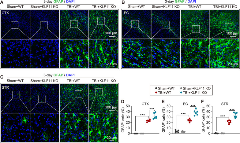
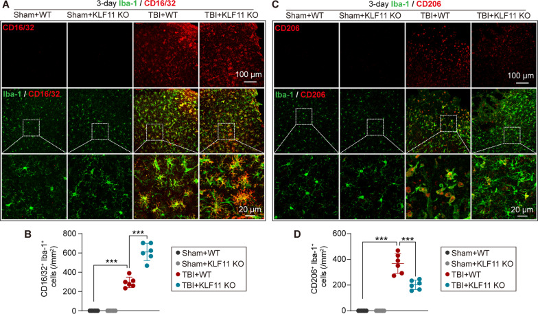
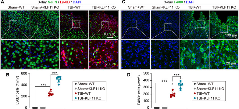
Methods: KLF11 knockout (KO) and wild-type (WT) mice were subjected to experimental TBI, and sensorimotor and cognitive functions were evaluated by rotarod, adhesive tape removal, foot fault, water maze, and passive avoidance tests. Brain tissue loss/neuronal death was examined by MAP2 and NeuN immunostaining, and Cresyl violet staining. White matter injury was assessed by Luxol fast blue staining, and also MBP/SMI32 and Caspr/Nav1.6 immunostaining. Activation of cerebral glial cells and infiltration of blood-borne immune cells were detected by GFAP, Iba-1/CD16/32, Iba-1/CD206, Ly-6B, and F4/80 immunostaining. Brian parenchymal inflammatory cytokines were measured with inflammatory array kits.

Results: Genetic deletion of KLF11 worsened brain trauma-induced sensorimotor and cognitive deficits, brain tissue loss and neuronal death, and white matter injury in mice. KLF11 genetic deficiency in mice also accelerated post-trauma astrocytic activation, promoted microglial polarization to a pro-inflammatory phenotype, and increased the infiltration of peripheral neutrophils and macrophages into the brain parenchyma. Mechanistically, loss-of-KLF11 function was found to directly increase the expression of pro-inflammatory cytokines in the brains of TBI mice.

Conclusion: KLF11 acts as a novel protective factor in TBI. KLF11 genetic deficiency in mice aggravated the neuroinflammatory responses, grey and white matter injury, and impaired long-term sensorimotor and cognitive recovery. Elucidating the functional importance of KLF11 in TBI may lead us to discover novel pharmacological targets for the development of effective therapies against brain trauma.

Keywords: Cytokines; Grey matter injury; Krüppel-like factor 11; Neurobehavioral deficits; Neuroinflammation; White matter injury.


Conflict of interest statement

The author(s) declared no potential conflicts of interest with respect to the research, authorship, and/or publication of this article.

Figures

Fig. 1
Fig. 1
Genetic deletion of KLF11 aggravates long-term sensorimotor deficits in mice after TBI. Experimental TBI was induced in KLF11 KO and WT mice by unilateral controlled cortical impact, followed by a 30-d survival period. Long-term sensorimotor function was examined by the rotarod test, adhesive tape removal test, and foot fault test at the indicated time points (− 1, 3, 5, 7, 14, 21, and 28 days after operation). A The latency to fall in the rotarod test. B The time to touch and C time to remove the tape in the adhesive tape removal test. D The forepaw foot fault rate and E hindpaw foot fault rate in the foot fault test. Data are presented as mean ± SD, n = 11–12/group. Statistical analyses were performed by two-way ANOVA with Tukey’s post hoc test. *p < 0.05, **p < 0.01, and ***p < 0.001 versus TBI + WT group
Fig. 2
Fig. 2
Genetic deletion of KLF11 exacerbates long-term cognitive impairment in mice after TBI. KLF11 KO and WT mice were subjected to TBI or sham operation, followed by a 30 d survival period. Long-term cognitive function was evaluated by the Morris water maze test at 23–28 d after TBI and the passive avoidance test at 29–30 d after TBI. A Swim paths of learning and memory phases during the water maze test. B The latency to find the hidden platform in the place navigation phase (learning). C The swim time in the target quadrant in the probe test (memory). D Average swimming speed in the water maze test. E Graphic protocol of the passive avoidance test. F The latency of entering into the dark box in the passive avoidance test. G Body weight changes 28 d after TBI or sham operation. Data are presented as mean ± SD, n = 11–12/group. Statistical analyses were performed by two-way ANOVA with Tukey post hoc test (B, G) and one-way ANOVA with Tukey post hoc test (C, D, F). *p < 0.05, **p < 0.01, and ***p < 0.001 versus TBI + WT group
Fig. 3
Fig. 3
Genetic deletion of KLF11 aggravates brain tissue loss in mice after TBI. KLF11 KO and WT mice were subjected to TBI or sham operation, followed by a 30-d survival period. Tissue loss was examined in brain sections at 30 d after operation by using MAP2 immunostaining. A Representative images of MAP2 immunostaining (Bregma from + 0.98 mm to − 1.58 mm). B, C Quantitative analysis of total volume of brain atrophy and tissue atrophy area in each brain slice (n = 11–12/group, unpaired t-test). Neuronal loss was examined by Cresyl Violet (CV) staining and NeuN immunostaining in brain sections at 30 d after TBI. D Representative images of CV staining in the peri-lesional cerebral cortex (CTX), hippocampal CA1, and hippocampal CA3 regions. E Peri-lesional brain areas (rectangles) in the CTX, CA1, and CA3 where images in D and I were captured. FH Quantitative analysis of CV-stained neurons in the peri-lesional CTX, CA1, and CA3 regions (n = 6/group, one-way ANOVA with Tukey post hoc test). I Representative images of NeuN immunostaining in the peri-lesional CTX, CA1, and CA3 regions. JL Quantitative analysis of NeuN-immunopositive neurons in the peri-lesional CTX, CA1, and CA3 regions (n = 6/group, one-way ANOVA with Tukey post hoc test). M Correlation analysis between sensorimotor or cognitive outcome and CV-stained/NeuN-positive neurons in the peri-lesional CTX, CA1, and CA3 regions (n = 6/group, Pearson correlation analysis). Data are presented as mean ± SD. *p < 0.05, **p < 0.01 or ***p < 0.001 versus TBI + WT group
Fig. 4
Fig. 4
KLF11 genetic deficiency exacerbates white matter injury in mice after TBI. Luxol fast blue (LFB) histological staining and MBP/SMI32 double-immunostaining were applied to detect white matter integrity in KLF11 KO and WT mouse brains at 30 d after TBI. A Representative LFB images of the pericontusional CTX, external capsule (EC), and striatum (STR) regions. B Pericontusional brain areas (rectangles) in the CTX, EC, and STR where images in A, D, and H were captured. C Quantitative analysis of relative OD values of LFB in pericontusional CTX, EC, and STR regions (n = 6/group, one-way ANOVA with Tukey post hoc test). D Representative images of MBP (green) and SMI32 (red) double-immunostaining in pericontusional CTX, EC, and STR regions. Quantitative analysis of MBP fluorescence intensities (E), SMI32 fluorescence intensities (F), and the ratio of SMI32/MBP (G) in the pericontusional CTX, EC, and STR regions (n = 6/group, one-way ANOVA with Tukey post hoc test). The nodes of Ranvier (NOR) were double-immunostained by Caspr/Nav1.6. H Representative images of Caspr (Red)/Nav1.6 (green) in the pericontusional EC region (rectangles: enlarged area of low-magnification images). I An image showing the composition and normal structure of the nodes of Ranvier. Quantitative analysis of the number of NORs (J), paranode length (K), and length of paranode gaps (L). M Correlation analysis of sensorimotor or cognitive outcome and white matter integrity (n = 6/group, Pearson correlation analysis). Data are presented as mean ± SD. *p < 0.05, **p < 0.01 or ***p < 0.001 versus TBI + WT group
Fig. 5
Fig. 5
KLF11 genetic deficiency promotes astrocytic activation in the white and grey matter of mice after TBI. KLF11 KO and WT mice were subjected to TBI or sham operation and reactive astrocytes were detected in brain sections at 3 d after surgery by GFAP immunostaining. AC Representative images of GFAP (green) and DAPI (blue) in the peri-lesional CTX (A), EC (B), and STR (C) regions (rectangles: enlarged area of low-magnification images). DF Quantitative analysis of GFAP-positive astrocytes in the peri-lesional CTX (D), EC (E), and STR (F) regions. Data are presented as mean ± SD, n = 6/group. Statistical analyses were performed by one-way ANOVA with the Tukey post hoc test. *p < 0.05, **p < 0.01 or ***p < 0.001 versus TBI + WT group
Fig. 6
Fig. 6
Genetic deletion of KLF11 increases pro-inflammatory microglia/macrophage polarization in mice after TBI. KLF11 KO and WT mice were subjected to TBI or sham operation and the activity of microglial polarization was examined in pericontusional brain regions at 3 d after TBI by double-immunostaining Iba-1 with CD16/32 (M1-phenotype) or CD206 (M2-phenotype). A Representative images of Iba-1 (green) and CD16/32 (red) double-immunofluorescence staining (rectangles: enlarged area of low-magnification images). B Quantitative analysis of Iba-1 and CD16/32 co-immunostained microglia/macrophages in experimental groups. C Representative images of Iba-1 (green) and CD206 (red) double-immunofluorescence staining (rectangles: enlarged area of low-magnification images). D Quantitative analysis of Iba-1 and CD206 co-immunostained microglia/macrophages in experimental groups. Data are presented as mean ± SD, n = 6/group. Statistical analyses were performed by one-way ANOVA with Tukey’s post hoc test. *p < 0.05, **p < 0.01 or ***p < 0.001 versus TBI + WT group
Fig. 7
Fig. 7
KLF11 genetic deletion increases TBI-induced infiltration of peripheral neutrophils and macrophages. KLF11 KO and WT mice were subjected to TBI or sham operation and the peripheral infiltration of neutrophils and macrophages was assessed by double-immunostaining in brain sections 3 d after operation. A Representative images of NeuN (green), Ly-6B (red, a marker of neutrophils), and DAPI (blue) immunostaining in peri-lesional brain regions. Rectangles: enlarged area of low-magnification images. B Quantitative analysis of Ly-6B-positive neutrophils from peri-lesional brains or sham mouse brains. C Representative images of F4/80 (green) and DAPI (blue) immunostaining in peri-lesional brain regions. Rectangles: enlarged area of low-magnification images. D Quantitative analysis of F4/80-positive macrophages/microglia from peri-lesional or sham mouse brains. Data are presented as mean ± SD, n = 6/group. Statistical analyses were performed by one-way ANOVA with Tukey’s post hoc test. *p < 0.05, **p < 0.01 or ***p < 0.001 versus TBI + WT group
Fig. 8
Fig. 8
KLF11 genetic deficiency in mice increases the inflammatory burden in post-trauma brains. KLF11 KO and WT mice were subjected to TBI or sham operation and a panel of 40 inflammatory mediators was measured in mouse brains at 3 d after CCI operation. A Array map of 40 inflammatory mediators. B Representative blots of the inflammatory array in experimental groups. CK Quantitative analysis showed significantly increased inflammatory mediators in the brains of KLF11 KO mice in comparison with WT controls 3 d after TBI, including Eotaxin-2, FAS ligand, MIP-1-α, MIP-1-γ, RANTES, SDF-1, TNF-α, sTNF RI, and sTNF-RII. Data are presented as mean ± SD, n = 3–5/group. Statistical analyses were performed by one-way ANOVA with Tukey post hoc test. L A heatmap showing the mean expression levels of 40 brain inflammatory mediators in experimental groups. *p < 0.05, **p < 0.01 or ***p < 0.001 Sham + WT versus TBI + WT group. &p < 0.05, &&p < 0.01 or &&&p < 0.001 TBI + KO versus TBI + WT group

Similar articles

Cited by

References

    1. Capizzi A, Woo J, Verduzco-Gutierrez M. Traumatic brain injury: an overview of epidemiology, pathophysiology, and medical management. Med Clin N Am. 2020;104:213–238. - PubMed
    1. Ghajar J. Traumatic brain injury. Lancet. 2000;356:923–929. - PubMed
    1. Thapa K, Khan H, Singh TG, Kaur A. Traumatic brain injury: mechanistic insight on pathophysiology and potential therapeutic targets. J Mol Neurosci. 2021;71:1725–1742. - PubMed
    1. Sato M, Chang E, Igarashi T, Noble LJ. Neuronal injury and loss after traumatic brain injury: time course and regional variability. Brain Res. 2001;917:45–54. - PubMed
    1. Kinnunen KM, Greenwood R, Powell JH, Leech R, Hawkins PC, Bonnelle V, Patel MC, Counsell SJ, Sharp DJ. White matter damage and cognitive impairment after traumatic brain injury. Brain. 2011;134:449–463. - PMC - PubMed

Substances