. 2022 Nov:82:100973.
doi: 10.1016/j.resmer.2022.100973. Epub 2022 Nov 17.

Fibrotic-like abnormalities notably prevalent one year after hospitalization with COVID-19

Affiliations

Fibrotic-like abnormalities notably prevalent one year after hospitalization with COVID-19

Bas F M van Raaij et al. Respir Med Res. 2022 Nov.

Abstract

Background: We investigated whether COVID-19 leads to persistent impaired pulmonary function, fibrotic-like abnormalities or psychological symptoms 12 months after discharge and whether severely ill patients (ICU admission) recover differently than moderately ill patients.

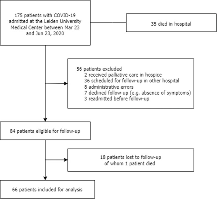
Methods: This single-centre cohort study followed adult COVID-19 survivors for a period of one year after discharge. Patients underwent pulmonary function tests 6 weeks, 3 months and 12 months after discharge and were psychologically evaluated at 6 weeks and 12 months. Computed tomography (CT) was performed after 3 months and 12 months.

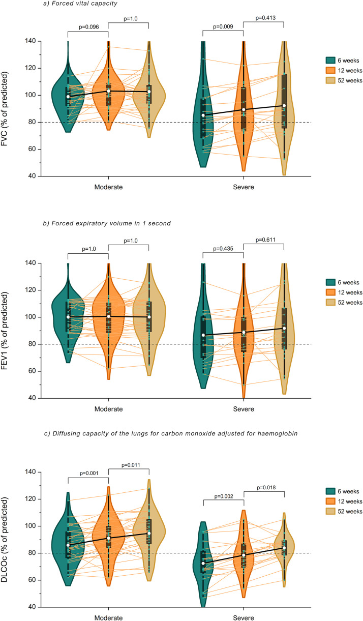
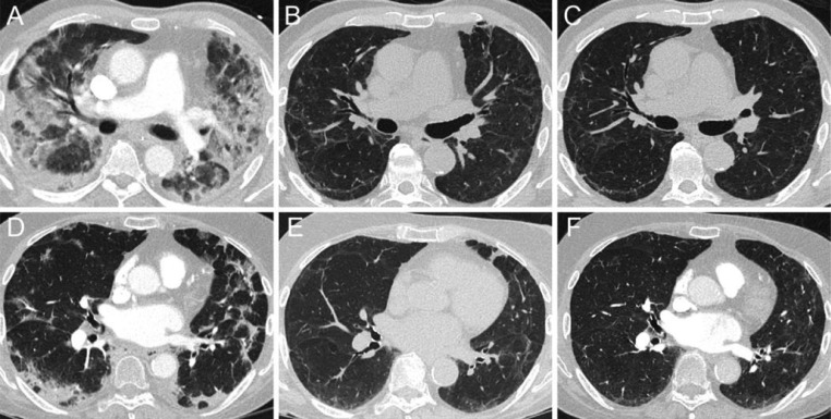
Results: 66 patients were analysed, their median age was 60.5 (IQR: 54-69) years, 46 (70%) patients were male. 38 (58%) patients had moderate disease and 28 (42%) patients had severe disease. Most patients had spirometric values within normal range after 12 months of follow-up. 12 (23%) patients still had an impaired lung diffusion after 12 months. Impaired pulmonary diffusion capacity was associated with residual CT abnormalities (OR 5.1,CI-95: 1.2-22.2), shortness of breath (OR 7.0, CI-95: 1.6-29.7) and with functional limitations (OR 5.8, CI-95: 1.4-23.8). Ground-glass opacities resolved in most patients during follow-up. Resorption of reticulation, bronchiectasis and curvilinear bands was rare and independent of disease severity. 81% of severely ill patients and 37% of moderately ill patients showed residual abnormalities after 12 months (OR 8.1, CI-95: 2.5-26.4). A minority of patients had symptoms of post-traumatic stress disorder, anxiety, depression and cognitive failure during follow-up.

Conclusion: Some patients still had impaired lung diffusion 12 months after discharge and fibrotic-like residual abnormalities were notably prevalent, especially in severely ill patients.

Keywords: COVID-19; Chest imaging; Pulmonary fibrosis; Pulmonary function; Respiratory infection.


Conflict of interest statement

Conflict of interest None.

Figures

Fig. 1
Fig. 1
Flowchart of this study.
Fig 2
Fig. 2
Change in pulmonary function during follow-up in COVID-19 patients with moderate or severe disease. Mean FVC, FEV1 and DLCOc (white dots) of COVID-19 patients with moderate and severe disease at 6 weeks, 12 weeks and 52 weeks are illustrated with violin plots and box plots. Individual values are shown with connected lines. A mixed model for repeated measures demonstrates that FVC only improved in severely ill patients between 6 and 12 weeks (p = 0.009). There was no change in FEV1 in both groups during follow-up. DLCOc improved between 6 and 12 weeks and continued to improve afterwards in both groups. Severely ill patients had lower spirometric values during follow-up compared to moderately ill patients (all p<0.001). There was no interaction between disease severity and time (FVC: pinteraction=0.160, FEV1: pinteraction =0.061, DLCOc: pinteraction =0.273).
Fig. 3
Fig. 3
Temporal evolution of typical radiological abnormalities in COVID-19 patients with moderate or severe disease. Axial CT-images illustrating the typical evolution of radiological abnormalities over time in a severely ill (A, B, C) and moderately ill (D, E, F) COVID-19 patient at baseline, 3 months and 12 months (left to right). During the acute phase of SARS-CoV-2-infection both patients predominantly demonstrate parenchymal consolidation in a pattern most resembling organizing pneumonia (pronounced peripheral involvement, bronchocentric distribution, perilobular pattern) (A, D). In time, parenchymal abnormalities also resolve in a manner similar to that of organizing pneumonia. At 3 months follow-up, resolution of prior consolidations leaves residual GGO, peripheral curvilinear bands and architectural distortion (B, E). At 12 months follow-up, these residual abnormalities have failed to fully resolve (C, F).

References

    1. Wang D., Hu B., Hu C., Zhu F., Liu X., Zhang J., et al. Clinical characteristics of 138 hospitalized patients with 2019 novel coronavirus-infected pneumonia in Wuhan, China. JAMA. 2020;323(11):1061–1069. - PMC - PubMed
    1. Chen N., Zhou M., Dong X., Qu J., Gong F., Han Y., et al. Epidemiological and clinical characteristics of 99 cases of 2019 novel coronavirus pneumonia in Wuhan, China: a descriptive study. Lancet. 2020;395(10223):507–513. - PMC - PubMed
    1. Shi H., Han X., Jiang N., Cao Y., Alwalid O., Gu J., et al. Radiological findings from 81 patients with COVID-19 pneumonia in Wuhan, China: a descriptive study. Lancet Infect Dis. 2020;20(4):425–434. - PMC - PubMed
    1. Polak S.B., Van Gool I.C., Cohen D., von der Thusen J.H., van Paassen J. A systematic review of pathological findings in COVID-19: a pathophysiological timeline and possible mechanisms of disease progression. Mod Pathol. 2020;33(11):2128–2138. - PMC - PubMed
    1. Antonio G.E., Wong K.T., Chu W.C., Hui D.S., Cheng F.W., Yuen E.H., et al. Imaging in severe acute respiratory syndrome (SARS) Clin Radiol. 2003;58(11):825–832. - PMC - PubMed