. 2023 Jan;12(6):e2202511.
doi: 10.1002/adhm.202202511. Epub 2022 Dec 4.

Surface Engineering of Auxetic Scaffolds for Neural and Vascular Differentiation from Human Pluripotent Stem Cells

Affiliations

Surface Engineering of Auxetic Scaffolds for Neural and Vascular Differentiation from Human Pluripotent Stem Cells

Xingchi Chen et al. Adv Healthc Mater. 2023 Jan.

Abstract

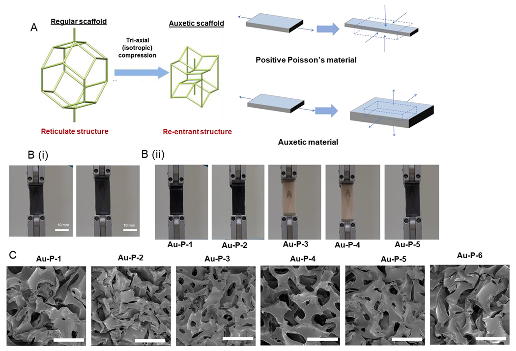
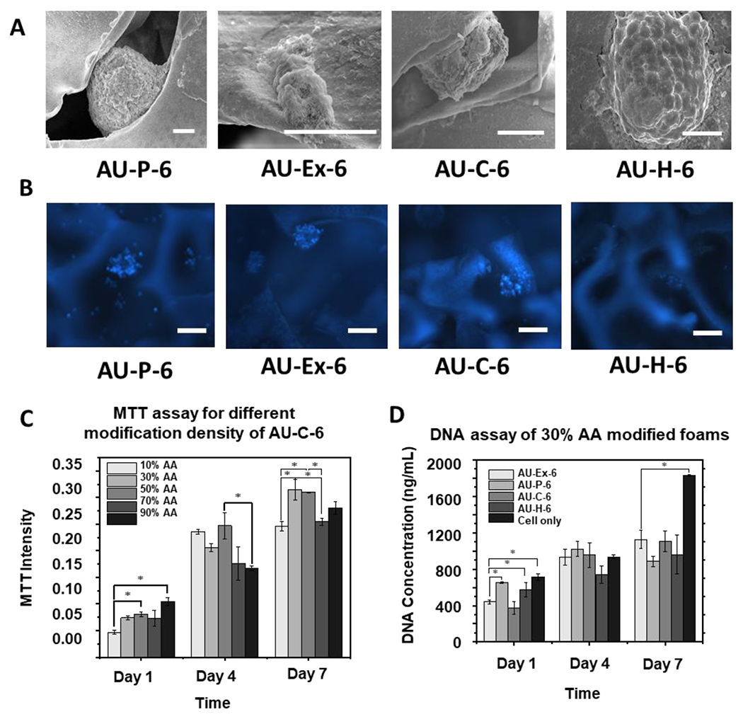
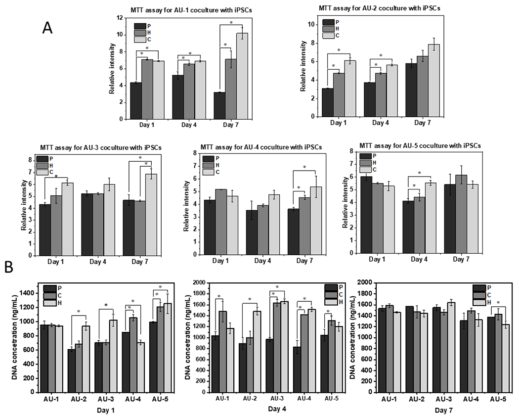
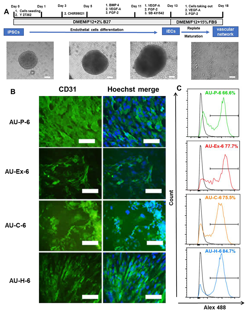
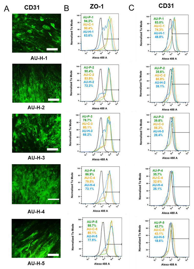
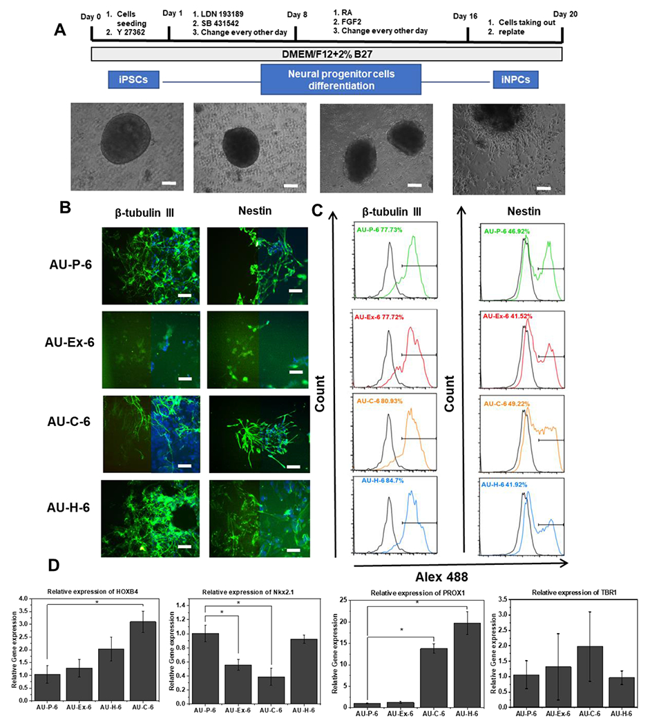
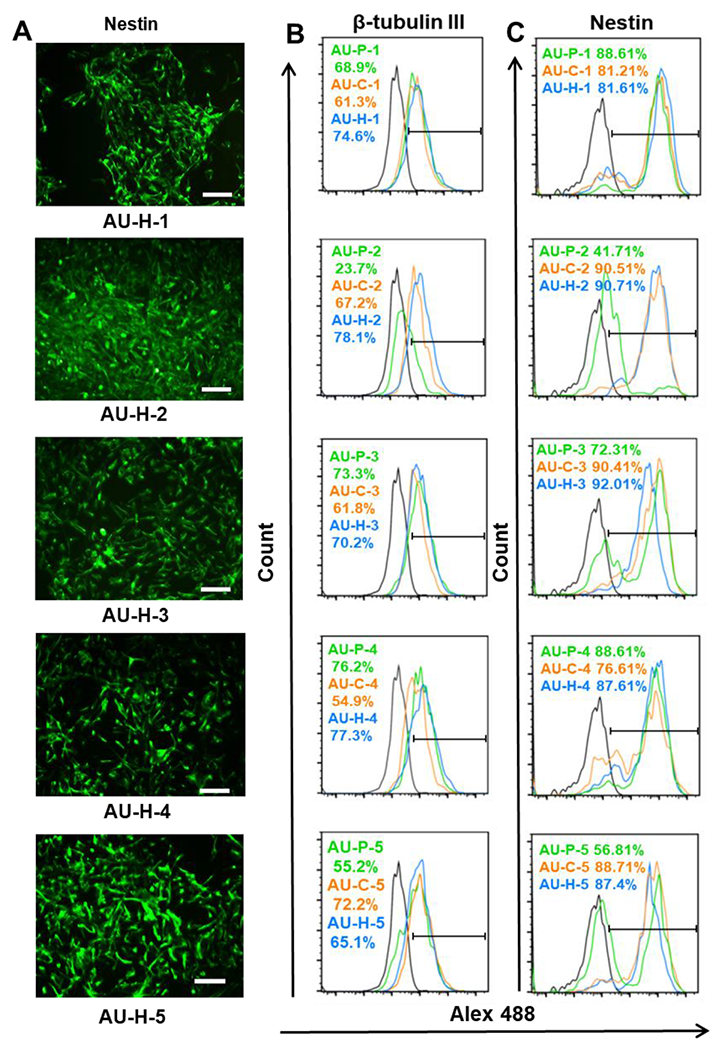
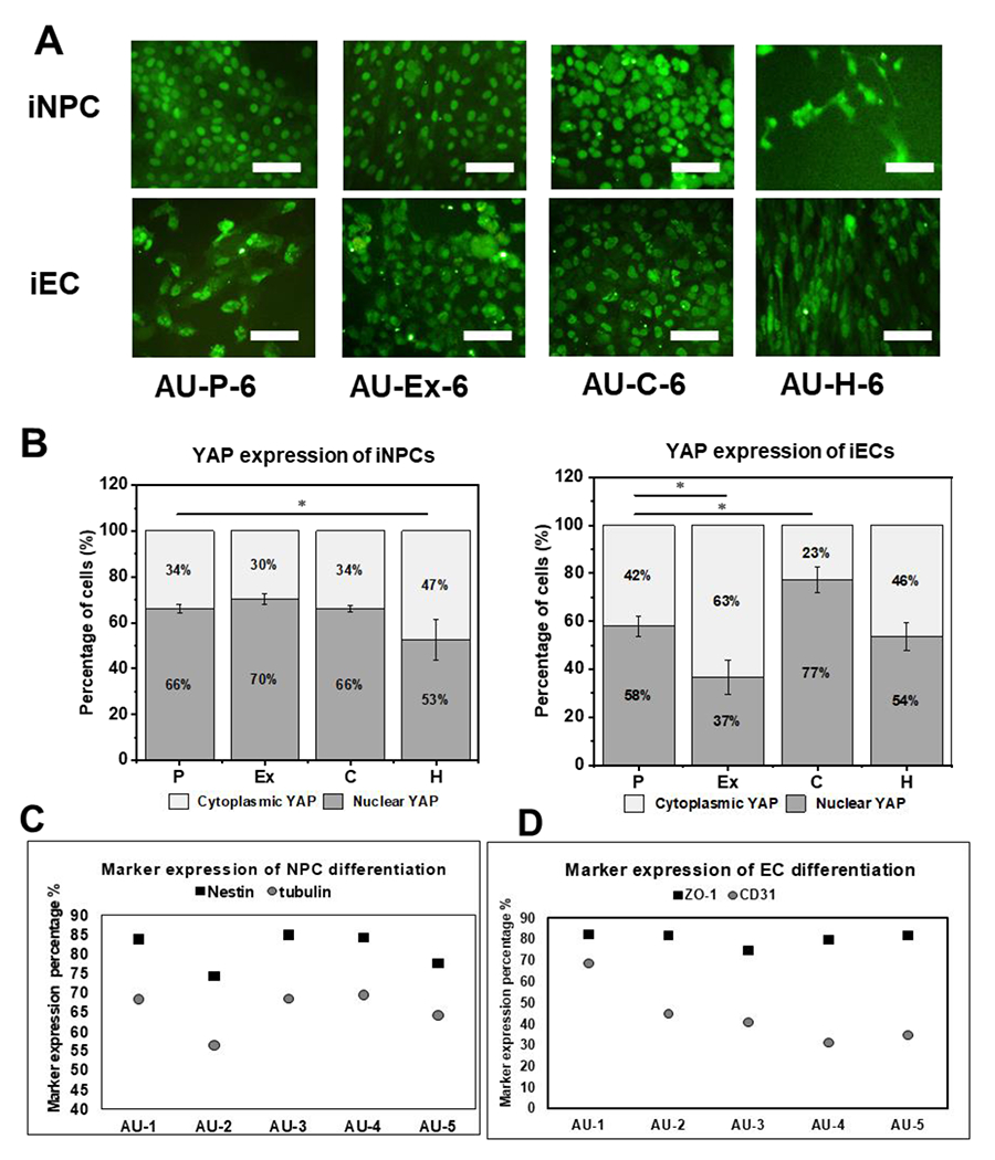
Auxetic materials are the materials that can display negative Poisson's ratio that describes the degree to which a material contracts (or expands) transversally when axially strained. Human stem cells sense the mechanical properties of the microenvironment, including material surface properties, stiffness, and Poisson's ratio. In this study, six different auxetic polyurethane (PU) foams with different elastic modulus (0.7-1.8 kPa) and Poisson's ratio (-0.1 to -0.5) are used to investigate lineage specification of human induced pluripotent stem cells (hiPSCs). The surfaces of the foams are modified with chitosan or heparin to enhance the adhesion and proliferation of hiPSCs. Then, the vascular and neural differentiation of hiPSCs are investigated on different foams with distinct elastic modulus and Poisson's ratio. With different auxetic foams, cells show differential adherent density and differentiation capacity. Chitosan and heparin surface functionalization promote the hindbrain and hippocampal markers, but not forebrain markers during neural patterning of hiPSCs. Properly surface engineered auxetic scaffolds can also promote vascular differentiation of hiPSCs. This study represents a versatile and multifunctional scaffold fabrication approach and can lead to a suitable system for establishing hiPSC culture models in applications of neurovascular disease modeling and drug screening.

Keywords: Poisson's ratio; auxetic scaffolds; elastic modulus; human pluripotent stem cells; neural differentiation; vascular differentiation.


Conflict of interest statement

Conflict of Interest:

There is no conflict of interest.

Figures

Figure 1.
Figure 1.. Characterization of physical properties of different auxetic PU foams.
(A) Schematics of auxetic foam fabrication and the deformation behavior; (B) Photographs illustrating the auxetic behavior of the AU foam under tension; (C) Scanning electron microscopy (SEM) images of the porous structure of the auxetic foams. Scale bar = 500 μm
Figure 2.
Figure 2.. Surface modification and characterization of the modified surfaces.
(A) Schematic illustration of PU auxetic foam modification. (B) Contact angle of the surface of the foam at different stages. (C) Fluorescence test for BSA-FITC conjugated with activated carboxylic group. Scale bar = 100 μm.
Figure 3.
Figure 3.. Biocompatibility with the extended foam under different surface modifications.
(A) Scanning electron microscopy (SEM) images of the hiPSC aggregates (day 7) in the AU foams. (B) Images of auxetic foam and extended foam. Scale bar: 100 μm; (C) MTT assay of different AA concentrations with chitosan modified foams. Scale bar: 100 μm; (D) DNA assay of 30% AA modified foams. n =3. * indicates p <0.05. AU-P: pristine auxetic scaffolds; AU-Ex: extended auxetic scaffolds; AU-C: Chitosan modified auxetic scaffolds; AU-H: heparin modified auxetic scaffolds.
Figure 4.
Figure 4.. Metabolic activity and DNA content for hiPSCs grown in different AU scaffolds.
(A) MTT activity of hiPSCs grown in AU-1 to AU-5 scaffolds with heparin and chitosan modifications; (B) DNA assay of hiPSCs grown in AU-1 to AU-5 scaffolds with heparin and chitosan modifications for 1, 4, and 7-day culture. n=3. * indicates p<0.05. P: pristine auxetic scaffolds; C: Chitosan-modified auxetic scaffolds; H: heparin-modified auxetic scaffolds.
Figure 5.
Figure 5.. iEC differentiation of hiPSCs grown in AU-6 scaffolds with different surface modifications.
(A) Schematic illustration and images of differentiation of hiPSCs into endothelial cells (hiPSC-ECs), or vascular organoids. Scale bar: 100 μm. (B) Fluorescent images of the markers expression of the replated cells from different scaffold were taken. Vascular maker, CD31 (green, day 20). Scale bar = 100 μm. (C) Flow cytometry histograms of CD31 expression. Black line: negative control. Color line: CD31 expression. AU-P: pristine auxetic scaffolds; AU-Ex: extended auxetic scaffolds; AU-C: Chitosan modified auxetic scaffolds; AU-H: heparin modified auxetic scaffolds.
Figure 6.
Figure 6.. Characterization of endothelial differentiation in AU scaffolds with different elastic modulus and Poisson’s ratio.
(A) Representative CD31 expression for cells grown in different AU foams modified by heparin. Scale bar = 100 μm. Flow cytometry histograms for (B) ZO-1 and (C) CD31 expression of cells grown in AU-1 to AU-5 scaffolds with different surface modifications. The blank line is the negative control. AU-P: pristine auxetic scaffolds; AU-Ex: extended auxetic scaffolds; AU-C: Chitosan modified auxetic scaffolds; AU-H: heparin modified auxetic scaffolds.
Figure 7.
Figure 7.. iNPC marker expression in auxetic foams with different surface modifications.
(A) Schematic illustration and images of differentiation of hiPSCs into neural progenitor cells (hiPSC-NPCs), or forebrain cortical organoids. Scale bar: 100 μm. (B) Fluorescent images of the marker expression of the replated cells from different scaffolds ay day 21. Left side: marker of interest only; right side: overlay with nuclear images. Beta III Tubulin (green) and nestin (green). Blue: Hoechst 33342. Scale bar: 100 μm. Flow cytometry histograms of the two markers (C) Beta III Tubulin and Nestin. The black line is the negative control. (D) mRNA expression determined by RT-PCR for brain regional identity markers. * indicates p<0.05. AU-P: pristine auxetic scaffolds; AU-Ex: extended auxetic scaffolds; AU-C: Chitosan modified auxetic scaffolds; AU-H: heparin modified auxetic scaffolds.
Figure 8.
Figure 8.. Characterization of iNPC differentiation in AU scaffolds with different elastic modulus and Poisson’s ratio.
(A) Representative Nestin expression for cells grown in different AU foams modified by heparin. Scale bar = 100 μm. Flow cytometry histograms for (B) Beta III Tubulin and (C) Nestin expression of cells grown in AU-1 to AU-5 scaffolds with different surface modifications. The blank line is the negative control. AU-P: pristine auxetic scaffolds; AU-Ex: extended auxetic scaffolds; AU-C: Chitosan modified auxetic scaffolds; AU-H: heparin modified auxetic scaffolds.
Figure 9.
Figure 9.. Effects of different modified AU scaffolds on YAP nuclear localization with hiPSC differentiation into iECs and iNPCs.
(A) Fluorescent images of YAP expression (day 20) of cells from different scaffolds. Scale bar: 100 μm. (B) Quantification of cytoplasmic YAP and nuclear YAP localization for cells from different scaffolds. (n=3). (C) Level of iNPC marker expression for cells from different scaffolds. (D) Level of iEC marker expression for cells from different scaffolds. * indicates p<0.05.

Similar articles

Cited by

References

    1. Lakes R, Science 1987, 235, 1038. - PubMed
    1. He C, Liu P, McMullan PJ, Griffin AC, physica status solidi (b) 2005, 242, 576
    2. Nkansah M, Evans K, Hutchinson I, Modelling and Simulation in Materials Science and Engineering 1994, 2, 337
    3. Gatt R, Zammit V, Caruana C, Grima JN, physica status solidi (b) 2008, 245, 502.
    1. Lee JJ, Ng HY, Lin YH, Lin TJ, Kao CT, Shie MY, Cells 2022, 11. - PMC - PubMed
    1. Kim Y, Son KH, Lee JW, Materials (Basel) 2021, 14. - PMC - PubMed
    1. Brazhkina O, Park JH, Park HJ, Bheri S, Maxwell JT, Hollister SJ, Davis ME, J Cardiovasc Dev Dis 2021, 8. - PMC - PubMed

Publication types