Review
. 2023 Feb;33(1):11-41.
doi: 10.1016/j.nic.2022.07.001.

Neuroimaging Patterns of Intracranial Infections: Meningitis, Cerebritis, and Their Complications

Affiliations
Review

Neuroimaging Patterns of Intracranial Infections: Meningitis, Cerebritis, and Their Complications

Michael Tran Duong et al. Neuroimaging Clin N Am. 2023 Feb.

Abstract

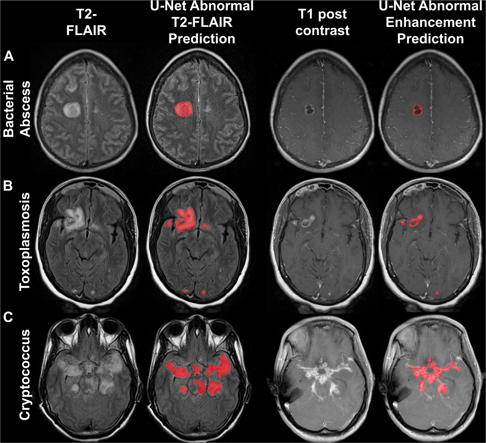
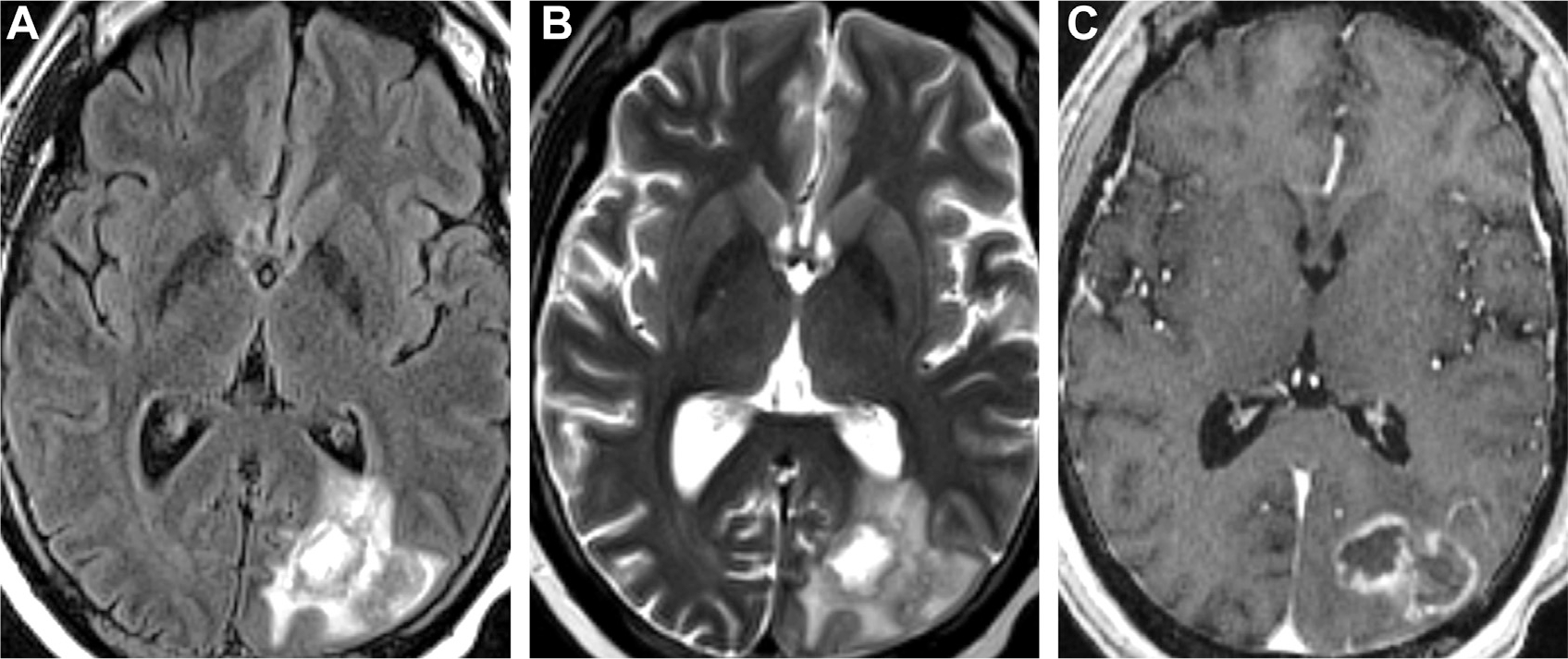
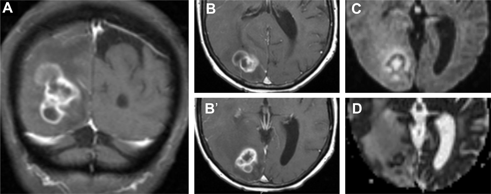
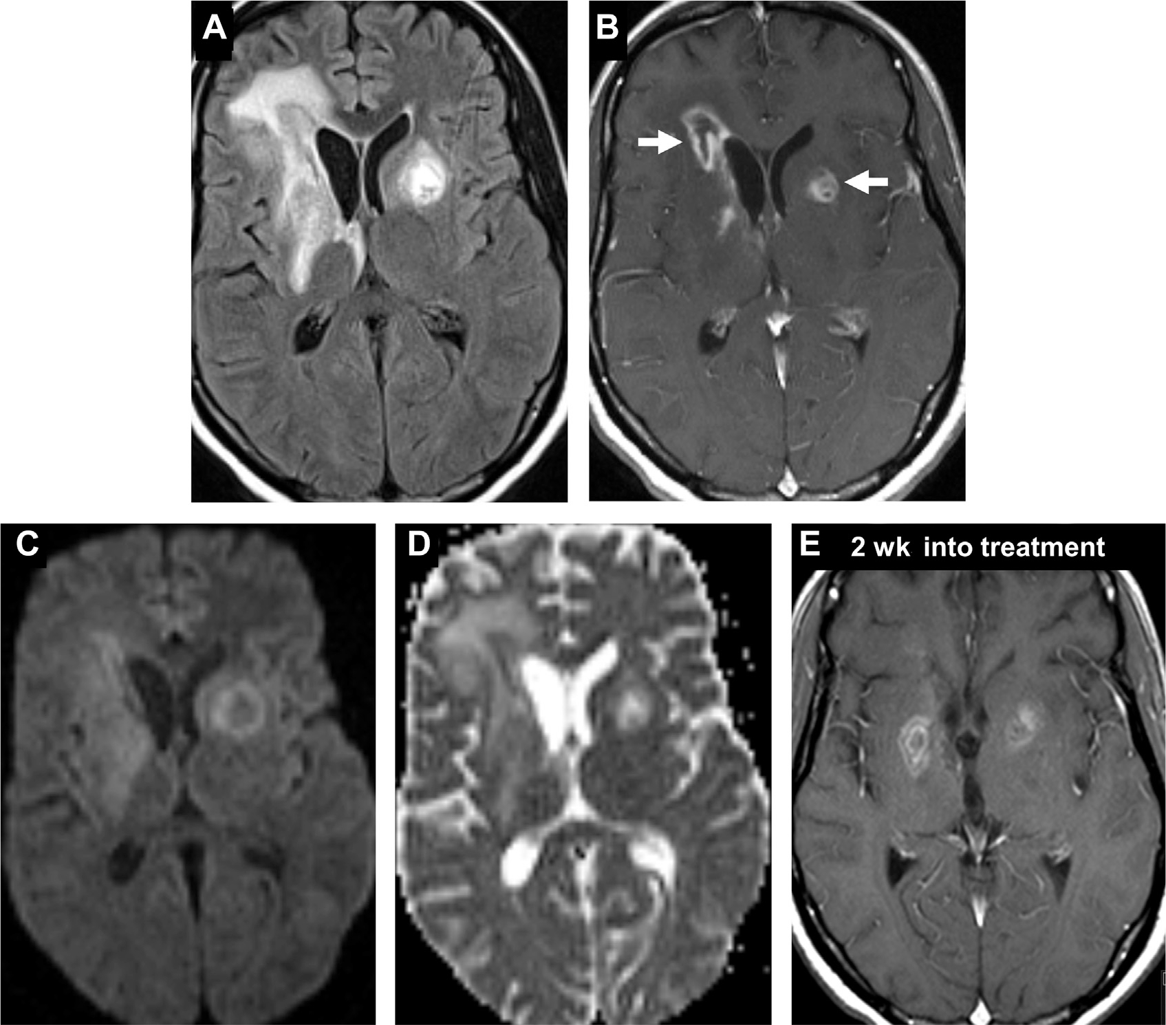
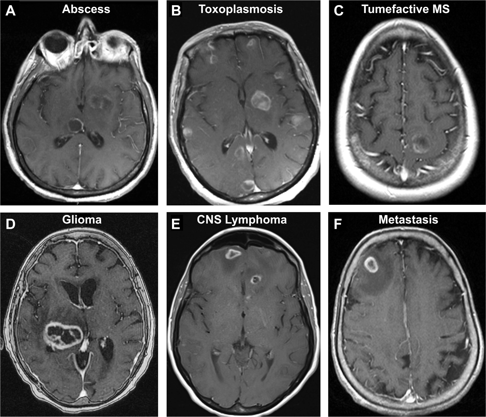
Neuroimaging provides rapid, noninvasive visualization of central nervous system infections for optimal diagnosis and management. Generalizable and characteristic imaging patterns help radiologists distinguish different types of intracranial infections including meningitis and cerebritis from a variety of bacterial, viral, fungal, and/or parasitic causes. Here, we describe key radiologic patterns of meningeal enhancement and diffusion restriction through profiles of meningitis, cerebritis, abscess, and ventriculitis. We discuss various imaging modalities and recent diagnostic advances such as deep learning through a survey of intracranial pathogens and their radiographic findings. Moreover, we explore critical complications and differential diagnoses of intracranial infections.

Keywords: Abscess; Central nervous system; Cerebritis; Glymphatics; Infection; Meningitis; Neuroimaging; Ventriculitis.


Conflict of interest statement

Disclosure S. Mohan has grant funding from the National Cancer Institute (NCI R01 CA262584), Galileo CDS and Novocure, USA. M. T. Duong has research funding from the National Institute on Aging (NIA F30 AG074524). J. D. Rudie has research support from the National Institute of Biomedical Imaging and Bioengineering (NIBIB T32 EB004311) and Radiological Society of North America (RSNA RR1778) research grant. There are no additional disclosures to report.

Figures

Fig. 1.
Fig. 1.
Coronal T1-weighted MR imaging shows the meningeal layers and spaces in an older adult. Fat in scalp (A) and outer table (B). Bone marrow (C). Epidural space (D). Dura mater (E), including the superior sagittal sinus and falx cerebri. Subdural space (F). Arachnoid mater is not visualized on imaging. Subarachnoid space including the lateral ventricles (G). Cerebral cortex (H).
Fig. 2.
Fig. 2.
Postsurgical CNS infection. T2-weighted MR imaging shows a low-grade cystic glioma abutting the left frontal horn (asterisk, A) before craniotomy/resection (A). Postoperative day one, axial T1 postcontrast image shows bilateral pneumocephalus with no enhancement around the resection cavity (B). Two months later, the patient presented with a seizure. T1 postcontrast MR imaging now depicts peripheral enhancement around the cavity (arrow, C) meningitis and ependymitis (arrowheads, D) and diffusion restricting, gravity-dependent pus-fluid meniscus levels (arrowheads, E, F) in the ventricles on DWI (E) and ADC (F), suggestive of meningitis, ventriculitis, and abscess.
Fig. 3.
Fig. 3.
Mastoiditis with intracranial complications of CNS infection in a patient presenting with headaches. Opacification of right mastoid air cells on CT (yellow arrow, A) is consistent with mastoiditis. Evidence of intracranial complications includes an FLAIR hyperintense lesion (arrowhead, B) that does not diffusion restrict on DWI (arrowhead, C). On T1 postcontrast (D), there is enhancement in the opacified mastoid air cells (yellow arrow, D) with adjacent pachymeningitis (multiple white arrows, D). Note the faintly ring-enhancing lesion in the posterior temporal lobe (arrowhead, E) on T1 postcontrast, indicative of cerebritis. Venous sinus thrombosis and thrombophlebitis of the right transverse sinus is seen on 3D shaded surface display volume rendering of CT venogram (F).
Fig. 4.
Fig. 4.
Patterns of normal enhancement in precontrast (A) and postcontrast MR imaging (B) along the anterior falx (arrows) and normal meningeal vessels/cortical veins running in the cerebral sulci (arrowheads).
Fig. 5.
Fig. 5.
Patterns of meningeal contrast enhancement (white outlines). Normal meninges (A). Diffuse pachymeningeal (B). Diffuse leptomeningeal (C) and localized leptomeningeal (D). Gyriform cortical (E). Ependymal (F).
Fig. 6.
Fig. 6.
Pachymeningeal enhancement in noninfectious granulomatosis with polyangiitis. (arrowheads, A, B). Diffuse pachymeningeal enhancement on MR imaging was complicated with chronic dural venous sinus thrombosis of the superior sagittal sinus (empty green box, C) and right transverse sinus (empty blue box, C) on an oblique view of CT venogram 3D reconstructions (C).
Fig. 7.
Fig. 7.
Pachymeningeal enhancement in idiopathic hypertrophic meningitis (A, B) and Tolosa-Hunt syndrome (C, D) on axial T1 postcontrast images. A patient with painless vision loss shows pachymeningeal enhancement (arrows, A, B) of the right cavernous sinus, consistent with idiopathic hypertrophic meningitis. Another patient with painful ophthalmoplegia, sudden diplopia, and right proptosis (C) demonstrates pachymeningeal enhancement along the right middle cranial fossa and cavernous sinus (arrows, C) and orbital apex (arrow, D) with orbital edema and fat stranding (not shown), consistent with Tolosa-Hunt syndrome.
Fig. 8.
Fig. 8.
Leptomeningeal and ependymal enhancement in bacterial meningitis. FLAIR (A, B) and T1 postcontrast MR imaging (C, D) in a patient status-post craniotomy shows leptomeningeal enhancement around the midbrain, adjacent sulci and cisterns (arrowheads, C) and ependymal surface of the left lateral ventricle (arrows, D). Note the ring-enhancing lesion (long arrow, D) consistent with abscess.
Fig. 9.
Fig. 9.
Progressive effacement of the basal cisterns on CT in an older adult with altered mental status at initial presentation (A, B, C) with interval changes on follow-up CT (D, E, F) and MR imaging (G, H, I). On admission, there is prominence of bilateral lateral ventricles (asterisks, A) with narrower yet still visible CSF-density in perimesencephalic cisterns (white arrows, B), cerebral aqueduct (white arrowhead, B), perimedullary cistern (black arrow, C) and cisterna magna (black arrowhead, C). Within 1.5 hours of admission, patient deteriorated clinically and a follow-up head CT demonstrated similar appearance of lateral ventricles (D) but interval marked effacement of perimesencephalic and perimedullary cisterns (E, F). By 5 hours after admission, T1 postcontrast MR imaging noted diffuse enhancement of cerebral sulci (arrows, G), around the brainstem and posterior fossa (arrows, H). Notice significant worsening mass effect and cerebral swelling with tonsillar herniation on sagittal T1-weighted MR imaging (black arrowhead, I). Mental status decline corresponded to progressive cerebral swelling and herniations and the patient subsequently passed away.
Fig. 10.
Fig. 10.
Abscess in a patient with primary ciliary dyskinesia (Kartagener syndrome). The left temporal lesion shows restricted diffusion on DWI (A) and ADC (B) and peripheral-enhancement on T1 postcontrast (C). A close-up of the T2-weighted MR imaging portrays a capsule with 2 T2 hypointense layers suggestive of the “dual rim sign” (black arrowheads, D) and vasogenic edema (asterisk, D). On axial T2 image, notice the left maxillary sinus inflammatory disease (white arrow, E) and underdeveloped bilateral mastoid air cells (white arrowheads, E) from ciliary dysfunction. Chest X-ray (F) portrays situs inversus totalis, typical of primary ciliary dyskinesia.
Fig. 11.
Fig. 11.
Fungal abscess on MR imaging and MRS. An FLAIR hyperintense (A), peripherally enhancing abscess (B) in the left occipital lobe. The orange square denotes placement of voxel including the abscess cavity while the green square depicts an unaffected region adjacent to the edematous abscess. Comparison of MRS peaks from the abscess cavity (C) and perilesional edematous region (D) reveals elevated cytosolic amino acids (0.9 ppm) and lactate/lipids (1.3 ppm) with decreased N-acetyl-aspartate (NAA, 2.0 ppm), choline (Cho, 3.0 ppm) and creatine (Cr, 3.2 ppm), consistent with an abscess.
Fig. 12.
Fig. 12.
Two patients with bacterial abscesses displaying acetate peaks on MRS. T1 postcontrast shows a left frontal abscess with (voxel, A). MRS (B) depicts elevated succinate (2.4 ppm), acetate (1.9 ppm), amino acids (0.9 ppm), and lactate/lipid (1.3 ppm) peaks and low NAA (2.0 ppm). In a second patient, T1 postcontrast MR imaging depicts a right thalamic abscess (voxel, C) with acetate peak and low NAA on MRS (D).
Fig. 13.
Fig. 13.
Predictions of T2 FLAIR (left) and T1 postcontrast (right) MR imaging lesions of CNS infections with a deep learning U-net model. Abscess (A), toxoplasmosis (B), cryptococcosis (C).
Fig. 14.
Fig. 14.
Aspergillosis in a patient with graft-versus-host disease presenting with right inferior quadrantanopia. A bilobed abscess in the left occipital region seems FLAIR hyperintense (A) with T2 hypointense capsule (B) and peripheral enhancement (C) and perilesional edema (A, B).
Fig. 15.
Fig. 15.
Nocardiosis in a patient with leukemia complicated by graft-versus-host disease. T1 postcontrast MR imaging in coronal (A) and axial (B, B’) planes visualize a multiloculated ring enhancing nocardial abscess in the right parietooccipital region with restricted diffusion on DWI (C) and ADC (D).
Fig. 16.
Fig. 16.
Toxoplasmosis in an HIV-positive patient. Ill-defined heterogenous lesions on FLAIR with surrounding vasogenic edema (A) and peripheral enhancement (B) are evident in the bilateral basal ganglia (arrows, B). Unlike typical abscess, there is facilitated diffusion on DWI (C) and ADC (D). Two weeks after initiating antitoxoplasma treatment (E), there is interval reduction in enhancement of the lesions, with decreased vasogenic edema (not shown). Note, “eccentric target sign” (arrow, B).
Fig. 17.
Fig. 17.
Tuberculoma with meningitis. A thalamic lesion with central T2 hypointensity (arrow, A), characteristic for tuberculoma, with no restricted diffusion on DWI (B) and ADC (C) but with FLAIR hyperintensity (arrow, D). On T1 postcontrast, there is ring enhancement (arrow, E) and associated meningitis (arrowheads, E, F).
Fig. 18.
Fig. 18.
Cryptococcosis with meningitis and acute infarction. Multifocal FLAIR hyperintense cryptococcomas are seen in the basal ganglia (arrows, A) and corpus callosum with enhancement in the right caudate nucleus (arrow, B) and bilateral cerebral sulci (B). Diffusion restriction in a right splenium of corpus callosum suggesting acute infarct (arrowhead, C, D) and faint diffusion abnormality in the basal ganglia (arrow, C, D) on DWI (C) and ADC (D).
Fig. 19.
Fig. 19.
Neurocysticercosis in a patient with seizures. A right posterior frontal cystic lesion seen along the pial surface seems with a T2 hypointense inner “dot in a hole” sign (arrows, A, B) on T2-weighted (A) and T2-weighted thin-section 3D constructive interference in steady state (B) images, pathognomonic for the scolex in neurocysticercosis. This cyst is FLAIR hyperintense (arrow, C) with central and ring enhancement (arrow, D). T1 postcontrast MR imaging reveals leptomeningeal enhancement (D, E, F) and additional ring enhancing cysts in the right parietal lobe (arrowhead, E), left frontal operculum (arrowhead, E) and right lateral temporal lobe (arrowhead, F).
Fig. 20.
Fig. 20.
Ventriculitis associated with ruptured abscess (long arrow, B–D). Axial T1-weighted precontrast (A) and postcontrast MR images (B) demonstrate a peripherally enhancing left caudate abscess (long arrow, B) with subtle linear enhancement of the ependyma of the left frontal horn and midline septum (short arrows, B) and diffuse leptomeningeal enhancement. The left caudate abscess is surrounded by vasogenic edema (short blue arrows, C), and has ruptured in the ventricles, susceptibility from blood products on GRE (long arrow, D) and restricted diffusion (long arrows, E, F) on DWI (E) and ADC (F). Notice a lack of susceptibility of the dependent debris in the occipital horns on GRE, with corresponding FLAIR hyperintensity (arrowheads, C) and restricted diffusion (arrowheads, E, F), consistent with layering pus.
Fig. 21.
Fig. 21.
Cryptococcosis and hydrocephalus. T1 postcontrast MR imaging visualizes subtle perimesencephalic and bilateral posterior leptomeningeal enhancement (A). Enhancement along an obstructed foramen of Monro (B). Ependymal enhancement with enlarged left lateral ventricle (C). The right lateral ventricle is reduced in volume due to left ventricle compression and drainage by a ventriculostomy catheter.
Fig. 22.
Fig. 22.
Subdural empyema in a patient with bacterial meningitis and cerebritis. A crescentic subdural collection in the right middle cranial fossa (arrows) is FLAIR hyperintense (A) with enhancement (B) and restricted diffusion on DWI (C) and ADC (D). In the left anterior convexity (arrows), a thin FLAIR hyperintense collection (E) with leptomeningeal enhancement (F) shows diffusion restriction on DWI (G) and ADC (H). Note also diffuse leptomeningitis (F) and frontal cerebritis, right worse than left (E–H).
Fig. 23.
Fig. 23.
Epidural empyema in bacterial meningitis. This seems as a biconvex collection with thick peripheral enhancement (A) subjacent the craniotomy site with FLAIR hyperintensity (B) and diffusion restriction on DWI (C) and ADC (D) maps.
Fig. 24.
Fig. 24.
Cranial neuropathy in a patient with bacterial leptomeningitis presenting with dilated, fixed pupils lacking several cranial nerve (CN) reflexes. T1 postcontrast images demonstrate enhancement of bilateral trigeminal nerves (CN V, arrowheads, A), facial/vestibulocochlear nerves (CN VII, VIII, arrowheads, A, B) and glossopharyngeal/vagus nerves (CN IX, X, arrowheads, C). Thick ependymal enhancement is noted (arrow, A).
Fig. 25.
Fig. 25.
Venous sinus thrombosis in an immunocompromised patient with MRSA bacteremia, sinusitis, and orbital cellulitis. Axial T1 postcontrast MR imaging (A) shows leptomeningeal and irregular peripheral enhancement, suggestive of meningitis. Diffusion restriction DWI (B) and ADC (C) maps indicate multiple abscesses. Time-of-flight venogram MIP images (D, E) demonstrate loss of flow-related enhancement of the anterior superior sagittal sinus, consistent with venous sinus thrombosis (arrowheads, D, E). Notice extensive preseptal and postseptal inflammation in the left orbit (arrowhead, F) and opacified ethmoid air cells (F). MIP, maximum intensity projection.
Fig. 26.
Fig. 26.
Vasculitis in tuberculous meningitis. In a patient with HIV and left-sided retroorbital headaches, T1 postcontrast axial (A) and coronal MR images (B) reveal a peripherally enhancing lesion in the left perisylvian region (arrows, A, B) and faint basilar enhancement, consistent with tuberculous meningitis with tuberculoma. CT angiography (CTA) via axial (C) and coronal thin MIP images (D) show focal narrowing and irregularity of the distal M1, M2, and proximal M3 segments of the left middle cerebral artery (arrowheads, C, D), suggestive of vasculitis secondary to tuberculous meningitis.
Fig. 27.
Fig. 27.
Carcinomatous meningitis with dural enhancement in a patient with acute myeloid leukemia. Notice FLAIR hyperintensity (arrow, A), and thick dural-based enhancement on T1 postcontrast (arrows, B, C) with restricted diffusion (arrow, D).
Fig. 28.
Fig. 28.
Neurosarcoidosis. T1 postcontrast MR imaging demonstrates enhancement of both prechiasmatic optic nerves (arrows, A) with patchy linear and nodular enhancement around the brainstem and cerebellum (B) and bilateral enhancement of the glossopharyngeal/vagus nerves (arrowheads, C).
Fig. 29.
Fig. 29.
Assortment of ring/peripherally enhancing lesions on T1 postcontrast MR imaging. Abscess in an immunocompromised patient (A). Multicentric toxoplasmosis (B). New demyelinating lesion in tumefactive multiple sclerosis (C). Necrotic high-grade glioma (D). Multifocal primary CNS lymphoma in an immunocompromised patient with congenital lymphangiectasia (E). Junctional metastasis with surrounding edema (F).

References

    1. Mohan S, Jain KK, Arabi M, et al. Imaging of Meningitis and Ventriculitis. Neuroimaging Clin N Am 2012;22(4):557–83. - PubMed
    1. Rath TJ, Hughes M, Arabi M, et al. Imaging of Cerebritis, Encephalitis, and Brain Abscess. Neuroimaging Clin N Am 2012;22(4):585–607. - PubMed
    1. Global Burden of Disease 2016 Meningitis Collaborators. Global, regional, and national burden of meningitis, 1990–2016: a systematic analysis for the Global Burden of Disease Study 2016. Lancet Neurol 2018;17(12):1061–82. - PMC - PubMed
    1. Patel K, Clifford DB. Bacterial Brain Abscess. Neurohospitalist 2014;4(4):196–204. - PMC - PubMed
    1. Mortazavi MM, Tubbs RS, Riech S, et al. Anatomy and pathology of the cranial emissary veins: a review with surgical implications. Neurosurgery 2012;70(5):1312–8. - PubMed