

Microscopic observations of SARS-CoV-2 like particles in different oral samples
- PMID: 36404273
- PMCID: PMC10099536
- DOI: 10.1111/eos.12903
Microscopic observations of SARS-CoV-2 like particles in different oral samples
Abstract
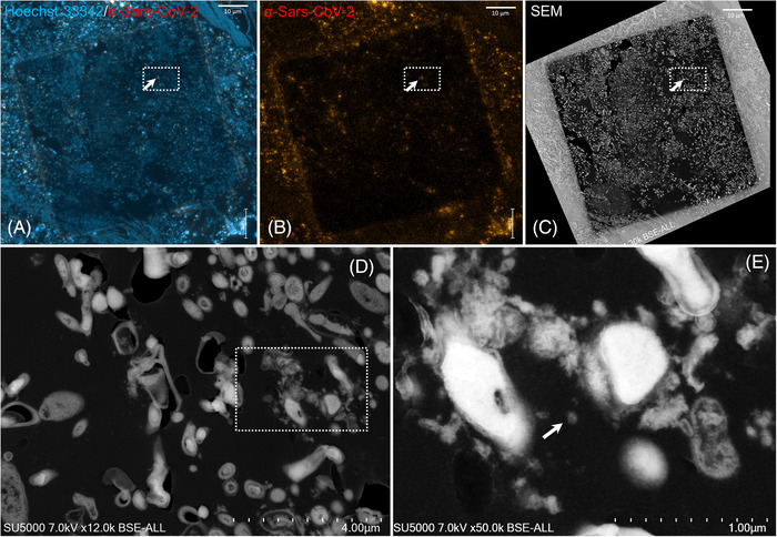
The emerging coronavirus pneumonia epidemic caused by the SARS-CoV-2 infection has spread rapidly around the world. The main routes of transmission of SARS-CoV-2 are currently recognised as aerosol/droplet inhalation. However, the involvement of the oral cavity in coronavirus disease 2019 (COVID-19) is poorly known. The current data indicates the presence of viral RNA in oral samples, suggesting the implication of saliva in SARS-CoV-2 transmission, however, no direct observation of SARS-CoV-2 particles in different oral samples has been reported. In this study, we investigated whether particles of SARS-CoV-2 were present in oral samples collected from three symptomatic COVID-19 patients. Using scanning electron microscopy (SEM), the correlative strategy of light microscopy and electron microscopy and immunofluorescence staining, we showed the presence of SARS-like particles in RT-qPCR SARS-CoV-2-positive saliva, dental plaque and gingival crevicular fluid (GCF) samples. In the saliva samples, we demonstrated the presence of epithelial oral cells with morphogenetic features of SARS-CoV-2 infected cells. Inside those cells, vacuoles filled with nascent particles were observed, suggesting the potential infection and replication of SARS-CoV-2 in oral tissues. Our results corroborate previous studies and confirm that the oral cavity may be a potential niche for SARS-CoV-2 infection and a potential source of transmission.
Keywords: COVID-19; dental plaque; saliva; scanning electron microscopy; virology.
© 2022 Assistance Publique Hopitaux de Marseille (MARSEILLE, PROVENCE-ALPES-CÔTE D'AZUR, FR). European Journal of Oral Sciences published by John Wiley & Sons Ltd on behalf of Scandinavian Division of the International Association for Dental Research.
Conflict of interest statement
The authors declare that the research was conducted in the absence of any commercial or financial relationships that could be construed as a potential conflict of interest.
Figures


Similar articles
-
SARS-CoV-2 Detection in Gingival Crevicular Fluid.J Dent Res. 2021 Feb;100(2):187-193. doi: 10.1177/0022034520970536. Epub 2020 Nov 2. J Dent Res. 2021. PMID: 33138663 Free PMC article.
-
Microscopic Observation of SARS-Like Particles in RT-qPCR SARS-CoV-2 Positive Sewage Samples.Pathogens. 2021 Apr 24;10(5):516. doi: 10.3390/pathogens10050516. Pathogens. 2021. PMID: 33923138 Free PMC article.
-
Saliva is a non-negligible factor in the spread of COVID-19.Mol Oral Microbiol. 2020 Aug;35(4):141-145. doi: 10.1111/omi.12289. Epub 2020 May 31. Mol Oral Microbiol. 2020. PMID: 32367576 Free PMC article. Review.
-
SARS-CoV-2 Infection and Significance of Oral Health Management in the Era of "the New Normal with COVID-19".Int J Mol Sci. 2021 Jun 18;22(12):6527. doi: 10.3390/ijms22126527. Int J Mol Sci. 2021. PMID: 34207046 Free PMC article. Review.
-
The "oral" history of COVID-19: Primary infection, salivary transmission, and post-acute implications.J Periodontol. 2021 Oct;92(10):1357-1367. doi: 10.1002/JPER.21-0277. Epub 2021 Sep 7. J Periodontol. 2021. PMID: 34390597 Free PMC article. Review.
Cited by
-
COVID-19 in Dental Practice Is Prevented by Eugenol Responsible for the Ambient Odor Specific to Dental Offices: Possibility and Speculation.Med Princ Pract. 2024;33(2):83-89. doi: 10.1159/000535966. Epub 2023 Dec 26. Med Princ Pract. 2024. PMID: 38147833 Free PMC article. Review.
-
COVID-19 on Oral Health: A New Bilateral Connection for the Pandemic.Biomedicines. 2023 Dec 26;12(1):60. doi: 10.3390/biomedicines12010060. Biomedicines. 2023. PMID: 38255167 Free PMC article. Review.
-
Severe acute respiratory syndrome coronavirus 2 pathology and cell tropism in tongue tissues of COVID-19 autopsies.Front Cell Infect Microbiol. 2024 Jun 21;14:1394721. doi: 10.3389/fcimb.2024.1394721. eCollection 2024. Front Cell Infect Microbiol. 2024. PMID: 38975331 Free PMC article.
References
-
- World Health Organization. WHO Coronavirus (COVID‐19) Dashboard. https://covid19.who.int/ Accessed 4 October 2021
Publication types
MeSH terms
LinkOut - more resources
Full Text Sources
Medical
Miscellaneous
