

Detection and evaluation of myocardial fibrosis in Eisenmenger syndrome using cardiovascular magnetic resonance late gadolinium enhancement and T1 mapping
- PMID: 36404313
- PMCID: PMC9677680
- DOI: 10.1186/s12968-022-00880-2
Detection and evaluation of myocardial fibrosis in Eisenmenger syndrome using cardiovascular magnetic resonance late gadolinium enhancement and T1 mapping
Abstract
Background: Myocardial fibrosis is a common pathophysiological process involved in many cardiovascular diseases. However, limited prior studies suggested no association between focal myocardial fibrosis detected by cardiovascular magnetic resonance (CMR) late gadolinium enhancement (LGE) and disease severity in Eisenmenger syndrome (ES). This study aimed to explore potential associations between myocardial fibrosis evaluated by the CMR LGE and T1 mapping and risk stratification profiles including exercise tolerance, serum biomarkers, hemodynamics, and right ventricular (RV) function in these patients.
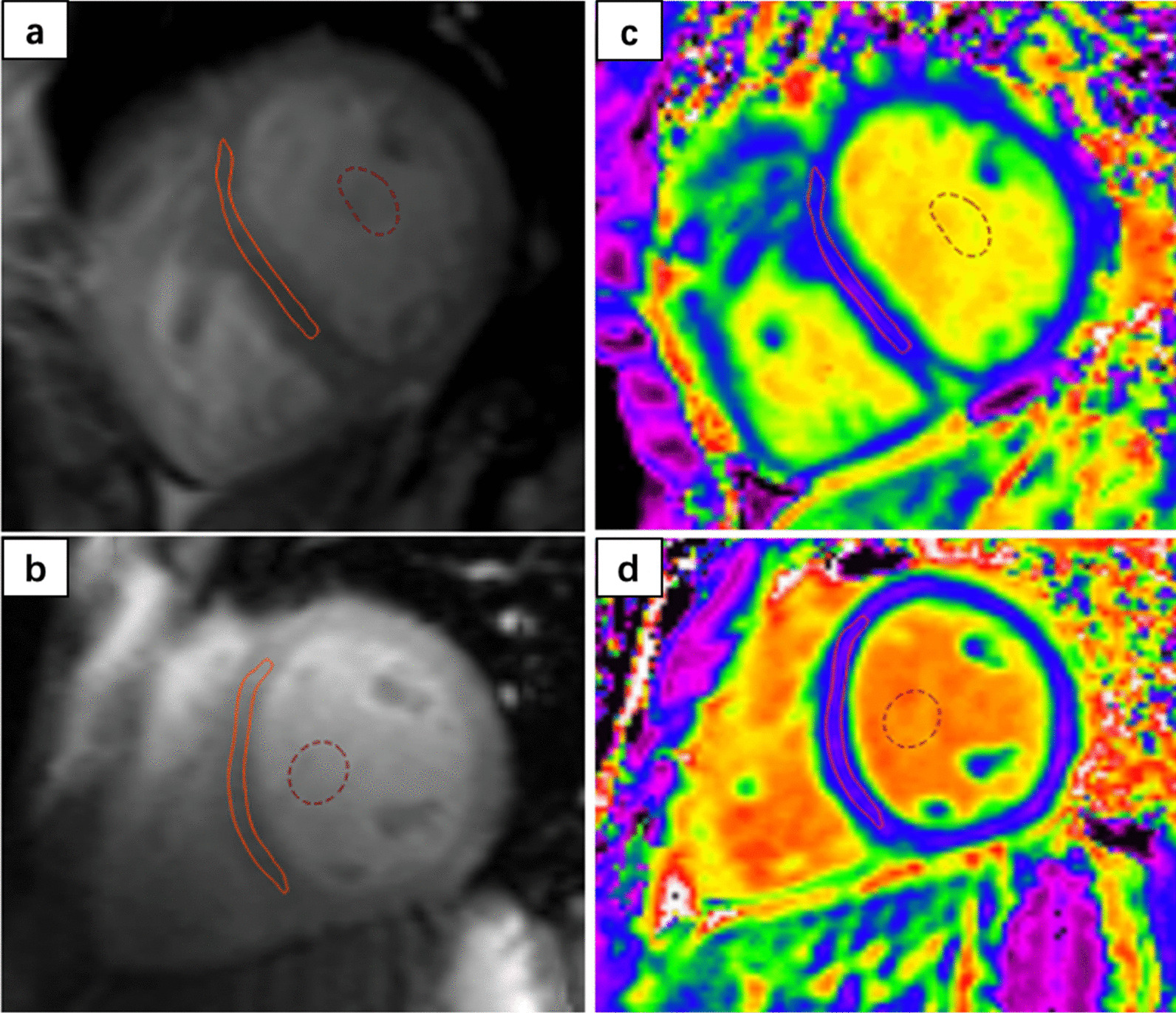
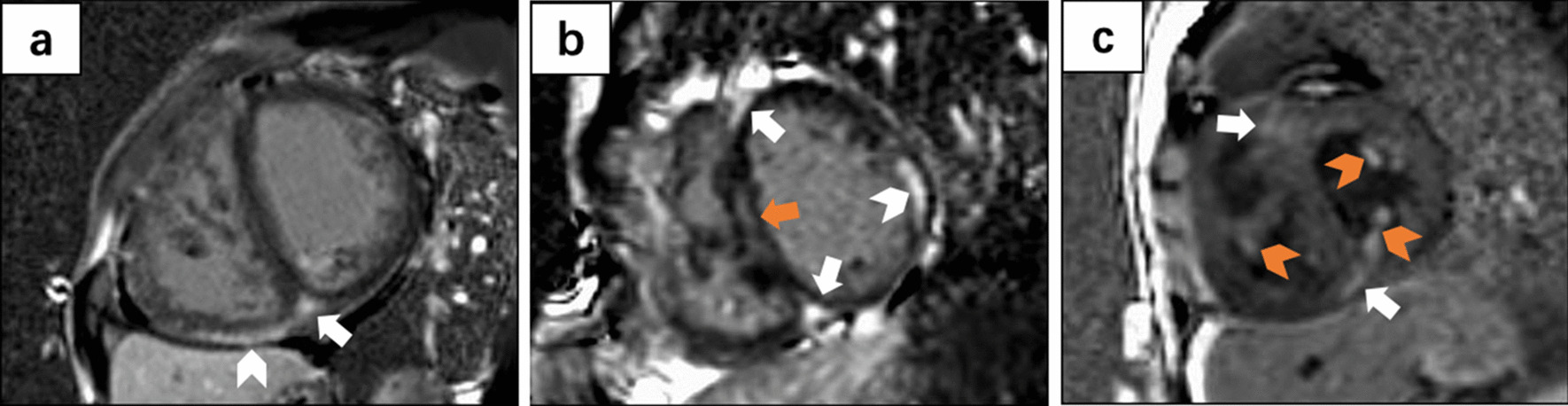
Methods: Forty-five adults with ES and 30 healthy subjects were included. All subjects underwent a contrast-enhanced 3T CMR. Focal replacement fibrosis was visualized on LGE images. The locations of LGE were recorded. After excluding LGE in ventricular insertion point (VIP), ES patients were divided into myocardial LGE-positive (LGE+) and LGE-negative (LGE-) subgroups. Regions of interest in the septal myocardium were manually contoured in the T1 mapping images to determine the diffuse myocardial fibrosis. The relationships between myocardial fibrosis and 6-min walk test (6MWT), N-terminal pro-brain natriuretic peptide (NT-pro BNP), hematocrit, mean pulmonary arterial pressure (mPAP), pulmonary vascular resistance index (PVRI), RV/left ventricular end-systolic volume (RV/LV ESV), RV ejection fraction (RVEF), and risk stratification were analyzed.
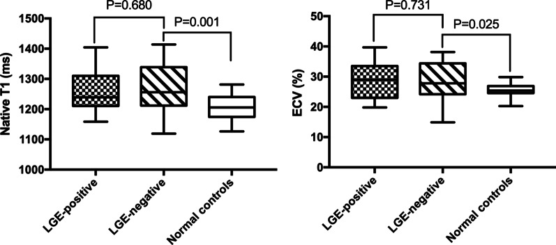
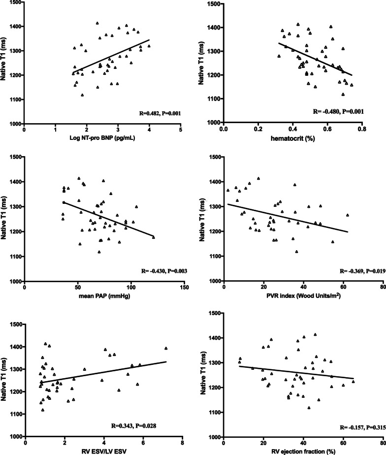
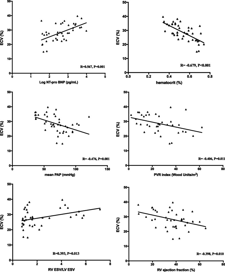
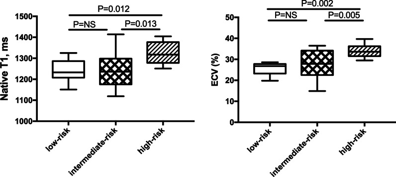
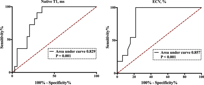
Results: Myocardial LGE (excluding VIP) was common in ES (16/45, 35.6%), and often located in the septum (12/45, 26.7%). The clinical characteristics, hemodynamics, CMR morphology and function, and extracellular volume fraction (ECV) were similar in the LGE+ and LGE- groups (all P > 0.05). ECV was significantly higher in ES patients (28.6 ± 5.9% vs. 25.6 ± 2.2%, P < 0.05) and those with LGE- ES (28.3 ± 5.9% vs. 25.6 ± 2.2%, P < 0.05) than healthy controls. We found significant correlations between ECV and log NT-pro BNP, hematocrit, mPAP, PVRI, RV/LV ESV, and RVEF (all P < 0.05), and correlations trends between ECV and 6MWT (P = 0.06) in ES patients. An ECV threshold of 29.0% performed well in differentiating patients with high-risk ES from those with intermediate or low risk (area under curve 0.857, P < 0.001).
Conclusions: Myocardial fibrosis is a common feature of ES. ECV may serve as an important imaging marker for ES disease severity.
Keywords: Cardiovascular magnetic resonance; Eisenmenger syndrome; Extracellular volume; Myocardial fibrosis; T1 mapping.
© 2022. The Author(s).
Conflict of interest statement
The authors declare that they have no competing interests.
Figures
Similar articles
-
T1-Mapping and extracellular volume estimates in pediatric subjects with Duchenne muscular dystrophy and healthy controls at 3T.J Cardiovasc Magn Reson. 2020 Dec 10;22(1):85. doi: 10.1186/s12968-020-00687-z. J Cardiovasc Magn Reson. 2020. PMID: 33302967 Free PMC article.
-
Cardiovascular magnetic resonance evidence of myocardial fibrosis and its clinical significance in adolescent and adult patients with Ebstein's anomaly.J Cardiovasc Magn Reson. 2018 Sep 27;20(1):69. doi: 10.1186/s12968-018-0488-1. J Cardiovasc Magn Reson. 2018. PMID: 30257686 Free PMC article.
-
Early detection of myocardial fibrosis in cardiomyopathy in the absence of late enhancement: role of T1 mapping and extracellular volume analysis.Eur Radiol. 2023 Mar;33(3):1982-1991. doi: 10.1007/s00330-022-09147-x. Epub 2022 Oct 14. Eur Radiol. 2023. PMID: 36241919
-
Native T1 Mapping, Extracellular Volume Mapping, and Late Gadolinium Enhancement in Cardiac Amyloidosis: A Meta-Analysis.JACC Cardiovasc Imaging. 2020 Jun;13(6):1299-1310. doi: 10.1016/j.jcmg.2020.03.010. JACC Cardiovasc Imaging. 2020. PMID: 32498919 Free PMC article.
-
Assessment of myocardial fibrosis with T1 mapping MRI.Clin Radiol. 2016 Aug;71(8):768-78. doi: 10.1016/j.crad.2016.02.013. Epub 2016 Mar 19. Clin Radiol. 2016. PMID: 27005015 Review.
Cited by
-
Recent Progresses in the Multimodality Imaging Assessment of Myocardial Fibrosis.Rev Cardiovasc Med. 2024 Jan 8;25(1):5. doi: 10.31083/j.rcm2501005. eCollection 2024 Jan. Rev Cardiovasc Med. 2024. PMID: 39077665 Free PMC article. Review.
-
Update on Eisenmenger syndrome - Review of pathophysiology and recent progress in risk assessment and management.Int J Cardiol Congenit Heart Dis. 2024 Jun 10;17:100520. doi: 10.1016/j.ijcchd.2024.100520. eCollection 2024 Sep. Int J Cardiol Congenit Heart Dis. 2024. PMID: 39711759 Free PMC article. Review.
-
Biventricular longitudinal strain analysis using cardiovascular magnetic resonance feature-tracking: Prognostic value in Eisenmenger syndrome.J Cardiovasc Magn Reson. 2024 Winter;26(2):101116. doi: 10.1016/j.jocmr.2024.101116. Epub 2024 Oct 28. J Cardiovasc Magn Reson. 2024. PMID: 39477153 Free PMC article.
References
-
- Nashat H, Kempny A, McCabe C, Price LC, Harries C, Alonso-Gonzalez R, et al. Eisenmenger syndrome: current perspectives. Res Rep Clin Cardiol. 2017;8:1–12.
-
- Diller GP, Alonso-Gonzalez R, Kempny A, Dimopoulos K, Inuzuka R, Giannakoulas G, et al. B-type natriuretic peptide concentrations in contemporary Eisenmenger syndrome patients: predictive value and response to disease targeting therapy. Heart. 2012;98:736–742. doi: 10.1136/heartjnl-2011-301522. - DOI - PubMed
-
- Buchhorn R, Ross RD, Bartmus D, Wessel A, Hulpke-Wette M, Bursch J. Activity of the renin-angiotensin-aldosterone and sympathetic nervous system and their relation to hemodynamic and clinical abnormalities in infants with left-to-right shunts. Int J Cardiol. 2001;78:225–230. doi: 10.1016/S0167-5273(01)00398-9. - DOI - PubMed
Publication types
MeSH terms
Substances
LinkOut - more resources
Full Text Sources
Medical
Research Materials
