Case Reports
. 2023;10(1):119-123.
doi: 10.3233/JND-221526.

A Multisystem Mitochondrial Disease Caused by a Novel MT-TL1 mtDNA Variant: A Case Report

Affiliations
Case Reports

A Multisystem Mitochondrial Disease Caused by a Novel MT-TL1 mtDNA Variant: A Case Report

Domenico Giannese et al. J Neuromuscul Dis. 2023.

Abstract

Background: Mitochondrial tRNA (MTT) genes are hotspot for mitochondrial DNA mutation and are responsible of half mitochondrial disease. MTT mutations are associated with a broad spectrum of phenotype often with complex multisystem involvement and complex genotype-phenotype correlations. MT-TL1 mutations, among which the m.3243A>G mutation is the most frequent, are associated with myopathy, maternal inherited diabetes and deafness, MELAS, cardiomyopathy, and focal segmental glomerulosclerosis.

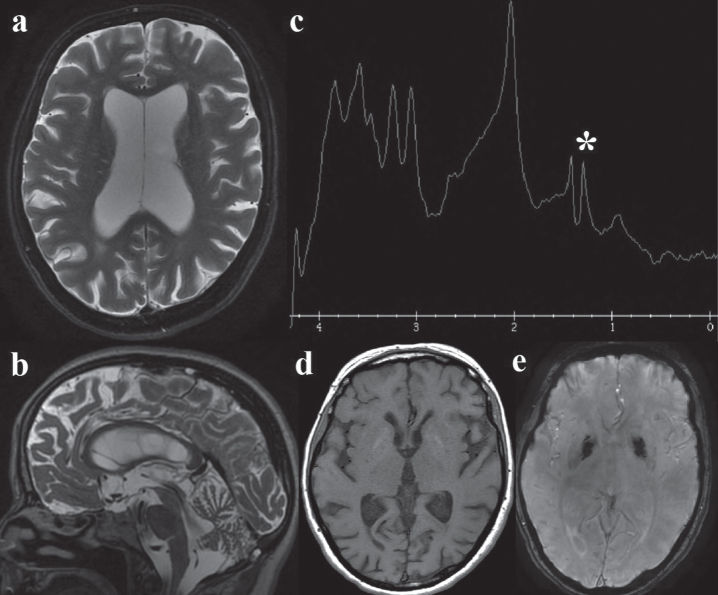
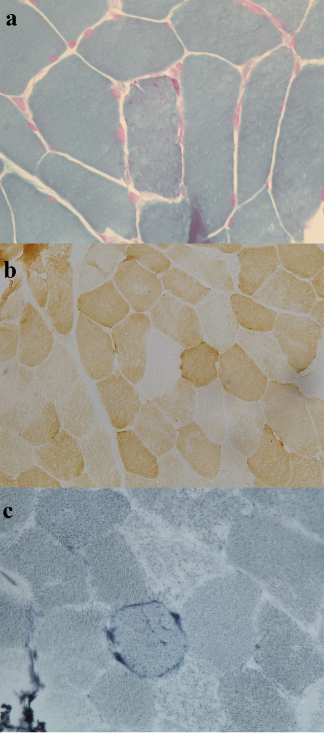
Case study: Here we report the case of an Italian 49-years old female presenting with encephalomyopathy, chronic proteinuric kidney disease and a new heteroplasmic m.3274_3275delAC MT-TL1 gene mutation.

Conclusions: Our case demonstrates a systemic mitochondrial disease caused by the heteroplasmic m.3274_3275delAC MT-TL1 gene mutation, not yet described in the literature. A mitochondrial disease should be suspected in case of complex multisystem phenotypes, including steroid-resistant nephrotic syndrome with multisystemic involvement.

Keywords: glomerulosclerosis; kidney disease; mitochondrial diseases; mitochondrial myopathies; mtDNA.


Conflict of interest statement

The authors have no conflict of interest.

Figures

Fig. 1
Fig. 1
Brain MRI: a. Axial T2, showing global cerebral atrophy with ex-vacuo lateral ventricles dilatation; b. Sagittal T2, cerebellar atrophy; c. MR spectroscopy: the white asterisk (*) marks the lactate peak; d. axial T1 and e. susceptibility-weighted angiography (SWAN): mild bipallidal calcifications.
Fig. 2
Fig. 2
Muscle biopsy reveals mild proliferation of mitochondria (a), COX negative fibers (b) and (c) one ragged blue fiber.

References

    1. Ng YS, Bindoff LA, Gorman GS, Klopstock T, Kornblum C, Mancuso M, et al.. Mitochondrial disease in adults: Recent advances and future promise. Lancet Neurol. 2021;20:573–584. 10.1016/S1474-4422(21)00098-3. - DOI - PubMed
    1. Guillausseau PJ, Massin P, Dubois-LaForgue D, Timsit J, Virally M, Gin H, et al.. Maternally inherited diabetes and deafness: A multicenter study. Ann Intern Med. 2001;134:721–728. 10.7326/0003-4819-134-9_part_1-200105010-00008. - DOI - PubMed
    1. Sonney S, Leipzig J, Lott MT, Zhang S, Procaccio V, Wallace DC, et al.. Predicting the pathogenicity of novel variants in mitochondrial tRNA with MitoTIP. PLoS Comput Biol. 2017;13. 10.1371/journal.pcbi.1005867. - DOI - PMC - PubMed
    1. Emma F, Pizzini C, Tessa A, Di Giandomenico S, Onetti-Muda A, Santorelli FM, et al.. ⪡Bartter-like⪢ phenotype in Kearns-Sayre syndrome. Pediatr Nephrol. 2006;21:355–360. 10.1007/s00467-005-2092-5. - DOI - PubMed
    1. Bhargava P, Schnellmann RG. Mitochondrial energetics in the kidney. Nat Rev Nephrol. 2017;13:629–646. 10.1038/nrneph.2017.107. - DOI - PMC - PubMed

Publication types

Substances

LinkOut - more resources