

Non-contrast MR dacryocystography for the evaluation of epiphora and recurrent dacryocystitis: A preliminary study
- PMID: 36404757
- PMCID: PMC10588605
- DOI: 10.1177/19714009221140484
Non-contrast MR dacryocystography for the evaluation of epiphora and recurrent dacryocystitis: A preliminary study
Abstract
Introduction: Obstruction of the lacrimal drainage represents a common ophthalmologic issue. The blockage may interest any level of the lacrimal drainage pathway, and it is important to find the site of obstruction to plan the most appropriate treatment. In this study, findings from magnetic resonance (MR) dacryocystography were compared with findings from endoscopic and surgical procedures to evaluate the accuracy of MR dacryocystography in localizing the site of nasolacrimal duct obstruction.
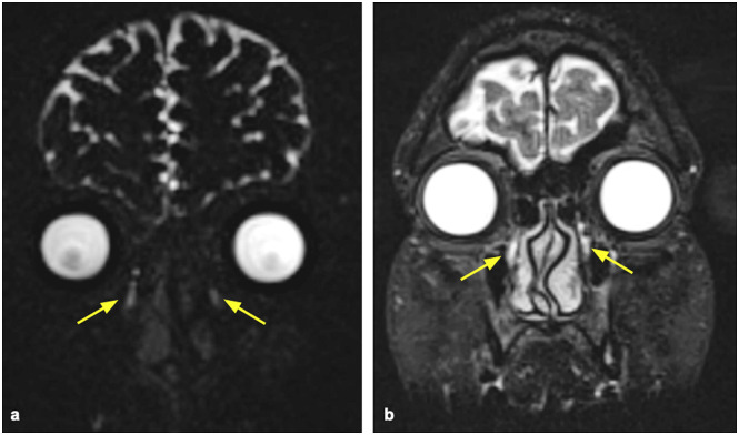
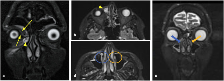
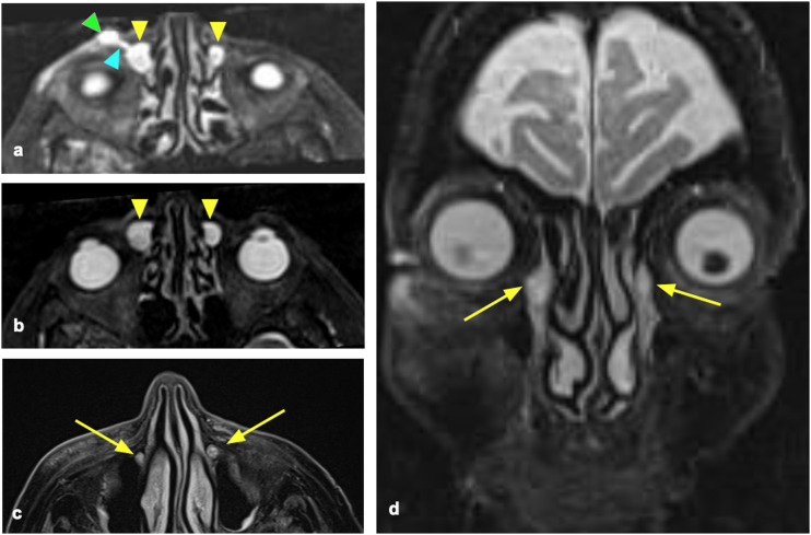
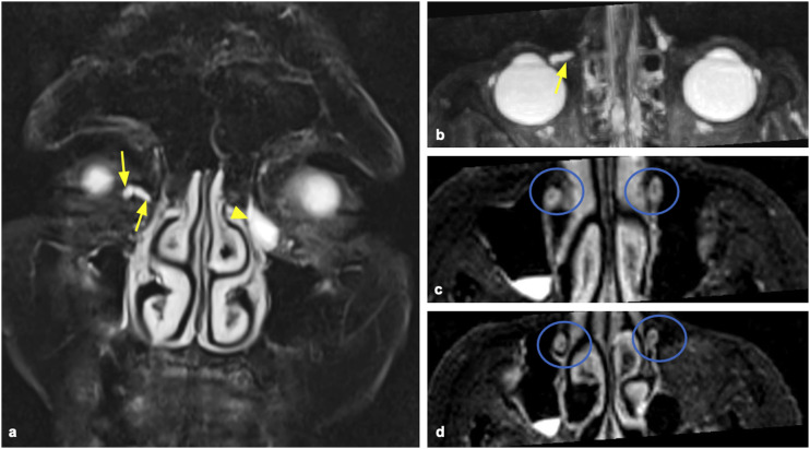
Methods: We enrolled twenty-one patients with clinical suspicion of nasolacrimal duct obstruction who underwent dacryoendoscopy and surgery. MR dacryocystography was performed with a heavily T2-weighted fast spin echo sequence in the coronal planes. Before the MRI was performed, a sterile 0.9% NaCl solution was administered into both conjunctival sacs. For each examination, two independent readers (with 8 and 10 years of experience in head and neck imaging) evaluated both heavily 3D space T2-weighted and STIR sequences.
Results: Stenosis/obstruction of nasolacrimal duct or lacrimal sac was diagnosed in all 21 patients who underwent MRI dacryocystography. In particular, the site of the obstruction was classified as lacrimal sac in 12 (57%) patients, nasolacrimal duct in 6 (29%) patients, and canaliculi in 3 (14%) patients by both readers. By comparison with the evidence resulting from the endoscopy, there were differences between MRI dacryocystography and dacryoendoscopy in the evaluation of the obstruction's site in three patients, with an overall accuracy of 85.7%.
Conclusion: MR dacryocystography allows a non-invasive evaluation of the lacrimal drainage pathway, valid for the planning of the most appropriate treatment.
Keywords: MR dacryocystography; dacryocystitis; lacrimal drainage pathway; lacrimal sac; magnetic resonance imaging.
Conflict of interest statement
The author(s) declared no potential conflicts of interest with respect to the research, authorship, and/or publication of this article.
Figures

References
-
- Weber A, Rodriguez-DeVelasquez A, Lucarelli M, et al. Normal anatomy and lesions of the lacrimal sac and duct: evaluated by dacryocystography, computed tomography, and MR imaging. Neuroimag Clin N Am 1996; 6: 199–217. - PubMed
-
- Sasaki T, Sugiyama K, Yuuko N. Nasolacrimal duct obstruction classified by dacryoendoscopy and treated with inferior meatal dacryorhinotomy. Part I: positional diagnosis of primary nasolacrimal duct obstruction with dacryoendoscope. Am J Ophthalmol 2005; 140(6): 1065–1070. DOI: 10.1016/j.ajo.2005.07.038 - DOI - PubMed
-
- Chan W, Malhotra R, Kakizaki H, et al. Perspective: what does the term functional mean in the context of epiphora? Clin experiment Ophthalmol 2012; 40: 749–755. - PubMed
MeSH terms
LinkOut - more resources
Full Text Sources
