. 2022 Aug 25;8(4):611.
doi: 10.18063/ijb.v8i4.611. eCollection 2022.

Recombinant Human Collagen-Based Bioinks for the 3D Bioprinting of Full-thickness Human Skin Equivalent

Affiliations

Recombinant Human Collagen-Based Bioinks for the 3D Bioprinting of Full-thickness Human Skin Equivalent

Yang Yang et al. Int J Bioprint. .

Abstract

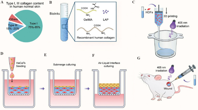
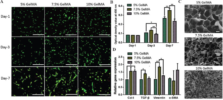
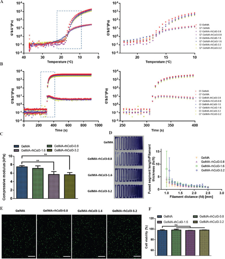
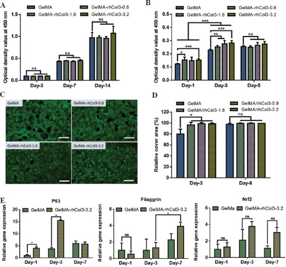
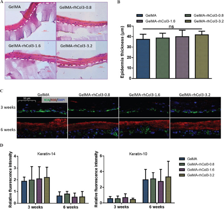
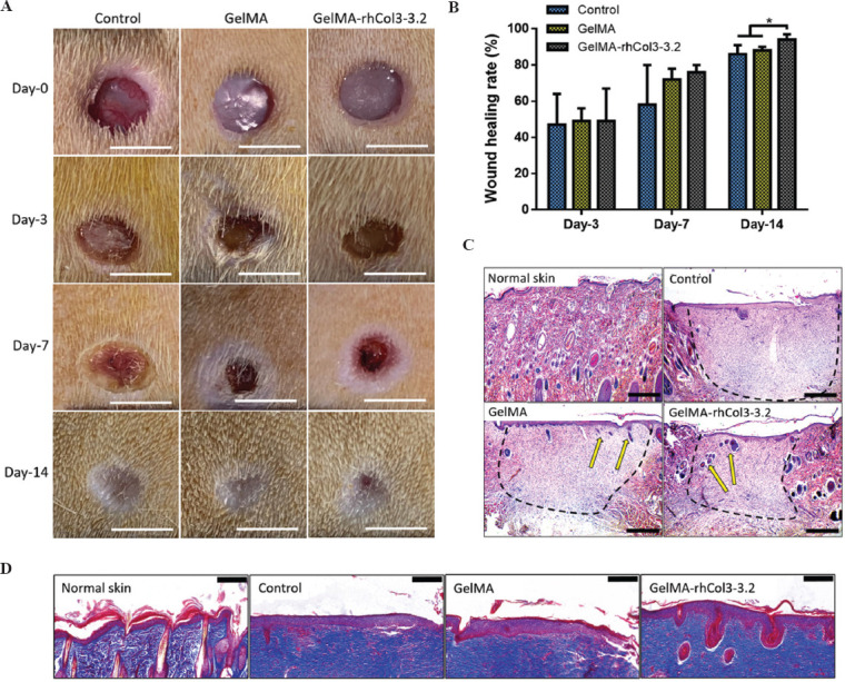
As a major extracellular matrix component within the skin, collagen has been widely used to engineer human skin tissues. However, most collagen is extracted from animals. Here, we introduced recombinant human type III collagen (rhCol3) as a bioactive component to formulate bioinks for the bioprinting of a full-thickness human skin equivalent. Human dermal fibroblasts were encapsulated in the gelatin methacryloyl-rhCol3 composite bioinks and printed on a transwell to form the dermis layer, on which human epidermal keratinocytes were seeded to perform an air-liquid interface culture for 6 weeks. After optimizing the bioink formulation and bioprinting process, we investigated the effect of rhCol3 on skin tissue formation. The results suggest that a higher concentration of rhCol3 would enhance the growth of both cells, resulting in a more confluent (~100%) spreading of the epidermal keratinocytes at an early stage (3 days), compared to the rhCol3-free counterpart. Moreover, in an in vivo experiment, adding rhCol3 in the hydrogel formulation would contribute to the skin wound healing process. Taken together, we conclude that rhCol3 could act as a functional bioink component to promote basic skin cellular processes for skin tissue engineering.

Keywords: 3D printing; Bioinks; Recombinant human collagen; Skin constructs.


Conflict of interest statement

The authors declare no conflicts of interest.

Figures

Figure 1
Figure 1
Schematic illustration of constructing the in vitro skin equivalent using rhCol3-based bioinks and 3D bioprinting, together with an in vivo test. (A) The content of type I and III collagen to the total collagens presented in the human skin. (B) Preparation of the rhCol3-based bioink formulations consisting of base component GelMA, bioactive rhCol3, and photoinitiator LAP. (C) 3D bioprinting of the HDFs-laden dermal constructs on a transwell followed by photocrosslinking. (D) Seeding of HaCaTs on top of printed dermal constructs. (E) Submerge culturing of the in vitro 3D skin tissue. (F) Air-liquid interface culturing process to obtain the differentiated epidermal layer. (G) In vivo test of the biocompatibility of the GelMA-rhCol3 bioinks.
Figure 2
Figure 2
Optimizing the base component in the bioink for the 3D bioprinting of the in vitro dermal constructs. (A) LIVE/DEAD™ staining of proliferated HDFs within 5, 7.5, and 10 wt% GelMA during 7-day culture. Scale bars: 50 µm. (B) Quantification of the proliferation activities of the HDFs in GelMA hydrogels using CCK-8 assay. Significance is indicated with *P < 0.05 and **P < 0.01, n = 3. (C) SEM images of lyophilized hydrogels, showing the microporous morphology. Scale bars: 100 mm. (D) Gene expressions of the skin ECM-associated proteins within 5, 7.5, and 10 wt% GelMA. Significance is indicated with *P < 0.05 and **P < 0.01, n = 3.
Figure 3
Figure 3
Assessing the rheology and printability of the bioinks. (A) Storage and loss moduli of the bioinks determined by an oscillatory temperature sweep from 37 to 4°C. (B) Storage and loss moduli of the bioinks determined during photocrosslinking at 37°C. (C) Compressive modulus of the photocrosslinked hydrogels with varied rhCol concentrations. Significance is indicated with *P < 0.05 and **P < 0.01, no significance is indicated with ns, n = 3. (D) Filament fusion test of the bioinks (pictures of printed constructs) and plot of fused filament length (fs) normalized by filament thickness (ft) as a function of the filament distance (fd) of the tested bioinks. (E) LIVE/DEAD™ fluorescent staining images of the printed HDFs in different bioinks post-photocrosslinking and (F) quantitative cell viability of the printed dermal constructs 1 h after printing. Scale bars: 50 mm.
Figure 4
Figure 4
Proliferation activities of HDFs and HaCaTs in printed dermal constructs. (A) Quantified proliferation of the HDFs within the dermal constructs and (B) optical density value of seeded HaCaTs on top of the dermal constructs at different time points. Significance is indicated with *P < 0.05, **P < 0.01, and ***P < 0.001, no significance is indicated with ns, n = 3. (C) LIVE/DEAD™ fluorescent images of the cell monolayers developed on dermal constructs at day 3. Scale bars: 200 mm. (D) Relative cover area formed by proliferated HaCaTs at days 3 and 6. Significance is indicated with *P < 0.05, none significance is indicated with ns, n = 3. (E) Gene expressions of P63, filaggrin, and Nrf2 in epidermal layers of GelMA-rhCol3-3.2 bioinks and the GelMA group. Significance is indicated with *P < 0.05, n = 3.
Figure 5
Figure 5
Histological examination of the prepared skin constructs and immunohistochemistry study of the epidermal layer. (A) H&E staining images of the epidermal layers of the in vitro skin constructs after 6 weeks of ALI culture, with the barrier of the epidermal layer (upper) to the bioprinted dermal constructs indicated with dotted lines. Scale bars: 50 mm. (B) Epidermis thickness of the skin constructs (n = 3). (C) Immunohistochemistry images of the epidermal layers on skin constructs showing merged DAPI (blue), K14 (green), and K10 (red). Scale bar: 50 mm. (D) Fluorescent intensity of the expression of the K14 and K10 from epidermal layers at weeks 3 and 6.
Figure 6
Figure 6
Closure of wounds treated with different bioinks. (A) Representative photographs of hydrogel-treated wounds at days 0, 3, 7, and 14. Scale bars: 5 mm. (B) The quantitative wound healing rates of the tests. Significance is indicated with *P < 0.05, n = 3. (C) H&E staining images of wound sections, with wound areas and new growth of hair follicles indicated with black dotted lines and yellow arrows, respectively. Scale bars: 500 mm. (D) Masson’s trichrome staining images of regenerated epidermal layers compared to normal skin. Scale bars: 200 mm.

Similar articles

Cited by

References

    1. Kanitakis J. Anatomy, histology and immunohistochemistry of normal human skin. Eur J Dermatol. 2002;12:390–9. - PubMed
    1. Gonzales KA, Fuchs E. Skin and its regenerative powers:An alliance between stem cells and their niche. Dev Cell. 2011;43:387–401. https://doi.org/10.1016/j.devcel.2017.10.001. - PMC - PubMed
    1. Fuchs E, Raghavan S. Getting under the skin of epidermal morphogenesis. Nat Rev Genet. 2002;3:199–209. https://doi.org/10.1038/nrg758. - PubMed
    1. Watt FM, Huck W. Role of the extracellular matrix in regulating stem cell fate. Nat Rev Mol Cell Biol. 2013;14:467–73. https://doi.org/10.1038/nrm3620. - PubMed
    1. Chung JH, Seo JY, Choi HR, et al. Modulation of skin collagen metabolism in aged and photoaged human skin in vivo. J Invest Dermatol. 2001;117:1218–24. https://doi.org/10.1046/j.0022-202x.2001.01544.x. - PubMed

LinkOut - more resources