. 2022 Sep 19;8(4):618.
doi: 10.18063/ijb.v8i4.618. eCollection 2022.

3D-Printed Gelatin-Alginate Hydrogel Dressings for Burn Wound Healing: A Comprehensive Study

Affiliations

3D-Printed Gelatin-Alginate Hydrogel Dressings for Burn Wound Healing: A Comprehensive Study

Fateme Fayyazbakhsh et al. Int J Bioprint. .

Abstract

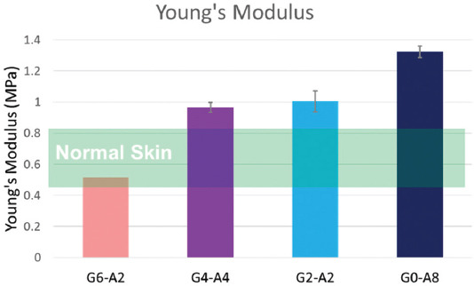
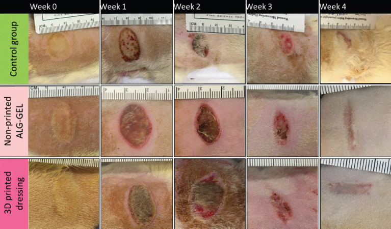
Burn wound treatment is still a clinical challenge due to the severity of tissue damage and dehydration. Among various wound dressings, hydrogel materials have gained significant attention for burn wound treatment in clinical practice due to their soothing and moisturizing activity. In this study, 3D-printed dressings were fabricated using clinically relevant hydrogels for deep partial-thickness burn (PTB) wounds. Different ratios of gelatin and alginate mixture were 3D-printed and examined in terms of rheological behavior, shear thinning behavior, mechanical properties, degradation rate, and hydration activity to tune the hydrogel composition for best functionality. The cell-laden dressings were bioprinted to evaluate the effect of the gelatin: alginate ratio on the proliferation and growth of human dermal fibroblasts. The present findings confirm that the higher alginate content is associated with higher viscosity and Young's modulus, while higher gelatin content is associated with faster degradation and higher cell viability. Together, the 3D-printed dressing with 75% gelatin and 25% alginate showed the best tradeoff between mechanical properties, hydration activity, and in vitro biological response. Findings from in vivo test using the most effective dressing showed the positive effect of 3D-printed porous pattern on wound healing, including faster wound closure, regenerated hair follicles, and non-traumatic dressing removal compared to the non-printed hydrogel with the same composition and the standard of care. Results from this research showed that 3D-printed dressings with an adequate gelatin: alginate ratio enhanced wound healing activity for up to 7 days of moisture retention on deep PTB wounds.

Keywords: 3D printing; Advanced dressings; Alginate; Burn wound; Gelatin; Moist wound healing.


Conflict of interest statement

The authors declare that they have no competing interests.

Figures

Figure 1
Figure 1
Schematic of the structure of sodium alginate, gelatin, and gelatin-alginate blend in 3D-printed cell-laden dressings. Gelatin and alginate are semi-interpenetrating networks (semi-IPN), whereby the linear chains of alginate are embedded within the gelatin network, which decreases the free volume[39].
Figure 2
Figure 2
Fabrication of 3D-printed wound dressings with alginate, gelatin, and human dermal fibroblasts for partial-thickness burn wounds followed by 10 min crosslinking with calcium chloride solution.
Figure 3
Figure 3
Animal test for the evaluation of deep partial-thickness burn wound healing using a rat model in three groups. Burn wounds covered with (A) petrolatum gauze, (B) non-printed hydrogel, and (C) 3D-printed hydrogel dressings.
Figure 4
Figure 4
(A) Complex shear moduli (G’ and G”) of the non-printed hydrogels. Alginate content is associated with higher G’ and G”, (B) shear thinning behavior of the non-printed hydrogels. Plain alginate and plain gelatin showed the lowest shear-thinning behavior, while G6-A2 exhibited the greatest shear-thinning behavior followed by G4-A4 and G2-A2.
Figure 5
Figure 5
Photographs of the 3D-printed dressings. G4-A4 and G6-A2 dressings showed the finest mesh structure and best shape fidelity. The G8-A0 showed poor printability and inconsistent pore shape fidelity, while G2-A6 and G0-A8 samples were too viscous and difficult to extrude with irregular pore shape and size.
Figure 6
Figure 6
Young’s modulus of 3D-printed dressings (n = 3). Mechanical stiffness is increased by alginate content; however, only G6-A2 samples are in the same range as normal skin. The Young’s Modulus of the normal skin is adopted from[41]. Wound dressings need to have the stiffness matched with normal skin to support body movement, non-adhesive coverage, and persistence on the wound site. The mechanical properties of the plain gelatin dressing are not measurable.
Figure 7
Figure 7
Fourier-transform infrared spectroscopy spectra of hydrogel samples. The characteristic IR bands associated with gelatin, sodium alginate, and water are shown by red arrows, blue arrows, and light blue boxes, respectively.
Figure 8
Figure 8
(A) Sodium alginate has linear chains composed of mannuronic acid and guluronic acid with carboxylate groups and hydroxyl groups. During cross-linking calcium ions replace the sodium ions in the guluronic acid monomers, resulting in intermolecular bonds between calcium ions and alginate chains that forms a linear and packed egg-box structure. (B) Gelatin is a bioactive derivative of collagen composed of amide groups with relatively high free volume and low viscosity.
Figure 9
Figure 9
(A) Swelling capacity and degradation of the 3D-printed dressings (n = 3). (B) One-week degradation rate of 3D-printed dressings (n = 3). Samples with higher gelatin content showed a faster degradation rate and higher swelling capacity. All samples could stay in PBS for at least 7 days (168 h). The G6-A2 dressings with the highest gelatin content showed significantly faster degradation (P < 0.05), which means that the permeability of this sample is higher than the other samples. (C) Total water content and hydration activity of the gelatin-alginate 3D-printed dressing on a super-absorbent surface to simulate dry burn wound surface (n = 3, P < 0.05). The higher permeability in this sample justifies its faster degradation and higher water donation.
Figure 10
Figure 10
Indirect in vitro biological evaluation of the 3D-printed dressings. MTT assay results showed that cell viability and proliferation increased significantly by increasing the gelatin content. G6-A2 samples showed higher cell viability than the control group on day 7. (n = 3; *, **, ***, and NS denote P < 0.05, P < 0.01, P <0.001, and non-significant difference, respectively).
Figure 11
Figure 11
Direct in vitro biological evaluation of the 3D-printed dressings. (A) Live/Dead confocal images of cell-laden dressings after 3 days of culture. Living cells are depicted in green, while dead cells are depicted in red. (B) Cell-laden dressing cultured in Dulbecco’s modified eagles medium for further Live/Dead assay. (C) Quantitative representation of cell viability based on the Live/Dead confocal images. Samples with higher gelatin content showed higher cell survival than those with higher alginate content. The G6-A2 dressings showed no significant difference with the control sample in terms of cell survival, while significantly higher cell survival compared to other samples. (n = 3, * and NS denote P < 0.05 and non-significant difference, respectively).
Figure 12
Figure 12
Wound healing analysis over 4 weeks (n = 6). Gross examination of wound healing. Wound images from control and treatment groups. 3D-printed dressings showed less necrotic tissue and smoother wound margins.
Figure 13
Figure 13
3D-printed dressings and non-printed G6-A2 dressings with 75% gelatin and 25% alginate showed significantly higher wound closure (i.e., smaller wound size) than the control sample, while the printed dressing showed slightly higher wound closure than the non-printed dressing. (n = 3; *, **, and *** denote P < 0.05, P < 0.01, and P < 0.001, respectively).
Figure 14
Figure 14
Representative H&E-stained slides for (A) control group: burn wound covered with petrolatum gauze, (B) wound covered with non-printed amorphous hydrogel composed of 75% gelatin and 25% alginate (G6-A2), (C) wounds covered with 3D-printed G6-A2. Pop-outs are regions of interest for further magnification. Guide: hyperkeratosis (black arrowhead), epidermal regeneration (dark purple outmost layer), dermal layer (white arrowhead), granulation tissue (red arrow), hair follicle (green arrowhead), and sweat glands (yellow arrowhead) labeled in the images.
Figure 15
Figure 15
Gross histology results based on the H&E grading scores (Table 3) regarding epidermal regeneration, dermal regeneration, and granulation tissue formation. The control group showed insufficient epidermal and dermal regeneration with the thickest granulation layer as an indicator of immature tissue treatment, while the 3D-printed dressing showed significantly higher regeneration of hair follicles. (n = 6; * and ** denote P < 0.05, P < 0.01, and P < 0.001, respectively).

Similar articles

Cited by

References

    1. Kowal S, Kruger E, Bilir P, et al. Cost-effectiveness of the Use of Autologous Cell Harvesting Device Compared to Standard of Care for Treatment of Severe Burns in the United States. Adv Ther. 2019;36:1715–29. https://doi.org/10.1007/s12325-019-00961-2. - PMC - PubMed
    1. James SL, Lucchesi LR, Bisignano C, et al. Epidemiology of Injuries from Fire, Heat and Hot Substances:Global, Regional and National Morbidity and Mortality Estimates from the Global Burden of Disease 2017 Study. Inj Prev. 2020;26:i36–45. https://doi.org/10.1136/injuryprev-2019-043299. - PMC - PubMed
    1. Price K, Lee KC, Woolley KE, et al. Burn Injury Prevention in Low-and Middle-Income Countries:Scoping Systematic Review. Burns Trauma. 2021;9:tkab037. https://doi.org/10.1093/burnst/tkab037. - PMC - PubMed
    1. Zavlin D, Chegireddy V, Boukovalas S, et al. Multi-institutional Analysis of Independent Predictors for Burn Mortality in the United States. Burns Trauma. 2018;6:24. https://doi.org/10.1186/s41038-018-0127-y. - PMC - PubMed
    1. Yu TC, Zhang X, Smiell J, et al. Healthcare Resource Utilization, Treatment Patterns, and Cost of Care Among Patients with Thermal Burns and Inpatient Autografting in Two Large Privately Insured Populations in the United States. Burns. 2020;46:825–35. https://doi.org/10.1016/j.burns.2019.10.019. - PubMed

LinkOut - more resources