

Thermo-sensitive Sacrificial Microsphere-based Bioink for Centimeter-scale Tissue with Angiogenesis
- PMID: 36404788
- PMCID: PMC9668484
- DOI: 10.18063/ijb.v8i4.599
Thermo-sensitive Sacrificial Microsphere-based Bioink for Centimeter-scale Tissue with Angiogenesis
Abstract
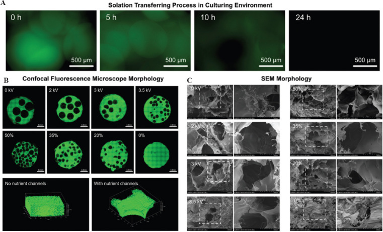
Centimeter-scale tissue with angiogenesis has become more and more significant in organ regeneration and drug screening. However, traditional bioink has obvious limitations such as balance of nutrient supporting, printability, and vascularization. Here, with "secondary bioprinting" of printed microspheres, an innovative bioink system was proposed, in which the thermo-crosslinked sacrificial gelatin microspheres encapsulating human umbilical vein endothelial cells (HUVECs) printed by electrospraying serve as auxiliary component while gelatin methacryloyl precursor solution mixed with subject cells serve as subject component. Benefiting from the reversible thermo-crosslinking feature, gelatin microspheres would experience solid-liquid conversion during 37°C culturing and form controllable porous nutrient network for promoting the nutrient/oxygen delivery in large-scale tissue and accelerate the functionalization of the encapsulated cells. Meanwhile, the encapsulated HUVECs would be released and attach to the pore boundary, which would further form three-dimensional vessel network inside the tissue with suitable inducing conditions. As an example, vascularized breast tumor tissue over 1 cm was successfully built and the HUVECs showed obvious sprout inside, which indicate the great potential of this bioink system in various biomedical applications.
Keywords: Angiogenesis; Bioprinting; Gelatin methacryloyl; Large-scale tissue; Microsphere; Thermo-sensitive material.
Copyright: © 2022 Xie et al.
Conflict of interest statement
All authors declare no financial/commercial conflicts of interest.
Figures


Similar articles
-
Reversible physical crosslinking strategy with optimal temperature for 3D bioprinting of human chondrocyte-laden gelatin methacryloyl bioink.J Biomater Appl. 2018 Nov;33(5):609-618. doi: 10.1177/0885328218805864. Epub 2018 Oct 25. J Biomater Appl. 2018. PMID: 30360677
-
Triaxial bioprinting large-size vascularized constructs with nutrient channels.Biomed Mater. 2023 Aug 30;18(5). doi: 10.1088/1748-605X/acf25a. Biomed Mater. 2023. PMID: 37604152
-
Synchronous 3D Bioprinting of Large-Scale Cell-Laden Constructs with Nutrient Networks.Adv Healthc Mater. 2020 Aug;9(15):e1901142. doi: 10.1002/adhm.201901142. Epub 2019 Dec 17. Adv Healthc Mater. 2020. PMID: 31846229
-
Platelet lysate functionalized gelatin methacrylate microspheres for improving angiogenesis in endodontic regeneration.Acta Biomater. 2021 Dec;136:441-455. doi: 10.1016/j.actbio.2021.09.024. Epub 2021 Sep 20. Acta Biomater. 2021. PMID: 34551330
-
A review on alginate-based bioinks, combination with other natural biomaterials and characteristics.J Biomater Appl. 2022 Aug;37(2):355-372. doi: 10.1177/08853282221085690. Epub 2022 May 5. J Biomater Appl. 2022. PMID: 35510845 Review.
Cited by
-
3D printing microporous scaffolds from modular bioinks containing sacrificial, cell-encapsulating microgels.Biomater Sci. 2023 Nov 21;11(23):7598-7615. doi: 10.1039/d3bm00721a. Biomater Sci. 2023. PMID: 37824082 Free PMC article.
-
Injectable Polyhydroxyalkanoate-Nano-Clay Microcarriers Loaded with r-BMSCs Enhance the Repair of Cranial Defects in Rats.Int J Nanomedicine. 2024 Dec 24;19:13839-13855. doi: 10.2147/IJN.S498950. eCollection 2024. Int J Nanomedicine. 2024. PMID: 39735323 Free PMC article.
-
The sculpting tool in bioprinting: research and application progress of sacrificial inks.Front Bioeng Biotechnol. 2025 Jun 25;13:1486459. doi: 10.3389/fbioe.2025.1486459. eCollection 2025. Front Bioeng Biotechnol. 2025. PMID: 40635697 Free PMC article. Review.
-
Light-based 3D bioprinting techniques for illuminating the advances of vascular tissue engineering.Mater Today Bio. 2024 Oct 2;29:101286. doi: 10.1016/j.mtbio.2024.101286. eCollection 2024 Dec. Mater Today Bio. 2024. PMID: 39435375 Free PMC article. Review.
-
Application of three-dimensional (3D) bioprinting in anti-cancer therapy.Heliyon. 2023 Sep 28;9(10):e20475. doi: 10.1016/j.heliyon.2023.e20475. eCollection 2023 Oct. Heliyon. 2023. PMID: 37800075 Free PMC article. Review.
References
-
- He Y, Gu Z, Xie M, et al. Why Choose 3D Bioprinting?Part II:Methods and Bioprinters. BioDesign Manuf. 2020;3:1–4. https://doi.org/10.1007/s42242-020-00064-w.
-
- Thakor J, Ahadian S, Niakan A, et al. Engineered Hydrogels for Brain Tumor Culture and Therapy. BioDesign Manuf. 2020;3:203–26. https://doi.org/10.1007/s42242-020-00084-6. - PMC - PubMed
-
- Lee M, Bae K, Guillon P, et al. Exploitation of Cationic Silica Nanoparticles for Bioprinting of Large-Scale Constructs with High Printing Fidelity. ACS Appl Mater Interfaces. 2018;10:37820–8. https://doi.org/10.1021/acsami.8b13166. - PubMed
-
- Ying GL, Jiang N, Maharjan S, et al. Aqueous Two-Phase Emulsion Bioink-Enabled 3D Bioprinting of Porous Hydrogels. Adv Mater. 2018;30:1805460. https://doi.org/10.1002/adma.201805460. - PMC - PubMed
-
- Shao L, Gao Q, Xie C, et al. Sacrificial Microgel-laden Bioink-enabled 3D Bioprinting of Mesoscale Pore Networks. BioDesign Manuf. 2020;3:30–9. https://doi.org/10.1007/s42242-020-00062-y.
LinkOut - more resources
Full Text Sources
Research Materials