. 2022 Sep 15;139(35):e52846.
doi: 10.1002/app.52846. Epub 2022 Jul 22.

Delivery of Bioactive Albumin from Multi-Functional Polyampholyte Hydrogels

Affiliations

Delivery of Bioactive Albumin from Multi-Functional Polyampholyte Hydrogels

Stephanie L Haag et al. J Appl Polym Sci. .

Abstract

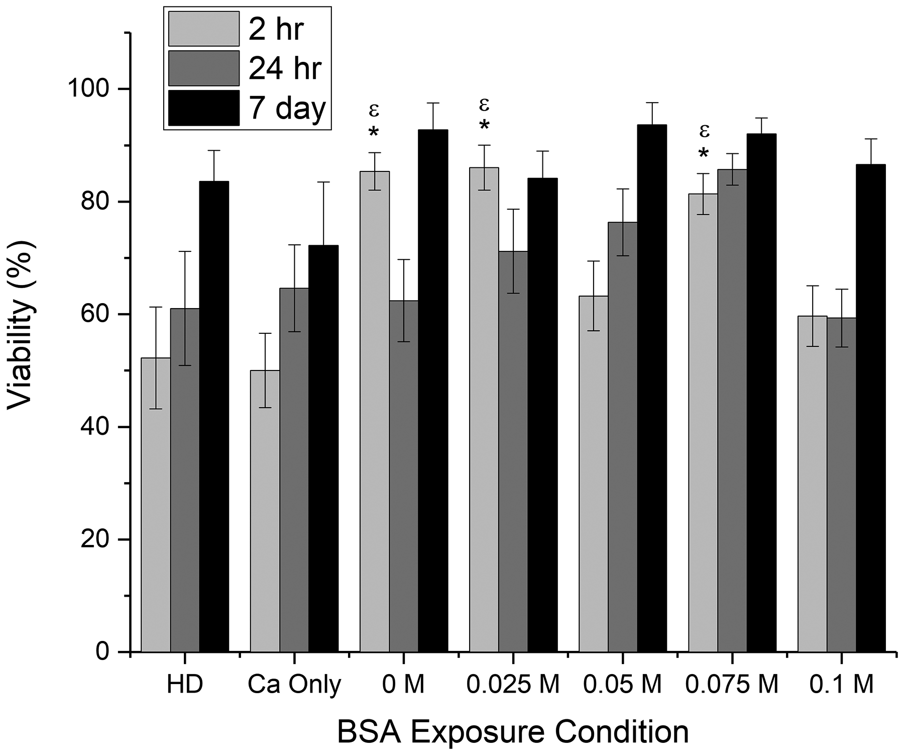
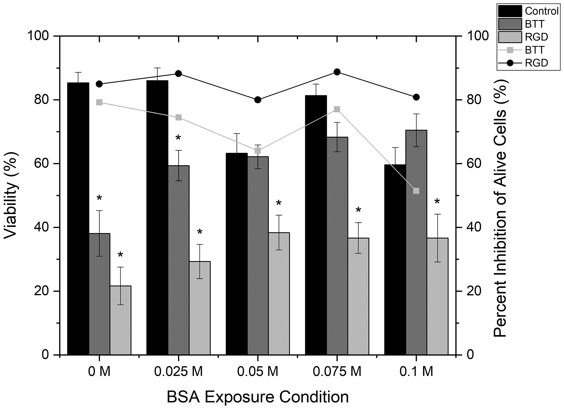
Tissue engineered scaffolds are currently being explored to aid in healing and regeneration of non-union fractures in bone. Additionally, albumin has been demonstrated to provide benefits to healing when applied to injury sites. This paper focuses on delivery of calcium modified, bioactive bovine serum albumin (BSA) from a multi-functional polyampholyte polymer scaffold. First, the inherent nonfouling and conjugation properties of the polyampholyte hydrogel were verified to determine the impact of calcium exposure. The polyampholyte hydrogel delivery platform was then assessed with calcium titrations and osteoblast-like cell (MC3T3-E1) adhesion, proliferation, and viability evaluations. Finally, integrin inhibitors were used to identify the binding mechanisms that mediate cell adhesion to the calcium-modified BSA-conjugated hydrogels. An increase in cell adhesion was observed following calcium exposure up to 0.075 M, although this and higher calcium concentrations affected hydrogel stability and cell growth. BSA exposed to 0.05 M calcium and delivered from polyampholyte hydrogels promoted the most promising viable cell adhesion over 7 days. Cell adhesion to the calcium-modified BSA-conjugated hydrogels appeared to be regulated by arginine-glycine-aspartic acid (RGD) and collagen specific integrins. These results demonstrate that the delivery of calcium modified BSA from an implantable polymer scaffold is promising for bone tissue engineering applications.


Conflict of interest statement

Conflicts of Interest The authors have no conflicts to declare.

Figures

Figure 1:
Figure 1:
Representative fluorescent microscopy images of 1 mg/mL FITC BSA adsorbed to TMA:CAA hydrogels following exposure to varying calcium concentrations. In each image, the left-hand side is a 0 M control while the right-hand side is a calcium exposed sample. The white vertical line is provided to distinguish the sample interface, and the scale bar represents 200 μm and is representative for all images.
Figure 2:
Figure 2:
Representative fluorescent microscopy images of 1 mg/mL FITC BSA conjugated to TMA:CAA hydrogels following BSA exposure to varying calcium concentrations. In each image, the left-hand side is a 0 M control while the right-hand side is a calcium exposed sample. The top row shows absolute fluorescence, while the bottom row is normalized to the darkest gel to highlight relative differences. The white line is provided to distinguish the sample interface, and the scale bar represents 200 μm and is representative for all images.
Figure 3:
Figure 3:
Mean ± SEM of calcium ion release from TMA:CAA hydrogels immediately following the conjugation procedure (burst) and after 2 hours of soaking (extended). A * represents a statistical difference between the indicated condition and all other conditions at the given time point (burst, extended, total). A # represents a statistically significant difference between indicated groups. Statistics are calculated at a 95% confidence level (p<0.05).
Figure 4:
Figure 4:
Representative confocal microscopy images of MC3T3-E1 cells after 2 hours of adhesion, and 24 hours or 7 days of culture on TMA:CAA hydrogels with conjugated HD BSA (negative control), BSA exposed to 0 M, 0.025 M, 0.05 M, 0.075 M, and 0.1 M calcium, or no protein (0.075 M Ca alone, as a negative control). The cells are stained with a live-dead stain, staining alive cells green and dead cells red. The scale bar is 100 μm and is representative for all images.
Figure 5:
Figure 5:
Mean ± SEM for (a) cell adhesion and (b) viability after 2 hours of adhesion, and 24 hours or 7 days of proliferation. A ε represents a statistically significant difference from the Ca Only control at the given time point. A * represents a statistically significant difference from the HD control at the given time point. A α represents a statistically significant difference from the indicated sample and all other groups except 0.075 M at the given time point. A β represents a statistically significant difference from the indicated sample and all other groups except 0.05 M at the given time point. A π represents a statistically significant difference from the indicated sample and all other groups at the given time point. Statistics are calculated at a 95% confidence level (p<0.05).
Figure 5:
Figure 5:
Mean ± SEM for (a) cell adhesion and (b) viability after 2 hours of adhesion, and 24 hours or 7 days of proliferation. A ε represents a statistically significant difference from the Ca Only control at the given time point. A * represents a statistically significant difference from the HD control at the given time point. A α represents a statistically significant difference from the indicated sample and all other groups except 0.075 M at the given time point. A β represents a statistically significant difference from the indicated sample and all other groups except 0.05 M at the given time point. A π represents a statistically significant difference from the indicated sample and all other groups at the given time point. Statistics are calculated at a 95% confidence level (p<0.05).
Figure 6:
Figure 6:
Mean ± SEM of calcium ion release from TMA:CAA hydrogels over 7 days. All groups are statistically significant from each other except those identified as n.s. (not statistical). Statistics are calculated at a 95% confidence level (p<0.05).
Figure 7:
Figure 7:
Mean ± SEM of MC3T3-E1 cell (a) adhesion to and (b) viability on calcium modified BSA conjugated to TMA:CAA hydrogels in the presence of either GRGDSP or BTT small molecule inhibitors. The data for cell adhesion in the absence of inhibitor molecules is replicated from Figure 5 to facilitate easier comparisons. The percent inhibition (lines) is shown based on the (a) total cell population and (b) viable cell population. The SEM for the percent inhibition is directly proportional to the cell adhesion SEM so it is not replicated for clarity. A * indicates a statistically significant difference between the indicated condition and the corresponding no inhibitor control at a 95% confidence level (p<0.05).
Figure 7:
Figure 7:
Mean ± SEM of MC3T3-E1 cell (a) adhesion to and (b) viability on calcium modified BSA conjugated to TMA:CAA hydrogels in the presence of either GRGDSP or BTT small molecule inhibitors. The data for cell adhesion in the absence of inhibitor molecules is replicated from Figure 5 to facilitate easier comparisons. The percent inhibition (lines) is shown based on the (a) total cell population and (b) viable cell population. The SEM for the percent inhibition is directly proportional to the cell adhesion SEM so it is not replicated for clarity. A * indicates a statistically significant difference between the indicated condition and the corresponding no inhibitor control at a 95% confidence level (p<0.05).

Similar articles

Cited by

References

    1. Hoerth RM, Seidt BM, Shah M, Schwarz C, Willie BM, Duda GN, Fratzl P, Wagermaier W, Mechanical and structural properties of bone in non-critical and critical healing in rat, Acta Biomater. 10(9) (2014) 4009–4019. - PubMed
    1. Chocholata P, Kulda V, Babuska V, Fabrication of Scaffolds for Bone-Tissue Regeneration, Materials (Basel) 12(4) (2019) 568. - PMC - PubMed
    1. Qu H, Fu H, Han Z, Sun Y, Biomaterials for bone tissue engineering scaffolds: a review, RSC Advances 9(45) (2019) 26252–26262. - PMC - PubMed
    1. Anderson JM, Rodriguez A, Chang DT, Foreign body reaction to biomaterials, Semin. Immunol 20(2) (2008) 86–100. - PMC - PubMed
    1. Ratner BD, Healing with medical implants: The body battles back, Sci. Transl. Med 7(272) (2015) 3. - PubMed

LinkOut - more resources