. 2022 Nov 4;25(12):105503.
doi: 10.1016/j.isci.2022.105503. eCollection 2022 Dec 22.

Biofabrication of synthetic human liver tissue with advanced programmable functions

Affiliations

Biofabrication of synthetic human liver tissue with advanced programmable functions

Rodrigo M Florentino et al. iScience. .

Abstract

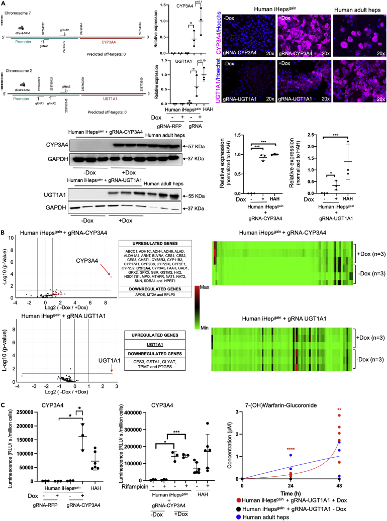
Advances in cellular engineering, as well as gene, and cell therapy, may be used to produce human tissues with programmable genetically enhanced functions designed to model and/or treat specific diseases. Fabrication of synthetic human liver tissue with these programmable functions has not been described. By generating human iPSCs with target gene expression controlled by a guide RNA-directed CRISPR-Cas9 synergistic-activation-mediator, we produced synthetic human liver tissues with programmable functions. Such iPSCs were guide-RNA-treated to enhance expression of the clinically relevant CYP3A4 and UGT1A1 genes, and after hepatocyte-directed differentiation, cells demonstrated enhanced functions compared to those found in primary human hepatocytes. We then generated human liver tissue with these synthetic human iPSC-derived hepatocytes (iHeps) and other non-parenchymal cells demonstrating advanced programmable functions. Fabrication of synthetic human liver tissue with modifiable functional genetic programs may be a useful tool for drug discovery, investigating biology, and potentially creating bioengineered organs with specialized functions.

Keywords: Bioengineering; Biological sciences; Tissue engineering.


Conflict of interest statement

A.C.-H., K.T., I.J.F., and A.S.-G. are inventors on a provisional international patent application that describes hepatic differentiation of human pluripotent stem cells and liver repopulation (PCT/US2018/018032) and a provisional international patent application that describes the use of human-induced pluripotent stem cells for highly genetic engineering (PCT/US2017/044719). A.S.-G., J.G.-L., A.C.-H., and I.J.F. are co-founders and have a financial interest in Von Baer Wolff, Inc. a company focused on biofabrication of autologous human hepatocytes from stem cells technology. I.J.F. and A.S.-G. are co-founders and have a financial interest in Pittsburgh ReLiver Inc, a company focused on programming liver failure and their interests are managed by the Conflict-of-Interest Office at the University of Pittsburgh in accordance with their policies.

Figures

None
Graphical abstract
Figure 1
Figure 1
Generation of custom-engineered hiPSCs-dCas9gain (A) Schematic diagram of the lentiviral vectors: pcLVi(3G)-SAM containing a TetOn system comprising of a tetracycline response element (pTRE-3g), a reverse tetracycline-controlled reverse transactivator (rtTA-3G), green fluorescent protein (GFP), a puromycin antibiotic selection cassette gene (PuroR), and the synergistic activation mediator (SAM). pcLVi(3G)-dCas9-VP64 containing a tetracycline response element (pTRE-3g), a neomycin antibiotic selection cassette gene (NeoR), and the deactivated CRISPR/Cas9 transcription activator VP64 (dCas9-VP64). Quantitative gene expression analysis of Cas9 and SAM normalized to ACTB in human fibroblasts transduced with lentiviral vector for dCas9-VP64 (HFs-dCas9gain) and non-transduced HF as control in the presence or absence of DOX (∗p = 0.0168, Welch’s t-test). Quantitative gene expression analysis of SAM normalized to ACTB transduced with lentiviral vector for SAM in HFs or HFs-dCas9gain (∗p = 0.0394, Welch’s t-test). In-live GFP-fluorescence of synergistic activation mediator in HFFs or HFFs-dCas9gain with and without DOX treatment. Data are represented as mean ± SD (B) Quantitative gene expression analysis of dCas9 and SAM normalized to ACTB in human iPSCs derived from fibroblasts transduced with lentiviral vector for dCas9-VP64 (Human iPSCs-dCas9gain) and non-transduced human iPSCs as control in the presence or absence of DOX (∗p < 0.0001, Welch’s t-test). Quantitative gene expression analysis of SAM normalized to ACTB transduced with lentiviral vector for SAM in Human iPSCs or Human iPSCs-dCas9gain (∗p < 0.05, Welch’s t-test). Immunofluorescence micrographs of dCas9 and GFP in Human iPSCs or Human iPSCs-dCas9gain with and without DOX treatment; HEK239-dCas9 was used as control. Western blot analysis of dCas9 and β-actin in human iPSCs and human iPSCs-Cas9gain in the presence or absence of DOX; HEK239-dCas9 was used as control. Also shown is the Quantification of GFP and Cas9 in human iPSCs, human iPSCs-Cas9gain, and HEK293-Cas9 in the presence or absence of DOX. Data are represented as mean ± SD (C) Immunofluorescence micrographs of pluripotency markers Nanog, Oct4, TRA-1-60, and SSEA-4 in human iPSCs-Cas9gain. Quantitative gene expression analysis of pluripotency markers c-myc, Lin28, Sox2, Nanog, and Oct3/4 normalized to ACTB of human iPSCs and human iPSCs-Cas9gain. (D) Human iPSCs-dCas9gain carries normal male karyotype in G-banding analysis. (E) Bright-field micrograph of human iPSCs-dCas9gain forming embryonic bodies after 20 days in culture. Immunofluorescence micrographs of the three germ layer markers: Ectoderm (Otx-2; SOX1), Mesoderm (HAND1; Brachuyry), and Endoderm (SOX17; GATA-4). (F) Genotyping results of human iPSCs-Cas9gain: PNPLA3 (rs738409) CT-heterozygous, MBOAT7 (rs641738) CC-major homozygous, and GCKR (rs780094) CT-heterozygous.
Figure 2
Figure 2
Characterization of hepatocyte-derived custom-engineered hiPSCs-Cas9gain (A) Schematic illustration of hiPSC-dCas9gain differentiation into hepatocytes like cells with the four main stages of differentiation by sequential addition of defined medium containing Activin A, BMP4, FGF2 (stage 1); Activin A (stage 2); dimethyl sulfoxide (DMSO), and hepatocyte growth factor (HGF) (stage 3); epidermal growth factor (EGF), dexamethasone (DEX), hydrocortisone (Hydro), free fatty acids (FFA), cholesterol (Chol), and bile acids (stage 4). (B) Immunofluorescence micrographs of hiPSC-dCas9gain for SOX17 show more than 80% of the cells positive for SOX17 after definitive endoderm induction stage 2. Immunofluorescence micrographs after hepatocyte differentiation stage 3 show protein expression of the adult isoform of HNF4a comparable to primary human hepatocytes (>80% respectively), expression of albumin comparable to primary human hepatocytes (>80% respectively), and no expression of α-fetoprotein (AFP); freshly isolated human fetal and adult hepatocytes were used as controls. Graphs showing quantification of cells expressing HNF4⍺, albumin, and alfa fetoprotein positive cells in iHepgain, human fetal hepatocytes, and human adult hepatocytes. (C) Quantitative gene expression analysis of undifferentiated hiPSC-dCas9gain (D0), Definitive endoderm (DE) (D4), Stage 3 of the hepatic differentiation (D14), Stage 4 of the hepatic differentiation (D18). qPCR is shown for genes encoding octamer-binding transcription factor 3/4 (OCT3/4), C-X-C motif chemokine receptor 4 (CXCR4), SRY-box 17 (Sox17), hepatocyte nuclear factor-4-alpha (HNF4a), hepatocyte nuclear factor-1-alpha (HNF1a), forkhead box A1 (FOXA1), forkhead box A2 (FOXA2), CCAAT enhancer-binding protein alpha (CEBPa), constitutive androstane receptor (CAR), liver X receptor (LXR), retinoid X receptor (RXR), peroxisome proliferator-activated receptor alpha (PPARa), Met, ATP binding cassette subfamily B member 11 (BSEP), ATP binding cassette subfamily C member 2 (MRP2), alpha-fetoprotein (AFP), UDP glucuronyltransferase family 1 member A1 (UGT1A1) and cytochrome P450 family 3 subfamily A member 4 (CYP3A4). Human adult hepatocytes and human fetal hepatocytes were included as controls. Data are represented as mean ± SD (D) Immunofluorescence micrographs of hiPSC-dCas9gain at stage 3 of hepatic differentiation (D14) stained for CYP3A4 and UGT1A1 compared to primary human adult and fetal hepatocytes. (E) Immunofluorescence micrographs for GFP and dCas9 of control human iPSCs and human iPSCs-Cas9gain the end of stage 2 (DE) and stage 3 (iHep) in the presence or absence of DOX. Quantification of GFP and Cas9 in human iPSCs and human iPSCs-Cas9gain at the end of stage 2 (DE) and stage 3 (iHep) in the presence or absence of DOX. (F) Quantitative gene expression analysis of dCas9 and synergistic activation mediator (SAM) normalized to ACTB at in hiPSCs and hiPSC-dCas9gain at different stages of hepatic differentiation: Definitive endoderm (DE), Stage 3 (iHeps) in the presence or absence of DOX (∗p < 0.05, Welch’s t test). Western Blot analysis of dCas9 in hiPSC and hiPSC-dCas9gain in the presence or absence of DOX at definitive endoderm (Stage 2) and in human iHepsgain (Stage 3). HEK-dCas9 was used as control.
Figure 3
Figure 3
Transcriptional activation of CYP3A4 and UGT1A1 in Custom-Engineered human iHep-dCas9gain (A) Schematic illustration of the gRNA binding site in the promoter region of CYP3A4 and UGT1A1 gene. No off-targets were predictable. Quantitative gene expression analysis of CYP3A4 and UGT1A1 in iHep-dCas9gain transduced with scrambled gRNA and gRNA-RFP or gRNA-CYP3A4 or gRNA-UGT1A1 in the presence and absence of DOX normalized to ACTB (∗p < 0.05, Welch’s t-test). Human adult hepatocytes (HAH) were used as control. Immunofluorescence micrographs CYP3A4 and UGT1A1 in iHep-dCas9gain under the presence or absence of DOX transduced with gRNA-CYP3A4 or gRNA-UGT1A1. Freshly isolated human hepatocytes were used as control. Western Blot analysis and quantification: on the left the relative expression of CYP3A4 (CYP3A4; ∗∗∗p < 0.0001, Ordinary one-way ANOVA with Turkey’s multiple comparisons) and UGT1A1 (UGT1A1; ∗p = 0.0427, unpaired t-test) in iHep-dCas9gain transduced with gRNA-CYP3A4 or UGT1A1 under the presence or absence of DOX. On the right, the expression of CYP3A4 and UGT1A1 in iHep-dCas9gain transduced with gRNA-CYP3A4 or UGT1A1 under the presence or absence of DOX normalized to human adult hepatocyte. The relative expression average of human adult hepatocytes was set up as 1. Data are represented as mean ± SD (B) Volcano plot and heatmap showing differential gene expression of iHep-dCas9gain transduced with gRNA-CYP3A4 under the presence or absence of DOX. Volcano plot and heatmap showing differential gene expression of iHep-dCas9gain transduced with gRNA-UGT1A1 under the presence or absence of DOX. (C) CYP3A4 activity of iHep-dCas9gain transduced with a scrambled gRNA and RFP or gRNA CYP3A4 in the presence or absence of DOX (∗p < 0.05, Welch’s t-test) and Rifampicin (∗p = 0.01, ∗∗∗p = 0.0008, Welch’s t-test). UGT1A1 activity assessed by 7(OH)Warfarin-Glucuronide production over time in iHep-dCas9gain transduced with gRNA-UGT1A1 in the presence or absence of DOX (∗∗∗∗p < 0.0001, ∗∗p = 0.0041, Welch’s t-test). Adult human hepatocytes were used as control. Data are represented as mean ± SD.
Figure 4
Figure 4
Bioengineering of hiPSC-derived dCas9gain tissue expressing CYP3A4 and UGT1A1 (A) Schematic representation of the cell types involved in the seeding process: Human iHepgain, human vascular endothelial cells, human fibroblasts, and human mesenchymal stem cells. (B) Schematic representation of Haemobrick production via a stereolithographic bioprinting process. The platform is submerged into a photosensitive bioink leaving a gap of defined layer thickness between itself and the transparent screen. A photomask is projected through the transparent screen onto the platform curing the bioink to a hydrogel layer (step 01). The size of the gap increases by raising the platform. The photomask of the next layer is projected onto the previous one accordingly (step 02). This process is repeated stacking hydrogel layers onto each other until the complete object has been created sequentially. Cells can be cultured in the culture area after application through the seeding socket. Immunofluorescence micrograph of human synthetic livergain tissue showing the human vascular endothelial cells (CD31 positive cells), human mesenchymal stem cells (CD44 positive cells), and human fibroblast (⍺SMA positive cells). (C) Immunofluorescence micrographs of human synthetic livergain tissue showing expression of GFP (Synergistic Activation Mediator) in the presence or absence of DOX. Moreover, also shown are Immunofluorescence micrographs of human synthetic livergain tissue showing expression of red fluorescent protein (RFP) demonstrating that high transduction of lentivirus containing gRNAs was achieved in human synthetic livergain tissue exposed to DOX and in the control group non-DOX treated. (D) Immunofluorescence micrographs of human synthetic livergain tissue for dCas9 and CYP3A4 transduced with gRNA-CYP3A4 in the presence or absence of DOX. CYP3A4 activity of human synthetic livergain tissue transduced with gRNA-CYP3A4 in the presence or absence of DOX (∗∗∗p < 0.0005, Welch’s-test). Data are represented as mean ± SD. (E) Immunofluorescence micrographs of human synthetic livergain tissue for UGT1A1 transduced with gRNA-UGT1A1 in the presence or absence of DOX.

Similar articles

Cited by

References

    1. Habka D., Mann D., Landes R., Soto-Gutierrez A. Future economics of liver transplantation: a 20-year cost modeling forecast and the prospect of bioengineering autologous liver grafts. PLoS One. 2015;10:e0131764. doi: 10.1371/journal.pone.0131764. - DOI - PMC - PubMed
    1. Kwong A.J., Kim W.R., Lake J.R., Smith J.M., Schladt D.P., Skeans M.A., Noreen S.M., Foutz J., Booker S.E., Cafarella M., et al. OPTN/SRTR 2019 annual data report: liver. Am. J. Transplant. 2021;21:208–315. doi: 10.1111/ajt.16494. - DOI - PubMed
    1. Haep N., Florentino R.M., Squires J.E., Bell A., Soto-Gutierrez A. The inside-out of end-stage liver disease: hepatocytes are the keystone. Semin. Liver Dis. 2021;41:213–224. doi: 10.1055/s-0041-1725023. - DOI - PMC - PubMed
    1. Hartog H., Hann A., Perera M.T.P.R. Primary non-function of the liver allograft. Transplantation. 2022;106:117–128. doi: 10.1097/TP.0000000000003682. - DOI - PubMed
    1. Ammori J.B., Pelletier S.J., Lynch R., Cohn J., Ads Y., Campbell D.A., Englesbe M.J. Incremental costs of post-liver transplantation complications. J. Am. Coll. Surg. 2008;206:89–95. doi: 10.1016/j.jamcollsurg.2007.06.292. - DOI - PubMed