. 2022 Nov 4:28:e20220002.
doi: 10.1590/1678-9199-JVATITD-2022-0002. eCollection 2022.

New multienzymatic complex formed between human cathepsin D and snake venom phospholipase A2

Affiliations

New multienzymatic complex formed between human cathepsin D and snake venom phospholipase A2

Jeane do Nascimento Moraes et al. J Venom Anim Toxins Incl Trop Dis. .

Abstract

Background: Cathepsin D (CatD) is a lysosomal proteolytic enzyme expressed in almost all tissues and organs. This protease is a multifunctional enzyme responsible for essential biological processes such as cell cycle regulation, differentiation, migration, tissue remodeling, neuronal growth, ovulation, and apoptosis. The overexpression and hypersecretion of CatD have been correlated with cancer aggressiveness and tumor progression, stimulating cancer cell proliferation, fibroblast growth, and angiogenesis. In addition, some studies report its participation in neurodegenerative diseases and inflammatory processes. In this regard, the search for new inhibitors from natural products could be an alternative against the harmful effects of this enzyme.

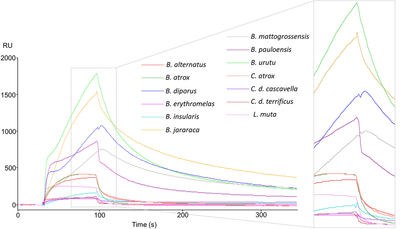
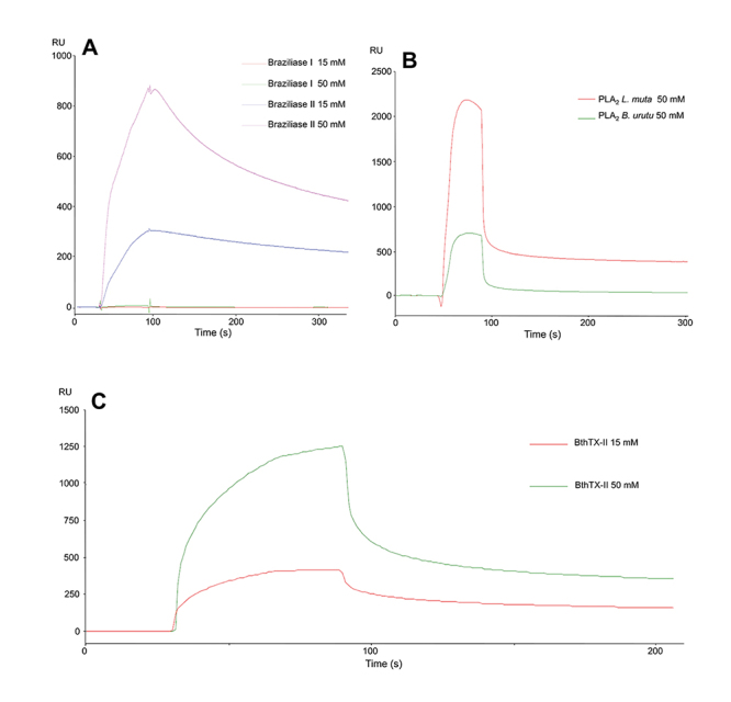
Methods: An investigation was carried out to analyze CatD interaction with snake venom toxins in an attempt to find inhibitory molecules. Interestingly, human CatD shows the ability to bind strongly to snake venom phospholipases A2 (svPLA2), forming a stable muti-enzymatic complex that maintains the catalytic activity of both CatD and PLA2. In addition, this complex remains active even under exposure to the specific inhibitor pepstatin A. Furthermore, the complex formation between CatD and svPLA2 was evidenced by surface plasmon resonance (SPR), two-dimensional electrophoresis, enzymatic assays, and extensive molecular docking and dynamics techniques.

Conclusion: The present study suggests the versatility of human CatD and svPLA2, showing that these enzymes can form a fully functional new enzymatic complex.

Keywords: Cathepsin D; Enzyme complex; Phospholipases A2; Snake venom.


Conflict of interest statement

Competing interests: The authors declare that they have no competing interests.

Figures

Figure 1.
Figure 1.. SPR assays between snake venoms and human cathepsin D (CatD). Sensorgrams were generated in a screening round of snake venoms against CatD. All interactions are plotted according to the response upon binding in RU (resonance units).
Figure 2.
Figure 2.. Chromatographic profile of B. jararaca snake venom, SDS-PAGE, and binding assays of the isolated fractions. (A) The chromatographic profile demonstrates fractionation on a CM-Sepharose column previously equilibrated with 50 mM AMBIC, pH 8.0, and fractions eluted with a 0-100% gradient of 500 mM AMBIC, pH 8.0, at a constant flow rate of 1 mL/min, monitored at 215 (red) and 280 nm (blue). The twelve fractions collected were numbered from 1 to 12, the fractions of interest 10, 11, and 12 being indicated with asterisks (*). SDS-PAGE of the 12 fractions from B. jararaca venom. (B) MM: molecular mass, V: crude venom, and eight fractions named F1 to F8. (C) MM: molecular mass, and fractions from F9 to F12. (D) Fraction interaction responses: 10 (blue), 11 (green) and 12 (red) with responses of 125, 10 and 12 RUs, respectively.
Figure 3.
Figure 3.. Binding assays between CatD and snake venom PLA2s. (A) Interactions of CatD with Braziliase-I and Braziliase-II (concentrations of 15 and 50 µM). (B) Responses were obtained from the interaction between CatD and svPLA2s from Bothrops neuwiedi urutu (BnuTX-I) and Lachesis muta (LmutTX). (C) Interaction test between CatD and BthTX-II (concentrations of 15 and 50 mM). The analyzed samples were submitted to salt removal in a 5 mL Hitrap desalting (GE) column.
Figure 4.
Figure 4.. Two-dimensional SDS-PAGE: CatD, BthTX-II, and enzymatic complex. (A) Two-dimensional SDS-PAGE of CatD showing a pI of 4.74 and approximate molecular mass of 35 kDa. (B) Two-dimensional SDS-PAGE of BthTX-ll with a pI of 8.74, with an approximate molecular mass of 14 kDa. (C) Two-dimensional SDS-PAGE of the complex with a pI of 5.79, and approximate molecular mass of 49 kDa.
Figure 5.
Figure 5.. Proteolytic activity of CatD and the CatD + BthTX-II complex. The evaluation was performed at pHs 3, 4, 5, 6, and 7, identified in the figure legend, highlighting CatD in white (positive control) and the CatD + BthTX-II complex in black. As a negative control, the buffer itself (sodium citrate) was used at different pHs. The toxin used in the tests (BthTX-II) was submitted to contamination analysis (described in the second section). Two-way analysis of variance (ANOVA) with Tukey’s multiple comparison post-test with significance level p < 0.05.
Figure 6.
Figure 6.. Proteolytic activity on casein. (A) Samples identified in the legend results: CatD, CatD + BthTX-II, CatD + BthTX-II + pepstatin A were considered negative controls; buffer (sodium citrate) was used at different pHs. (B) Proteolytic activities SDS-PAGE. Samples: (1) CatD; (2) pepstatin A (PepA); (3) casein; (4) CatD + PepA + casein; (5) CatD + casein; (6) CatD + BthTX-II Casein + PepA; (7) CatD + BthTX-II + casein (30 min); (8) CatD + BthTX-II + casein (15 min); (9) CatD + BthTX-II + casein (5 min). Samples 3 through 6 were incubated for 30 min at 27 ºC. Positive control: CatD; negative control: pepstatin A (PepA).
Figure 7.
Figure 7.. PLA2 enzymatic activity on artificial substrate 4N3OBA. Samples: (1) BthTX-II; (2) CatD + BthTX-II; (3) CatD + BthTX-II + PepA; (4) BthTX-I; (5) CatD; (6) PepA. Positive control: BthTX-II. Negative control: BthTX-I.
Figure 8.
Figure 8.. Molecular modeling of the interaction between three snake venom PLA2s (LmuTX, Braziliase-II and BthTX-II) and human CatD using different docking tools (HDOCK, pyDOCK, GRAM-X, ClusPro, and ZDOCK). The CatD surface is represented in dark gray (light chain) and light gray (heavy chain). The svPLA2s are colored according to the docking tool used.
Figure 9.
Figure 9.. Molecular modeling of the CatD + BthTX-II complex. The complex formed between human CatD is shown in gray (light chain in dark gray and heavy chain in light gray) and BthTX-II is shown in orange. The interactions were enlarged to show amino acid residues in the interface and their interactions. H-bonds are highlighted by green dashed lines, and hydrophobic interactions are depicted as protrusions colored to match each amino acid residue.
Figure 10.
Figure 10.. CatD + BthTX-II complex molecular dynamics. CatD is shown in gray (light chain in dark gray and heavy chain in light gray) and BthTX-II is shown in orange. The radius of gyration and backbone RMSD graphics are located on the left side of the figure. All five replicas are plotted in an overlapped manner in order to highlight all minor variations and overall stability throughout the 100 ns of each replica. The structures on the right end of the figure show a superposition respective to each of the three CatD + BthTX-II complexes representing the most predominant conformations during the total 500 ns simulated. These superposed complexes are the central structures extracted from the three most populated clusters generated in the clusterization analysis performed with the trajectories of all five replicas.

Similar articles

References

    1. Yadati T, Houben T, Bitorina A, Shiri-Sverdlov R. The ins and outs of cathepsins: Physiological function and role in disease management. Cells. 2020;9(7):1679. doi: 10.3390/cells9071679. - DOI - PMC - PubMed
    1. Turk V, Stoka V, Vasiljeva O, Renko M, Sun T, Turk B, Turk D. Cysteine cathepsins: From structure, function and regulation to new frontiers. Biochim Biophys Acta. 2012;1824(1):68–88. doi: 10.1016/j.bbapap.2011.10.002. - DOI - PMC - PubMed
    1. Patel S, Homaei A, El-Seedi HR, Akhtar N. Cathepsins: Proteases that are vital for survival but can also be fatal. Biomed Pharmacother. 2018;105:526–532. doi: 10.1016/j.biopha.2018.05.148. - DOI - PMC - PubMed
    1. Brix K, Dunkhorst A, Mayer K, Jordans S. Cysteine cathepsins: cellular roadmap to different functions. Biochimie. 2008;90(2):194–207. doi: 10.1016/j.biochi.2007.07.024. - DOI - PubMed
    1. Ward C, Martinez-Lopez N, Otten EG, Carroll B, Maetzel D, Singh R, Sarkar S, Korolchuk VI. Autophagy, lipophagy and lysosomal lipid storage disorders. Biochim Biophys Acta. 2016;1861(4):269–284. doi: 10.1016/j.bbalip.2016.01.006. - DOI - PubMed

LinkOut - more resources