

doi: 10.18240/ijo.2022.11.22.
eCollection 2022.
Choroidal folds associated with carotid cavernous fistula: a case report
Affiliations
- PMID: 36404968
- PMCID: PMC9631197
- DOI: 10.18240/ijo.2022.11.22
Item in Clipboard
Choroidal folds associated with carotid cavernous fistula: a case report
Int J Ophthalmol.
.
No abstract available
Figures

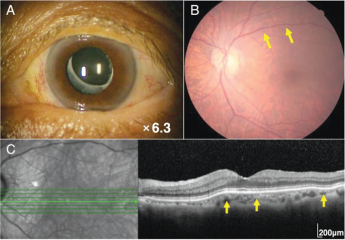
Tortuous corkscrew vessels on the left ocular surface (A). Fundus photograph showing retinal venous dilatation and vessel tortuosity in the left eye (B, yellow arrows). Spectral domain optical coherence tomography showing the choroidal folds in the left eye (C, yellow arrows).

Axial view showing swelling of the left medial, inferior, and superior rectus muscles (A, yellow arrows). Coronal view showing enlargement of the left superior ophthalmic vein (B, yellow arrow).

The left-sided carotid cavernous fistula (CCF) (A, B, red arrows) is supplied by the right external carotid artery (A, yellow arrow) and the right internal carotid artery (B, yellow arrow). After coil embolization of the left CCF (C, red arrow), blood flow from the right external carotid artery (D, yellow arrow) and the right internal carotid artery (E, yellow arrow) is undetectable (D, E, red arrows).
One month after embolization of the carotid cavernous fistula, tortuous corkscrew blood vessels on the left ocular surface are significantly reduced (A). Six months after embolization, retinal venous dilatation and vessel tortuosity are improved (B, yellow arrows). Six months after embolization, the choroidal folds are also improved (C, yellow arrows).
Similar articles
-
Worsening angle closure glaucoma and choroidal detachments subsequent to closure of a carotid cavernous fistula.BMC Ophthalmol. 2012 Jul 28;12:28. doi: 10.1186/1471-2415-12-28. BMC Ophthalmol. 2012. PMID: 22839357 Free PMC article.
-
Subfoveal choroidal thickness changes in carotid cavernous fistula following spontaneous resolution.BMC Ophthalmol. 2016 May 26;16:63. doi: 10.1186/s12886-016-0240-2. BMC Ophthalmol. 2016. PMID: 27230080 Free PMC article.
-
Choroidal detachment and dural carotid-cavernous sinus fistula--case report.Neurol Med Chir (Tokyo). 1997 Jun;37(6):459-63. doi: 10.2176/nmc.37.459. Neurol Med Chir (Tokyo). 1997. PMID: 9232097
-
Alteration of choroidal thickness in a case of carotid cavernous fistula: a case report and a review of the literature.BMC Ophthalmol. 2013 Dec 5;13:75. doi: 10.1186/1471-2415-13-75. BMC Ophthalmol. 2013. PMID: 24308366 Free PMC article. Review.
-
Spontaneous carotid-cavernous fistula supplied by the contralateral meningohypophyseal trunk: case report and literature review.J Neurosurg Sci. 2010 Mar;54(1):45-8. J Neurosurg Sci. 2010. PMID: 20436398 Review.
References
-
- Phelps CD, Thompson HS, Ossoinig KC. The diagnosis and prognosis of atypical carotid-cavernous fistula (red-eyed shunt syndrome) Am J Ophthalmol. 1982;93(4):423–436. - PubMed
-
- Ellis JA, Goldstein H, Connolly ES, Jr, Meyers PM. Carotid-cavernous fistulas. Neurosurg Focus. 2012;32(5):E9. - PubMed
-
- Barrow DL, Spector RH, Braun IF, Landman JA, Tindall SC, Tindall GT. Classification and treatment of spontaneous carotid-cavernous sinus fistulas. J Neurosurg. 1985;62(2):248–256. - PubMed
LinkOut - more resources
Full Text Sources